Note: Descriptions are shown in the official language in which they were submitted.
1 333528
-- 1 --
The present invention relates to a fiber optic
probe for measuring reflectance spectrum. More
specifically, the present invention relates to a reflected
spectrum measuring probe for measuring hemodynamics and
oxygen sufficiency such as volume of hemoglobin and oxygen
saturation in blood near the surface of a living body and
accordingly the diagnose state of organs such as liver by
irradiating the surface of a living body with light and by
analyzing the light transmitted through and reflected from
the organism in accordance with spectrophotometry.
A fiber optic probe for measuring the reflectance
spectrum has been used for analyzing the state of pigments
in the surface layer of a living body by measuring the
light irradiating and reflected from the surface of the
organism. Such fiber optic probe comprises a fiber bundle
including light illuminating optical fibers and light
receiving optical fibers in which a plurality of optical
fibers formed of transparent materials such as plastic or
glass are bundled in parallel. The fiber bundle is
enclosed by a cover on the outer periphery of the bundle.
Such a fiber bundle used as a measuring probe is known
from, for example, Japanese Utility Model Laying Open No.
59-113749.
The optical fiber generally comprises a core and
a jacket covering the core, with the diameter of the core
being several 10 ~m and the diameter of the jacket or
cladding being about 150 ~m.
The above mentioned Japanese Utility Model Laying
Open No. 59-113749 discloses a metal sleeve holder provided
at the outermost periphery so as to protect the probe and
to facilitate the use of the probe. However, what is
disclosed in that reference is a number of common bundled
optical fibers, each fiber consisting of a core and a
cladding. The diameter of the cladding is about 100 ~m and
the diameter of the core is about several 10 ~m, in a
generally used plastic fiber or a glass fiber.
~. ,
l't~
1 333528
The invention will now be described with
reference to the accompanying drawings in which:
Fig. 1 is vertical sectional view showing a
portion of a conventional probe on an enlarged scale;
Fig. 2 shows a reflected spectrum with the
distortion, measured by the conventional probe of Fig. 1;
Fig. 3 shows the relationship between the blood
volume and the optical density in a sample area;
Figs. 4 and 5 are vertical sectional views of one
embodiment of the present invention;
Fig. 6 is a vertical sectional view showing one
embodiment of the present invention during use;
Fig. 7 is a cross sectional view showing an end
portion of the probe in accordance with another embodiment
of the present invention; and
Fig. 8 is a perspective view showing the whole
structure of another embodiment of the present invention.
Fig. 1 is an enlarged view of a portion of a
conventional probe. Fig. 2 shows a reflectance spectrum
with distortions measured by the conventional probe of Fig.
1. Fig. 3 is a graph showing the relationship between the
blood volume and the optical density in a sample area.
The conventional probe shown in Fig. 1 includes
an illuminating light supplying optical fiber 1 and a light
receiving optical fiber 2 are arranged next to each other.
The illuminating light supplying optical fiber 1 comprises
a core la and a jacket or cladding lb, while the light
receiving optical fiber 2 comprises a core 2a and a jacket
or cladding 2b. Each of the cores la and 2a has a diameter
of about several 10 ~m and each of the jackets lb and 2b
has a diameter of about 100 ~m. Therefore, the distance e
between the centers of the illuminating light supply
optical fiber 1 and the light receiving optical fiber 2 is
about 100 ~m.
Now, when a portion of a tissue such as a liver
containing much blood, is to be measured by the
~, ,
,, ,
1 333528
conventional probe, the reflectance spectrum measured
should be as shown by the dotted line in Fig. 2. However,
actually the measured result is as shown by the solid line.
The reason for this is as follows. As shown by (1) in Fig.
3, the optical density linearly increases as the blood
volume increases. When the wavelength of the illuminating
light becomes shorter, the effect of scattering increases,
so that the increase of the optical density becomes non-
linear and is distorted as shown at (2) in Fig. 3. Such a
measuring probe is not suitable for measuring organ
functions, for example of the liver containing much blood.
It is an object of the present invention to
provide a reflectance spectrum measuring a probe capable of
the measuring condition of blood and the like with the
distortion in the optical density reduced as much as
possible.
Briefly stated, in the present invention, an
illuminating light supply fiber for transmitting light and
a light receiving fiber for receiving the light transmitted
through an organism to be tested are arranged in parallel
to form a fiber bundle, and a lens is provided over the tip
end surfaces of the light supply fiber and the light
receiving fiber.
Therefore, in accordance with the present
invention, the depth of incident light transmitted through
the living tissue, and reflected can be made shallower, so
that the measurement can be carried out with little
influence of the blood volume, whereby organs such as a
liver containing much blood, can be tested without
distortion. In addition, by providing a number of lenses
having different focal length to be attached and detached,
various tissue portions or blood in different organs having
different blood volumes can be directly measured.
In a preferred embodiment of the present
invention, the fiber bundle is covered with a coating layer
made of a heat shrinkable tube and the periphery of the
....
1 333528
-
lens is covered by an end portion of the coating layer.
Consequently, according to a preferred embodiment
of the present invention, the fiber bundle and the lens can
be as an integral component.
In another aspect of the present invention, a
plurality of illuminating light supply optical fiber cores
and a plurality of light receiving optical fiber cores are
arranged next to each other to form a matrix in a jacket,
thereby providing an image fiber device. Therefore, in
accordance with the present invention, the distance between
the centers of the illuminating light supplying fiber core
of the light receiving fiber core can be made smaller and
therefore the distortion in the reflected spectrum caused
by the scattering effect can be avoided, whereby organs
holding much blood, as the liver, can be tested by
spectrophotometry with a high reliability.
It has been found that the optical density, which
should increase linearly as shown by the line (1) in Fig.
3, as the volume of blood increases, does not increase
linearly due to a scattering effect when the wavelength
becomes shorter, as shown at (2) in Fig. 3, and that this
non-linearity is the cause of distortions in the
reflectance spectrum. The invention aims at avoiding the
effects of these distortions. More specifically the blood
volume shown in Fig. 3 is the blood volume in the sample
area of the measuring probe. Namely, when the sample area
is moved from the side (a) to (b) of Fig. 3, the
relationship between the volume of blood and the optical
density corresponding to that area, becomes linear,
enabling a measurement without distortion. The depth d of
the sample area shown in Fig. 1 depends on the distance e
in Fig. 1, so that the depth d becomes shallower when the
distance e is made smaller.
The blood volume that influences the distortion,
is the blood volume in the sample area of the probe being
measured or tested. The sample area in turn depends on the
1 333528
distance e between the light transmitting optical fiber 1
and the light receiving optical fiber 2, as is apparent
from Fig. 1 showing the state of use of the conventional
measuring probe. Therefore, when the distance e is made
smaller such that the sample area is moved from (a) to (b)
in Fig. 3 showing the volume of blood, a measurement
without distortion becomes possible. More specifically, in
the present embodiment, the distance e ~ shown in Fig. 6 is
smaller than e shown in Fig. 1 and therefore the depth d'
in Fig. 6 is shallower than d in Fig. 1, whereby the
distortion caused by the large blood volume can be made
smaller.
One embodiment of the present invention will now
be described based on this understanding.
Figs. 4 and 5 are vertical sectional views of one
embodiment of the present invention. Referring to Fig. 4,
an illuminating light supplying optical fiber 1 and a light
receiving optical fiber 2 are put together in parallel to
form a fiber bundle 3 and condenser means 4 formed of, for
example, a convex lens is provided on the end surfaces of
the illuminating light supply optical fiber 1 and the light
receiving optical fiber 2 at the end portion of the fiber
bundle 3. Each of the optical fibers 1 and 2 comprises a
bundle of a plurality of fibers formed of a transparent
material such as plastic or glass. As shown in Fig. 5, the
fiber bundle 3 is inserted into a heat shrinkable
polyethylene tube so as to fix the bundle of optical fibers
1 and 2 in a common jacket 6. The tube is shrunk by heat
to form the coating jacket 6 having the thickness of 0.3
mm. The coating jacket 6 extends beyond the end of the
fiber bundle 3 so that a jacket rim 7 covers the periphery
of the lens 4. The end portion of the coating jacket 6
forming the rim 7 is bent inwardly, so as to hold the lens
4 in its position and to prevent losing the lens 4.
The lens 4 is exchangeable, enabling an
adjustment of the degree of condensing the light
.~
- 1 333528
corresponding to different organs containing different
amounts of blood. When the lens 4 is to be exchanged, the
diameter of the jacket rim 7 is dilated for removing the
lens and replacing it by another lens. The coating jacket
6 is made of a polyolefine resin such as polyethylene or
rubber tube, and the fiber bundle 3 may be jacketed by
inserting the same into the heat shrinkable tube.
Fig. 6 is a vertical sectional view showing one
embodiment of the present invention during use. Referring
to Fig. 6, the light emitted from the tip end surface of
the light transmitting optical fiber 1 is condensed by the
lens 4 and enter the living tissue 5 as shown by the arrow
in Fig. 6. The incident light is scattered and reflected
to enter the light receiving optical fiber 2 again through
the lens 4. On this occasion, the distance between the
light transmitting optical fiber 1 and the light receiving
optical fiber 2 is e ~ in Fig. 6 and the depth of the light
penetration into the living tissue is d'. The depth d' is
shallower than in the case of the conventional measuring
probe, so that the measuring probe of the present invention
is capable of measuring organs containing much blood.
The lens 4 employed in this embodiment should
preferably be planar as shown in Fig. 6 in order to enable
a close contact between the living tissue 5 and the surface
of the lens 4 and to prevent a fluctuation of the measured
values caused by a vibration of the probe. In addition, in
order to realize a uniform contact and to reduce optical
losses, the contact surface of the lens 4 with the
illuminating light supplying fiber 1 and the light
receiving fiber 2 should be planar. An example of such
lens is a collimator lens.
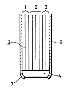
Fig. 7 is a cross section showing an end portion
of the probe in accordance with another embodiment of the
present invention. Fig. 8 is a perspective view showing
the whole structure. The embodiment shown in Fig. 7
reduces the distance e in Fig. 3 to further reduce the
1 3~3528
-- 7
depth d without providing a light condensing means.
Referring to Fig. 7, the measuring probe 10
comprises an image fiber 11. The image fiber 11 comprises
a number of cores 13 each having a diameter of several 10
~m arranged in parallel to each other to form a matrix.
All cores 13 in Fig. 7 are embedded in a common embedding
material 12 and the thickness of the embedding material
between neighboring cores is several ~m. At the tip end
portion lla of the probe shown in Fig. 8, one of the cores
is used for transmitting illuminating light and the other
one is used for receiving light. In Fig. 7, cores for
illuminating light are shown white and the cores for
receiving light are hatched so as to facilitate
understanding.
These cores 13 are divided into two groups, one
group is formed by the light transmitting cores and the
other group is formed by the light receiving cores. The
two groups are branched at an intermediate portion of the
measuring probe 10 as shown at llb and llc in Fig. 8. The
branched cores are respectively bundled, covered by jackets
and integrally arranged in a matrix in each jacket,
providing the branched image fibers llb and llc. The
branched image fiber llb is connected to a light source of
an analyzing apparatus and the branched image fiber llc is
connected to a spectrometer of the apparatus. The outer
periphery of the image fiber 11 is covered by a light
shading flexible coating 14 made of a non-transparent
material. In this embodiment, a black polyvinyl chloride
resin is used as the coating 14. A holder may be provided
on the outermost periphery of the probe tip end lla so as
to protect the probe and to facilitate handling of the
probe 10.
Assuming that the diameter of a core 13 is 30 ~m
and the embedding material 12 between cores 13 has a
thickness of 5 ~m in the sensing probe structured as
described above, the distance e 1 between the centers of the
1 333528
.
light illuminating core and the light receiving core, which
is represented by the sum of the core diameter and the
thickness of the embedding material 12 between neighboring
cores 13, can be made to be less than 40 ~m, while distance
e in the conventional probe was about 100 ~m. Therefore,
the value of the depth d described with reference to Fig.
1 is now not more than one half of the conventional probe.
Therefore, the sample area moves to the left of (b) in Fig.
3, namely, the area at which the optical density changes
linearly, free from the influence of the scattering effect.
A condensing means such as the selfoc lens 4 may
be provided at the end surface of the image fiber 11 also
in this embodiment as shown in Fig. 8.
Although the present invention has been described
and illustrated in detail, it is clearly understood that
the same is by way of illustration and example only and is
not to be taken by way of limitation, the spirit and scope
of the present invention being limited by the scope and
interpretation of the appended claims.