Note: Descriptions are shown in the official language in which they were submitted.
1333872
BGHOO I
Cancer Treatment
Brief Description of The Invention
The proce~s of perfusing a high concentration of anti-cancer a~ents
through a body organ cont~ining a tumor without cont~min~tin~ the Dody's
5 general circulation, removing them from the organ wi~h ef~luent blood,
transporting the cont~min~ted blood to an extracorporeal circuit, treating the
blood in the extracorporeal circuit to remove the cont~min~tion~ and returning
the treated blood to the body. The process prevents toxic levels of the agents
from entering the body's general circulation while delivering lethal doses of
10 them to the tumor. A variety of apparatus for effecting the intra- and extra- corporeal treatment of such cont~min~ted blood are described.
Background To The Invention
Primary cancer of the liver (hepatocellular t.lmor, hepatoma) is a
disease with a dismal prognosis due to its relentless pro~es~ion despite many
15 therapeutic modalities. Although uncommon in the United States (ap-
proximately 14,000 new cases per year), hepatoma is the most prevalent tumor
in the most populous countries of the world. It is quite common in sub-Sahara
Africa, Southeast Asia, Japan, the Pacific Islands, Greece and Italy. For those
patients not surgically resectable, median survival is approximately 8 weeks.
20 In North A~nerica, this m~ligntqncy most commonly arises in elderly patients
with alcoholic or postnecrotic cirrhosis. However, in other parts of the world,
it is epidemic and often occurs in young patients. This demographic variation
is correlated with a high incidence of early childhood infection with hepatitis B
virus in the geog;aphic areas where hepatocellular tumor is most common.
Although the incidence of primary liver cancer is not high in the
United States, cancer of the colon is a major health problem, and cancer of the
colon reaches the liver in about 50% of the patients. Over 140,000 new cases
are diagnosed yearly. Once the desease has spread, therapy is ineffective, with
approximately 50% of all patients dying from their rlice~ce within five years of
.,
1333872
BGHOOl
diagnosis. In 15% to 20% of the patients, the tumor will have spread to the
liver by the time of diagnosis, and in over 50~ of patients, colon cancer will
eventually spread to the liver me~astasis even when there is no tl:mor spread
elsewher~. Tumor cells reach the liver via the ~:~rtal ~in arld establi~h a
5 blood supply from the hepatic artery, perhaps through the elaboration of
tumor angiogenesis factor(s).
The impact of colon cancer on the liver is grim. When liver metas-
tases are diagnosed, the median survival time falls to 4-9 months without
treatment. While tumors that originate in other organs do not spread to the
10 liver as frequently, their prognosis is also significantly worsened uhen theyreach the liver. Much medical research assumes that effective treatnent of
tumors in the liver will extend survival, improve quality of life, and reduce the
financial and emotional irnpact of this desease.
It is a widely held view, and currently being acted upon, as noted
15 below, that effectiveness of chemotherapy is improved by intraarterial infu-
sion. However, systemic toxicity has limited drug tolerance. Detoxification of
blood cont~ining chemotherapeutic agents has not been developed until this
invention.
The current treatment modalities for colon tumor metastatic to the
20 liver are unsatisfactory. A solitary metastatic deposit of colon cancer is best
handled by surgical resection, which leads to a 1 year survival rate of 80% and
a 3-year survival rate of 40%. However, in 95% of the cases, multiple metas-
tatic lesions are present. Systemic chemotherapy has little lasting effect on
these metastatic lesions. Although certain drugs have shown activity in
25 various studies, when used at higher doses their effects are negated by theirsystemic toxicities. These same drugs may prove to be much more effective if
their systemic toxicities can be avoided. A treatment which exposes a tumor to
high antineoplastic drug concentrations and removes the drug from the blood
before systemic exposure occurs may be an effective therapy for cancer in the
30 liver.
-
1333~72
BGHOO~
At present surgical resection offers the only chance of cure of
h~patoma. For resection to be possible, at least one hepatic segment must be
spared. The uninvolved segment(s) of liver must be free of cirrhosis. Unfor-
tunately, the proportion of patients with potentially resectable tumors is
5 small.
Hepatic artery infusion (HAI) of chemotherapy has been widely
investigated. Arterial infusion of 5-FU and FUDR increases their effectiveness
by delivering the drug directly to liver tumor cells before its dilution by the
systemic circulation. This approach is attractive because hepatocellular
10 tumors frequently remains localized to the liver, and, like most
chemotherapeutic drugs, 5-FU display a dose-response effect, i.e., increasing
~- the dose can give a proportionately greater increase in ~ffect. Also, certain
drugs, including the fluorinated pyrimidines, doxorubicin and others, are
metabolized by the liver and excreted through the biliary tract thereby
15 reducing systemic drug toxicity.
Initially, chemotherapy was given via percutaneously placed catheters
with the use of external pumps. Response rates obtained with this form of
treatment in patients with colon cancer metastatic to the liver was generally
superior than those attained when identical drugs were given intravenously,
20 with objective responses seen in 34% to 83% of the patients. More recent
studies, employing surgically placed catheters and implanted pumps, have
yielded response rates in 50% to 60% (range 20% to 88%) of the patients with
colon cancer metastatic to the liver. The fluorinated pyrimidines (5-FU and
FUDR) are the drugs most commonb used for prolonged (over 1 to 2 weeks)
25 HAI, while Mitomycin C and other drugs have been given alone or in combina-
tion with these drugs as intermittent bolus injections into the hepatic artery.
To date, no randomized comparative studies have demonstrated that HAI
administration of fluorinated pyrimidines is therapeutically superior to
systemically ~tlminictered drug Local and systemic toxicities limit the amount
30 of therapy which can be delivered even by the arterial route.
1333372
- BGHOO 1
Local toxicities in the gastrointestinal tract h~ e included gastric
and duodenal ulceration, gastric bleeding and/or perforation, severe dyspepsia,
gastritis and diarrhea. Many of the ?atients ~ ho developed these local
toxicities were found to have had a misplaced or dislodged catheter tip. In
5 these cases, the drug was perfusing a large portion of the stomach and
duodenum via the gastric arteries. Gastrointestinal toxicities did not occur
when the gastric arteries were separated and ligated from the hepatic artery
or embolized at the time of catheter pl~celnent. Diarrhea, a systemic toxicity
of the fluorinated pyrimidines, occurs more commonly in patients with arterial
10 to venous (A-V) shunting of 30% or greater A-V shunting allow~ drug to
bypass functioning liver cells and avoid being metabolized by the liver thereby
increasing systemic drug exposure.
Local hepatobiliary toxicities (hepatitis, cholecystitis, biliary sclerosis,
stenosis and stricture) occur in up to 50% of the patients treated with conven-
15 tional HAI 5-FU or FUDR chemotherapy. The gallbladder and biliary tree
receive all of their blood supply from the hepatic artery whereas ~he liver
receives approximately one-fourth of its blood supply from the hepatic artery.
Biliary tract toxicity seems to be more common in patients who have had the
blood supply to the biliary tree disrupted by ligation of the gastric arteries.
20 Choliangiography, CT sc~nning and ~lk~line phosphatase elevations have been
shown to be effective monitoring tools for identifying patients with impending
biliary tract toxicity. Hepatitis, manifested by nausea, vomiting, abdominal
pain and jaundice in association with elevated serum concentrations of liver
tr~n~min~ces and bilirubin has occurred in patients receiving conventional
25 HAI 5-FU or FUDR. Hepatitis appears to be related to the dose and duration
of the hepatic arterial drug infusion.
Systemic toxicity of HAI chemotherapy has not been a major problem
when drugs with a high liver extraction ratio, such as 5-FU and FUDR, have
been given in conventional doses that are defined by systemic toxicity. Drugs
30 which are not substantially metabolized by the liver upon first pass often cause
1. Toxicity resulting from un;~ ti~n~ ection of drug into an arte~y other than a hepatic
artery, most often gastric or ~llo~er~nl branches.
- 4 -
1333872
- BGHOOl
system toxicities, primarily myelosuppression, when given intraarterially.
Obviously, without the use of a detoxif~ing system that removes unmetaboli-
zed drug, most drugs cannot be empl(,sed in higher, potentially more effective,
doses by the HAI route.
Systemic chemotherapy for hepatocellular tumor remains a
therapeutic challenge. Numerous agents have been tested in Phase II trials;
objective responses to therapy are uncommon. 5-FU and doxorubicin
(Adriamycin) are the only dru~s which have consistently been shown to have
significant activity. Initial reports of East African blacks treated with
doxorubicin, 75mg/m every 3 weeks, resulted in 22 patients attflinin~ an
objective response (3 complete) in 50 patients treated. Substantial toxicity
occurred with the use of doxorubicin at this dose. Hence, most other studies
report on the use of doxorubicin, 60mg/m every 3 weeks. At this dose,.
objective therapeutic responses occurred in appro~im~tely 20% of the patients.
.
Allsm~n, R. K (1961) Development of a technic for isolated perfusion
of the liver, NY State J. Med., vol. 61, p. 3993. discloses isolating the liver by
curgically separating the portion of the inferior vena cava which includes the
hepatic veins, infusing a chemotherapy agent to the liver through the splenic
and common hepatic arteries, and collecting the chemotherapy agent from the
20 isolated portion of the inferior vena cava. This reference does not disclose a
method for detoxifying blood of chemotherapeutic agent.
K Schwemmle and K Aigner, Recent Results in Cancer Research. vol.
100, pp. 229-233, pub. by Springer-Verlag, Berlin, 1986, utilized isolated
hepatic perfusion in two patients suffering from disseminated hepatic metas-
25 tases of colorectal cancer. They characterize their work, and that carried outprior to their efforts, as follows:
"Among the various treatment modalities for liver metastases such as
resection, intraarterial infusion, isolated perfusion, or chemoe~nboli7~-
tion, isolated perfusion enables chemotherapeutic agents to be added to
30 the perfusion circuit in dosages higher than could be tolerated by sys-
temic ?riministration. The upper limit of dosage is only the local toxicity.
Because hepatic metastases are mainly vascularized by the hepatic
artery, intraarterial infusion of anti-cancer agents provides a much
1333872
BGHOOI
higher concentration of the drug in these tumors than can be achieved by
systemic chemotherapy.
"To develop a method for intraarterial treatment with m~im~l doses of
chemotherapeutic drugs, we started with isolated perfusion of the liver
5 in P.nim~l experiments according to previously published methods [4-6'.
Optimal surgical techniques and drug toxicity were studied in dogs.
After these studies in ~nim.ql~ had proved that the method wa~ pr~c-
ticable and safe, in November 1981 we performed an isolAted h~l)atjc
perfusion in two patients suffering from disseminated hepatic met&s-
10 tases of colorectal cancer ~1]. After these two patients had survived 5months without complication, another 38 patients were submitted to
isolated hyperthermic perfusion of the liver with chemotherapeutic~ [2].
"In the isolated perfusion circuit both the hepatic artery and portal vein
are perfused and the hepatic venous return is collected via a single
15 venous line. During isolated perfusion a portocaval shunt is established
in which arnmonium ic filtered out of the portal blood (Fig. 1). Recentl~,
we have omitted the filtration unit.
"During the operative procedure through an abdominal midline incision
the liver, the hepatoduodenal li~ament, and the inferior caval vein are
20 exposed. Tourniquets are placed around the gastroduodenal arterv and
portal vein and around the caval vein below and above the renal veins as
well as intrapericardially.
"In order to collect the hepatic venous outflow a double-channel catheter
is inserted into the caval vein from below the renal veins. This special
25 catheter consists of a longer channel shunting the caval vein to maintain
cardiac venous return and a second shorter channel for the isolated
hepatic venous return. The portocaval shunt tube is inserted into the
caval vein channel, whereas two lateral openings collect the venous
return from the kidneys.
30 "After the perfusion catheter is inserted into the caval vein the portal
vein is cannulated in both directions. The peripheral catheter is con-
nected to the portocaval filtration unit consisting of a roller pump and a
hemofiltrating system. At flow rates of approximately 300-400 ml portal
venous blood has been filtered and with adequate volume substitution
35 returned to the caval shunt tube. Thus blood levels of ammoniurn have
been kept within normal ranges during the period of isolsted hepatic
perfusion. In case of leakage to the systemic circulation a part o~ the
anti-cancer drugs as well has been filtered out in that portacaval shunt.
As soon as the shunt is established, the common ~epatic artery is
40 clamped and perfusion is started via the central portal vem catheter in a
partial circuit. Then the arterial catheter is inserted into the
gastroduodenal artery and the liver is perfused via two arterial lines at a
flow rate of 200-350 ml/min in the hepatic artery and 150 ml/min in the
portal vein. Heating the perfusion circuit the temperature of the hepatic
45 tissue is increased. The temperature, which should not exceed 40C, is
measured with needle probes in the right and left liver lobes. In our first
31 isolated liver perfusions we only applied 5-fluorouracil (5-FU) in a
13338~2
BGHOO~
dosage between 600 and 1000 mg. In the last ei~..t patients a combina-
tior. consisting of mito!nycin C ~;nd 5-FU was used.
"Few com lications occurred after iso~ted liver perfusion. Three pa-
tients die~ a short tim~ after the operation. One patient died 2 hours
5 later from untreatable bleedin~ a~ter an isols~ed hepatic perfusion
combined with a hemihepatectomy. Th~ tumor had already infiltrated
the caval vein. In another patient sep-icemia and respir~.tory distr~ss
occurred 6 davs after the ~erfusion. The third patient died from ren~l
failure 2 weeks after the perfusion. His autopsv showed a 90% regres-
10 sion of the tumor Seventy percent of the hepatic tissue in thiC case hadbeerl involved in rnetastases. In the last 30 patients there were no fatal
outcomes.
"In spite of the increased survival time unfortunately in the combined
group the patients developed extrahepatic metastases after the treat-
15 ment. In ten cases (83%) metastases of the lung developed. Half of thepatients developed peritoneal carcinosis or at least positive Iymph nodes
at the hepatoduodenal ligament and in three cases (25%) recurrences at
the colorectal anastomoses or at the pcrineal scar developed."
"In conclusion we see the following advantages of liver perfusion:
20 1. We are able to use a high anti-cancer dosage which cannot be achieved
by other methods, for example, intermittent or continuous intraarterial
infusion.
2. The ~flministration of the anti-cancer agents is performed via both
the hepatic arteIy and the portal vein.
25 3. A combination with hyperthermia is given.
~The most important disadvantage of this method is the fact
that the perfusion cannot be repeated. However, it is possible
at any time to continue the therapy with intraarterial infusions
and/or with chemoembolization."
(Emphasis supplied)
The following publications relate generally to perfusing individual
organs with chemotherapy agents: Creech et al. , Healy, Healy, et al.
2. Annals of Surr~en, ~rol. 148, DO. 4, pp. 616-632 (October 1958), note summary at page 632:
'Ch~rnntl~Prapy of canoer has not been entirely ~ fr tory bccause the a~3mir~ie~ration of
doses large enough to ci~nifirnntly affect a tumor protuce serious to~ic ef~ects on the bone
marrow and ga_trointest~nal tract.~
3. Sur~en-~ynecolo~r Obstetrics, vol. 120, no. 6, pp. 1187-1193 (June, 1965)
4. ~,, vol. I, no. 2, pp. 111-116 (July, 1961) (deals sl,e :r,~ y with liver i~
- 7 -
133387~
BGH001
Pierpont et al. , and Shingleton, et al. None discloses detoxi~ying blood
which has passed through an isolated organ and returning the detoxified blood
to the patient.
The following publications disclose applying a chemotherapy agent t~
5 a specific organ, collecting blood generally from the patient, detoxifying theblood, and returning the blood to the patient: Kamidono, et al., and Agishi
Krementz, Cancer. vol. 57, no. 3, pp. 416 - 432 (1986), reviewed the
development of regional chemotherapy by perfusion. He stated the following
re~arding liver perfusion performed surgically in an ~tnim~tl:
10 "Techniques for perfusion of the liver have been complicated, and re-
present major abdominal ~-lr~y. Our techniques, developed for the
experimental ztnim~l and applicable to patients, mvolved isolation of the
liver by p~t~-cin~ a Foley balloon catheter (Bard Urological Division,
Murray Hill, NJ) with a ligated tip through the vena cava from the
15 femoral vein to a point proximal to the hepatic veins. The vena cava was
occluded above by the balloon that was positioned above the diaphra~rn
and below the hepatic veins by a snare placed above the renal veins (Fig.
7), and the hepatic vein drainage was returned to the pump reservoir
through the catheter. The hepatic artery was temporarily clamped, and
20 oxygenated blood, and oxygenated blood from the pump and the
chemotherapeutic agents were delivered to the liver through the pro-
ximal portal vein. Blood from the distal portal vein and vena cava was
returned to the heart through an stccessory bypass from the femoral vein
to the external jugular vein. We did not persist in our efforts to use
25 hepatic perfusion clinically, but other investigators have separatelY
developed techniques for perfusion of human livers. The approach
developed by Ai~ner and colleagues uses a double-lumen tube to collect
hepatic venous blood i~ the outer tube, with bypass of the distal caval
blood through the i~er tube. Arterial blood and chemotherapy are
5. J. Thoracic and Cardiovas. Sur~.. vol. 39, no. 2. pp. 159-165 (February, 1960)
6. Annals of Sureen, vol. 152, no. 4, pp. 583-593 (October, 1960)
7. The Journal of Urolonr, vol. 131, pp. 3640 (1984) ant Inv~ ;Y9~ UroloYv. vol. 19, No. 3,
pp. 176- 178
8. Said to have utilized ~selective delivery of the ~nt j~nr~. drug is achieved by perc~ttsnPo~
injection of 1 mg/kg of mitomycin C into a drug-cbamber of an ~y_.dti._l~ impj9nted
vascular access port, another end of which is insertet in the feeding artery of the
cancer-bearing organ.~
~Removal of the drug is performed by the usual method of charcoal hc.,.o~y~.ru;~io~ Dr.
Agishi stated. ~However, in order to augment the anticancer erfect, local hyperthermia is
fftAbli~l~ed utilizing a radiur~ .,./ wave - 13.56 MHz . emission ..pyar~.tus.~ (Miles
Pharm- .'ti~91 Oncology News Update, 1987)
-8-
1333872 BGHool
delivercd through the hepatic artelg and proximal protal ~sic: portal]
vein to the liver. This method is under study, particularly iri Germany,
and has been performed safely with acceptable mor~idit~."
Double balloon catheters in general are described in the following
5 references:
Weikl, et al., U.S. 4,573,966, patented ~iarch 4, 1986, and U.S. 4,610,662,
patented September 9, 1986, describe thi.~ use of double balloon
catheters to treat stenosis;
Solar, U.S. 4,546,759, patented October 15, 1985, is directed to a triple
balloon catheter to assist right ventricle functioning,
Hussein, et al., U.S. 4,445,892, patented May 1, 1984, relate to a dual
balloon catheter for insertion in blood vessels to provide an isolated
operating region in the vessel between the balloons which facilitates
the use of an optic system;
Baran, et al., U.S. 4,423,725, patented January 3, 1984, describe a
multiple surgical cuff alleged to have a variety of uses.
Betancourt, U.S. 4,180,076, December 25, 1979, describes a nasogastric
catheter contS inin~ two infl~tsthle vessels.
German Offenle~ln~schrift 28 34 956 and R~l~si~n Patents 651817 and
511951 describe the use of double balloon catheters for use in isolating
the liver for the purpose of blocking blood flow from the liver. The
catheters are provided with a bypass to allow blood flow to continue
through the ~ 9. Rn~si~n patent 511951 describes the use of a
perforated wall catheter for removing blood from the liver and isolating
it via a pump, and with respect to the perfusior of the liver with
medicants and coolants, the perfusate i9 collected and returned to the
liver via a pump.
1333872
BGHOOl
Implantable pumps have recentl~ ~olne into vogue. However, studies
have indicated that a large proportion of the patients developed toxicity due tothe systemic effects of chemotherapy.
In snmm~ry, chemotherapy has not made a dramatic impact on the
5 treatment of primary or metastuti^ liver cancer. Certain drugs and biologicalshave shown considerable activity in various studies, but their effects are
negated by systemic toxicity. Some of these may prove to be much more
ef~ective if their systemic toxicity can be eliminated.
A treatment which exposes tumors to high concentrations of antineop-
10 lastic drugs and biologicals and removes them from the blood before systemicexposure would be an advancein therapy for cancer in the liver. Moreover, it
would be desirable to have a method which allows the opportunity for explor-
ing HAI therapy with a variety of drugs and biologicals at dosage levels higher
than ever before found tolerable by the body A process which allows the
15 variations in the kind and dosage of chemotherapuetic agents to livers would
be a significant advance in the treatment of such cancers. A process that does
not require general anesthesia or surgery, an.l is suf~lciently non-invasive to
allow frequent repetition of therapy would be a significant advance in the art.
There is described herein a process which provides such advantages.
.
The Invention
This invention relates to a process of perfusing a high concentration of
anti-cancer agents through a body organ cont~ining a tumor without con-
t~min~ting the body's general circulation, removing them from the organ with
effluent blood, transporting the cont~rnin~ted blood to an extracorporeal
25 circuit, treating the blood in the extracorporeal circuit to remove the con-
t~min~tion, and returning the treated blood to the body. The process prevents
toxic levels of the agents from entering the body's general circ~ tion while
delivering lethal doses of them to the tumor. A variety of apparatus for
ef~ecting the intra- and extracorporeal treatment of such cont~min~ted blood
30 are described.
- 10 -
1333872
BGNOOI
The process of the invention embraces a system of non-operative an~
sufficiently non-invasive intracorporeal and extracorporeal means to allow
frequent repetition of therapy, if desired, which comprises
perfusing~lan anti-cancer agent to a tumor,
5 collecting and cont~ining the cont~min~ted blood emanating from the
tumor without general circulation of the cont~min~ted blood to the
body,
transporting the contQ~nin~teA blood from the body to an extracorporeal
treatment system,
10 removing anti-cancer agent from the blood in the extracorporeal treat-
ment system, and
returning the treated blood to the body.
There is described a technique by which anti-cancer agents, such as
chemotherapeutic agents, can be removed from the hepatic venous blood
15 before entering the systemic circulation. This permits safe infusion of greater
than usual concentrations of anti-cancer agents, such as cytotoxic levels of
chemotherapeutic agents, into the hepatic artery for treatment of turnors of
the liver. However, the invention in its broadest sense, allows the treatment
of a variety of tumor-bearing organs with anti-cancer agents, such as
20 chemotherapeutic agents, while avoiding systemic toxicity.
Tbe invention encornp~ses a process of treating organ site tumors
which comprises
a. exposing a turnor in a body organ to one or more anti-cancer agents in
higher than usual concentrations,
25 b. removing from the organ effluent blood cont~min~ted with the agent
provided to the organ, without systemic exposure to the body,
1333872
BGHOOI
c. passing the efnuent blood from tributa y veins in the organ into a larger
vein in which has been provided a catheter cont~ining
i. at least one inn~t~sble balloon provided to obstruct passage of the ef-
fluent blood to the heart and
5 ii. an avenue, such a plurality of openin~ or a large opening, in the
catheter sufficient to accommodate the volume of effluent blood
traversing the tribulary veins,
d. transporting the cont~smin~ted effluent blood through the catheter and
thence from the body into an extracorporeal circuit,
10 e. detoxifying the blood in the extracorporeal circuit, and
f. returning the detoxified blood to the body.
More particularly, the invention relates to the treatment of organ site
tumors which comprises
a. exposing a tumor in a body organ to one or more anti-cancer agents such as
15 antineoplastic drug and biological response modifiers in higher than usual
concentrations,
b. removing from the organ effluent blood contsmin~sted with the agent
provided to the organ, without systemic exposure to the body,
c. p~sccing the ef~uent blood from tributa~y veins in the organ into a larger
20 vein in which has been provided a catheter cont~ining
i. spaced-apart inflstLs.hle balloons provided to obstruct the large vein
above and below said tributary veins and
1 3 3 3 8 7 2 BGHOOl
ii. an avenue, such a plurality of openings or a large opening, in the
catheter between the balloons sufficient to accommodate the volume of
effluent blood traversing the tributaIy veins,
d. transporting the cont~min~ted effluent blood through the catheter and
5 thence from the body into an extracorporeal circuit,
e. detoxifying the blood in the extracorporeal circuit, and
f. returning the detoxified blood to the body.
In respect to the defined treatment, the extracorporeal circuit or
- treatment system comprises (a) means for transporting the cont~min~ted
10 blood to (b) means for separating the drug concentration from the blood and
also returning the decont~min~ted blood to the body.
The invention has particular importance in the antineoplastic treat-
ment of tumors of the liver since anti-cancer levels of antineoplastic agents can
be safely infused into the hepatic artery for treatment and systemic toxicity
15 can be avoided. The invention involves intraarterial infusion of the liver with
antineoplastic agents and removal of the antineoplastic agents from the
hepatic venous blood before it enters the systemic circulation. This invention
includes the percutaneous insertion of a special double balloon catheter into
the inferior vena cava The catheter contains two inflatable balloons ap~ro~-
20 riately spaced to obstruct the inferior vena cava above and below the hepaticveins. Hepatic venous blood is drawn through fenestrations in the catheter
wall and thence into an extracorporeal circuit that is openly connected with
the catheter's lumen. The blood is decont~min~ted in this circuit and then
returned to the systemic circulation via either a subclavian vein, an external
25 jugular veins, the superior vena cava or the right atrium.
The invention includes the use of detoyifi~qtion means, such as one or
more of: a hemoperfusion cartridge, hemodialysis, hemofiltration, and
hemoadsorbtion through antibodies or biological ligands or molecules able to
render them nontoxic and/or to clear the blood of the antineoplastic agent and
1333872 71746-4
allow the re-administration of the patient's own detoxified blood.
The invention embraces the passage of the contaminated blood from
the double balloon catheter through tubing into a pump that
assists the passage of the contaminated blood to a detoxification
means such as a hemoperfusion cartridge containing one of a
variety of substances, such as a sorbing solid, and/or a
hemodialysis unit that removes the drug from the blood. The
treated blood is returned to the body via an appropriate large
caliber vein.
The invention also includes a disposable kit, that may
be used, e.g., for inpatient hospital use with cancer patients,
comprising a double balloon catheter, a detoxification means,
piping and valves. The kit may include for guidewires, heparin,
and other related equipment.
The invention encompasses a double balloon catheter of
a. being percutaneously inserted into the inferior vena
cava,
b. closing off the flow of contaminated blood from the
hepatic veins, and
c. recovering the contaminated blood from the hepatic
veins.
A variety of novel catheters are described that may be
used for the recovery of contaminated blood derived from an organ
containing a tumor that has been perfused with a anti-cancer agent
for its treatment, and for the removal of the contaminated blood
from the body so that the blood can be detoxified outside of the
body.
14
133~872 71746-4
In one embodiment, the present invention provides a
catheter adapted for percutaneous insertion into a vein or artery,
comprising: a plastic tube defining a main lumen for outflowing
blood, two balloons, fixedly spaced apart about said plastic tube
and bonded thereto for inflation thereabout, one being contiguous
to the cranial end of said plastic tube, and said balloons, when
inflated, having a size sufficient to block the flow of blood in a
vein or artery into which said catheter is designed to be
inserted; fenestrations in said plastic tube between said balloons
to said main lumen; and second and third lumina within said
plastic tube, said second lumen connecting to the caudal of said
balloons and said third lumen connecting to the cranial of said
balloons for effecting inflation or deflation of said balloons,
the cranial end of said plastic tube being closed to any
appreciable inflow of blood.
In another embodiment the present invention provides a
kit for removing a treating agent from blood perfused through a
body organ of a patient, comprising: a catheter for isolating and
removing the blood issuing from said body organ and containing
said treating agent; a detoxification means for treating said
blood so removed to remove said treating agent; and return
catheter means for returning the blood so treated to said patient;
said catheter comprising: a plastic tube defining a main lumen
for outflowing blood, two balloons, fixedly spaced apart about
said plastic tube and bonded thereto for inflation thereabout, one
being contiguous to the cranial end of said plastic tube and said
balloons, when inflated, having a size sufficient to block the
. 14a
,~`
~3338~2 71746-4
flow of blood in a vein into which said catheter is designed to be
inserted; fenestrations in said plastic tube between said balloons
to said lumen and second and third lumina within said plastic
tube, said second lumen connecting to the caudal of said balloons
and said third lumen connecting to the cranial of said balloons
for effecting inflation or deflation of said balloons, the cranial
end of said plastic tube being closed to any appreciable inflow of
blood.
In yet a further embodiment, the present invention
provides a process for removing a treating agent from blood
perfused through a body organ of a patient comprising:
isolating blood issuing from said body organ with a catheter;
removing said blood containing said treating agent from said
patient through said catheter; detoxifying the blood so removed to
remove said treating agent; and returning the blood so detoxified
to the patient; said catheter comprising: a plastic tube defining
a main lumen for outflowing blood, two balloons, fixedly spaced
apart about said plastic tube and bonded thereto for inflation
thereabout, one being contiguous to the cranial end of said
plastic tube, and said balloons when inflated having a size
sufficient to block the flow of blood in a vein into which said
catheter is designed to be inserted; fenestrations in said plastic
tube between said balloons to said main lumen and second and third
lumina within said plastic tube, said second lumen connecting to
the caudal of said balloons and said third lumen connecting to the
cranial of said balloons for effecting inflation or deflation of
said balloons, the cranial end of said plastic tube being closed
14b
133387~
14c - 71746-4
to any appreclable inflow of blood.
In yet another embodiment the present invention provides
an apparatus for perfusing a hlgh concentration of an agent
through a body organ requiring treatment without contaminating the
body's general circulation, said apparatus comprising
- means for removing the agent from the organ with effluent
blood;
- means for transporting the contaminated blood to an
extracorporeal circuit;
- means for treating the blood in the extracorporeal circuit
to remove the contaminatlon; and
- meahs to return the treated blood to the body.
In a further embodlment the present invention provides
apparatus for isolatlon and treatment of a portion of the body
comprising a first catheter adapted for percutaneous insertion
into a vein or artery, comprising (a) a plastlc tube havlng a
cranlal end and a caudal end, sald plastlc tube defining a maln
lumen for outflowing blood; two balloons, fixedly spaced apart
about said plastic tube and bonded thereto for inflation
thereabout, one being contiguous to said cranlal end, and sald
balloons, when inflated, havlng a slze sufflclent to block the
flow of blood in a vein or artery into which said catheter is
designed to be lnserted; fenestratlons ln sald plastlc tube
between said balloons to sald main lumen; second and third lumlna
withln sald plastlc tube, sald second lumen connectlng to one of
sald balloons and sald third lumen connecting to the other of said
balloons for effecting inflation or deflation of said balloons,
said cranial end of said plastic tube being closed to any
14d 13338~ 2 71746-4
appreclable lnflow of blood and (b) a second catheter for
returning blood removed through sald maln lumen to the patlent.
Also withln the scope of the present inventlon ls a use
of the apparatus of the present invention to treat a body organ
without contaminating the body's general circulation.
Brief Description of the Drawlng
Flgure 1 shows a diagrammatic and schematic view of
signlflcant apparatus features ln relatlonshlp to the body for
carrylng out the process of the inventlon.
Flgure 2 shows a diagrammatlc and seml-schematlc vlew of
an apparatus assembly for carrylng out the process of the
lnventlon.
1333872
~:;HOOl
rigure 3 shows a partial cross-se^tional side view of one design of a
double balloon cathete. useful in the process of the invention.
Figure 4 shows a cross-sectional end view of th~ shaft of the double
balloon catheter of Figure 3.
Figure 5 shows a cross-sectional end view of the midsection of a
modification of the double balloon catheter of Figure 3.
Figure 6 shows a partial cross-sectional side view of another design of
double balloon catheter useful in the process of the invention.
Figure 7 shows a cross-sectional end view of the shaft of the double
10 balloon catheter of Figure 6.
Figure 8 shows a cutaway cross-sectional side view of the interior of a
double balloon catheter encompassed by the invention.
Details Of The Invention
The process of the invention avoids the use of surgery to isolate the
15 flow of cont~min~ted blood and returns the same~~lood but in a more purified
condition to the patient. As a result, the process of the ir:vention may be usedfor extended periods of time, indeed, for periods of time far longer than
previously used in the treatment of the same tumors.
The process of the invention is applicable to the treatment of a
20 number of tumors such as those of the kidney, liver, pancreas, bladder and
pelvis. Primary and metastatic liver tumors are especially tre~t~ble by the
process of this invention. As pointed out above, many of them have shown
responsiveness to chemotherapeutic drugs.
In the most preferred case, the invention is directed to the treatment
25 of tumors in the liver by the use of one or more antineoplastic agents, such as
chemotherap .etic agents and/or biologicals, and the purification of venous
- 16-
1333872
BGHOO~
blood from the liver to avoid systemic circulation of the agent(s). This may
involve the use of balloon catheters that are suitable for insertion in the
inferior vena cava to isolate venous outflow from the liver and permit the
removal of blood cont~min~ted with antineoplastic agent from the body with a
5 pump. The cont~min~ted blood will be filtered through detoxifying means and
then returned to the patient via a large caliber systemic vein at a point above
the diaphragm. Because primary hepatocellular and metastatic hepatic
tumors derive their blood supply from the hepatic artery, the tumor will be
perfused by high concentrations of, for example, a chemotherapeutic agent
10 such as 5-FU or biologicals. Because a normal liver receives three-fourths ofits blood supply from the portal vein the drug or biological will be diluted by a
factor of about three before it reaches normal, uninvolved liver cells, thereby
protecting them against hepatotoxicity.
r
As noted above, si~nific~nt elements of this process have been tested
15 in man. In a manner, each component of the system (HAI arterial catheter
infusion of chemotherapy, hemodynamic isolation of the hepatic veins by
balloon catheters, hemofiltration of ~-FU, and subclavian vein insertion) has
been used in h-lm~nc. However, an extracorporeal drug removal approach
with hepatic isolation has only been performed by laparotomy.
The Catheters
As mentioned above, the ~rocess of the invention involves the per-
cutaneous placement of unique double balloon catheter desi~ns. These
catheters may use a variety of designs and sizes depending on the organ whose
veins are isolated and the sizes and locations of it's veins. The primary
25 function of the double balloon catheter is to isolate the flow of blood from the
veins carrying the effluent blood from the organ cont~ining the tumor that is
under treatment. Venous isolation precludes systemic perfusion of the
cont~min~ted blood. Thus the tip of the double balloon catheter is to be placed
in the body so that the venous effluent from the organ being treated is pre-
30 vented from flowing to the heart. The space between the two balloons ispredetermined to ensure removing the full quantity of cont~rnin~ted blood
from the treated organ. The space between the balloons is large enough that
- 16-
13~3872
BGHOO 1
the balloon central in position can be located in a position in the most centraldraining vein to block cont~min~ted venous blood flow to the h~ ~rt and the
balloon peripheral in position can be located; ~ripheral in the most central
draining vein to block the flow of uncont~min~ed blood to the cont~min~ted
5 venous blood flow. Veins from organs not under treatment can enter the
segment between balloons without detrimental efrect as long as the ~ump and
filtration system can accommodate the additional volume. The ~ cnous
anatomy of the organ under treatment or of adjacent organs can be altered
where necessary by obstruction using angiographic embolisation or ablation
10 techniques and materials, including detachable balloons or stainless steel coils.
The lumen of the catheter between the balloons is openly connected,
or can be made openly connected, to the surrounding vein In addition,- the
same lumen of the catheter is also openly connected, or can be made openly
connected, to the extracorporeal circuit, thereby providing free flow of the
15 cont~min~ted blood from the veins to the extracorporeal circuit. Thus the
catheter has a main lumen to act as a conduit for the cont~min~ted blood flow
from the venous effluent(s) to the extracorporeal circuit.
The size of the main lumen is determined by the material of which it
is made, the volume of blood to be transported through it and the diarneter of
20 the vein in which it will be located. The main lumen may be an open annulus
or semi-annulus located within the peripheral balloon that is openly colmected
to the extracorporeal circuit. In this type of catheter, a central rod or rodlike
axis is provided for support for the balloons.
The catheter may also have supplemental l~min~ The supplemental
25 lumina are smaller in size, i.e., in diameter or cross-sectional area, than the
main lumen. They may serve any of a number of ancillary functions in the
process. For example, in one lesiFn, a supplemental lumen courses through
the full length of the catheter for the purpose of accommodating a guidewire
that is desirable for percutaneous insertion of the catheter. Each balloon may
30 be provided with a supplemental lumina to be used for its inflation, or one
supplemental lumen may be used for supplying nuid for the inflation of both
balloons. A~ additional supplemental lumen may be provided for connection
1333872
BGHOOI
to a pressure monitor to continuously measure the pressure of the venous
emuent. This lumen can also be used to inject contrast medium, if provided
with a connector that can accommodate an injection device. In some ~1esign~,
the main lumen may be used for one or more of the above functions. This
5 multifunctionality can serve to reduce the cost in m~king the catheter and
simplify the apparatus. The main and/or supplemental lumina may be made
from separate tubing threaded into the catheter or from channels molded into
the structure of the catheter. Another supplemental lumen can be used to
return detoxified blood to the general circulation and avoid puncture of
10 another vein.
The wall(s) of the segment of the catheter between the balloons
is/are provided with fenestrations to allow entry of venous blood into the main
lumen. The number, shape and size of the fenestrations may vary according to.
the size of the catheter, the rate and volume of blood they must transmit, and
15 the materials of construction of the catheter. The shape and size of the
fenestrations should take into consideration turbulence effects as the blood
courses though the fenestrations and into the main lumen. Fenestrations that
are too small can elevate hepatic sinusoidal pressure and fenestrations that aretoo large may weaken the catheter walls and compromise the integrity of the
20 catheter.
One practical double balloon catheter design would have one large
central lumen, 2 smaller lumina and 2 inflatable balloons that are separated by
about 9 to 10 cm. in the length of catheter that contains perforations. The
catheter is designed to be positioned (under fluoroscopic guidance) in the
25 inferior vena cava (IVC) such that the central balloon, when inflated, occludes
the IVC just above the hepatic veins. The peripheral balloon, when inflated,
occludes the IVC just below the hepatic veins, thus isolating hepatic venous
blood from the systemic circulation. Perforations in the catheter between the
two inflated balloons convey blood through the large central catheter lumen to
30 a variable speed pump and filtering device. An inferior vena cavagram throughthe main lumen can be used to document complete obstruction of the inferior
vena cava proarim~l and distal to the hepatic veins. The effectiveness of
passage of blood from the liver through the extracorporeal circuit can be
1333872
BGHOOl
monitored by pressure measurement in the central catheter lumen. The
variable speed pump is adjusted to maintain normal hepatic vein pressure and
flow. The detoxifying means reduce the chemotherapeutic agent such as 5-FU
in the blood to nontoxic levels before the blood is returned to the ~ stemic
5 circulation.
In another design an independent return lumen courses through the
main lumen. One end to the return lumen is connected to the outlet of the
extracorporeal circuit and the other end openly outlets into a vein at a location
superior to the diaphragrn. When the double balloon catheter is located in the
10 IVC, this return lu~nen extends beyond the end of the main catheter to the
right atrium. In this construction, the return lumen consists of a separate
piece of tubing threaded inside the main lumen and through the end hole of
the catheter. The return lumen is large enough to carry the full volume of the.
blood being returned to the patient from the detoxifying apparatus. In
15 another embodiment of the invention, part of the return flow of the effectively
detoxified blood is fed through the return lumen and the remainder is
separately fed to the patient via a separate feed system, such as through a
separate catheter feed to one of the subclav~an veins, as described by
Krementz, supra.
The double balloon catheter, once properly located in the body,
extends through the skin to the outside of the body. It terrninates in a Luer
fitting and a valve cutoff such as a stopcock. The extracorporeal circuit can beseparated from the double balloon catheter and reconnected at will. When the
balloons are not inflated, blood flow through the IVC is maintained. When the
25 balloons are inflated, the blood below the peripheral balloon will find
secondary pathways to the heart.
This convenience may be duplicated on the supply side of the process,
where the chemotherapuetic agent is supplied to the arterial side of the liver,
via the hepatic artery, by the percutaneous insertion of a feed catheter to the
30 hepatic artery, leaving the tubular ending of the feed catheter in a plastic
reservoir surgically implanted just below the patient's skin and surgically tiedtherein below the skin. The plastic reser~oir contains a resealing membrane
- 19 -
1333872
BGHOOl
of a type simil~r to those used in multi-dose vials that can be percutaneously
r)enetrated from the outside of the body by one or more needles to reinitiate
the flow of chemotherapuetic agent to the diseased organ. Illustrative of such
devices is Implantof~ Drug Delivery System, sold by Burron Medical Inc., 824
5 Twelfth Avenu~ Bethlehem, PA 18018.
The double balloon catheter can be introduced into the femoral vein
using the Seldinger technique. A guidewire made of stainle~s steel is first
passed through a needle that has been inierted percutaneously into the vein.
A catheter with a single balloon is inserted over the guidewire and the balloon
10 is innated to dilate the percutaneous tract to the diameter of the sheath that
will transmit the double balloon catheter. A plastic sheath tubing is passed
over the guidewire when the single balloon catheter is removed. After the
sheath is properly located in the vein the double balloon catheter is inserted
within the sheath and over the guidewire and advanced to the proper position
15 relative to the organ to be treated. All manipulations of the double balloon
catheter are done under fluoroscopic control. An inferior vena cavagram can
be performed prior to catheter insertion or prior to balloon inflation with the
patient lying on an opaque ruler, parallel to the IVC. The hepatic veins and
renal veins can be identified and their location determined according to the
20 opaque ruler.
, . . ~
Under fluoroscopic guidance, the catheter is positioned so that the
central balloon, when inflated, occludes the IVC just above the hepatic veins.
The peripheral balloon, when inflated, occludes the IVC just below the hepatic
veins. Dilute contrast medium such as saline solution is used to inflate the
25 balloons and reference to the ruler insures their accurate positioning.
In a specific embodiment of the invention, the double-balloon catheter
contains three lllmin~ One lumen transmits an angiographic guidewire and is
used for percutaneous insertion. A main lumen carries hepatic venous blood
from the fenestrations between the balloons to the extracorporeal circuit. The
30 third lumen terminates at the fenestrations and is used to measure pressure
or inject contrast medium. A pressure monitor, attached to this lumen,
measures pressure within the isolated segment of the vena cava before and
- 20 -
1333872
BGHOol
during balloon infl~tion The pres ure measured before balloon inflation is the
systemic venous pressure. The pressure measured after balloon inflation but
before opening the extracorporeal circuit is equal to the wedge hepatic venous
pressure, which is assumed to be equal to portal pressure. This measurement
5 can determine the presence or absence of portal hypertension. The pressure
measured after balloon inflation and during flow through the extracorporeal
circuit is the hepatic venous pressure. The hepatic venous pressure can be
monitored continuously during drug infusion. The speed of the pump in the
extracorporeal circuit can be adjusted to maintain hepatic venous pressure
10 above systemic venous pres~lre but below portal pressure. Thi~ prevents
hepatic sinusoidal congestion. The caliber of the balloon catheter and of the
tubing in the extracorporeal circuit are calculated to ensure that they are of
sufficient size to transmit the necessary volumes of blood with minim~l
resistance.
After infl~tion of the balloons, an inferior vena cavagrarn (contrast
medium injected into the inferior vena cava) is typically performed through
the double balloon catheter prior to infusion to document complete obstruction
of the vena cava proxirnal and distal to the hepatic veins and to demonstrate
the anatomy of the hepatic veins. Samples of hepatic venous blood are
20 generally aspirated through the pressure port of the double balloon catheter
immediately after the beginning of infusion, and, in the typical case, at inter-vals not to exceed one hour during infusion, and for at least three hours after
infusion, the samples are analyzed for chemotherapy agent concentrations.
Simultaneous blood samples are taken from the extracorporeal circuit after
25 detoxification and analyzed for drug concentrations in order to document the
efficiency of the detoxification means in removing the drug from the blood
before returning the blood to the systemic circulation. In addition, blood
samples are obtained from a peripheral vein to evaluate drug concentrations
re~rhing the systernic circulation. Systemic drug concentrations are then
30 measured over 24 to 48 hours following the infusion.
Another double balloon catheter design may utilize only 2 supplemen-
tal lumina and one main lumen for blood transfer to the extracorporeal circuit.
Each supplemental lumen can supply fluids to one of the balloons.
- 21 -
13~3~
BGHOO I
Th~ Pump
The venous pressure provides the pressure for ~assaFe of b, ~od to ti.e
extracorporeal circuit. The function of the pump i~ to continue the movement
of blood though the extracorF)oreal circuit and return it to the patient. Th~
5 blood is removed from the body by a combination of gravitational displacement
and the venous blood pressure. The pump does not generate a negative
pressure and pull blood from the body. The pressure of the return flow of the
blood from the extracorporeal circuit to t~.;? systemic veneous system should beless than about 300 mm Hg.
A variety of suitable pumps are comm;~rcially available. They come in
a number of designs. A preferred design is a centrifugal cardiopulmonar~r
bypass pump that utilizes smooth surface rotators without relying on rotatin~
vanes. These pumps have been used in long term support of cardiac bypass
and in liver transplants. Such designs are shown in the following U.S. Patents:
3,487,784, patented Jan. 6, 1970
Reissue 28,742, reissued Mar. 23, 1976
3,647,324, patented Mar. 7, 1972
3,864,055, patented Feb. 4, 1975
3,957,389, patented May 18,1976
3,970,408, patented July 20, 1976
4,037,984, patented July 26, 1977
Such pumps are obt~in~hle from Bio-Medicus, Inc., Minneapolis, MN
55344. This pump reacts to pressure changes automatically, and it has several
inherent safety features. The centrifuge is volume dependent and the pump
25 can decrease the flow rate if the venous drainage is interrupted. The pllmp
can also slow itself down in instances of too much resistance to flow. For
example, at a resistance of 700 mm Hg., the smooth rotator pump reduces its
output flow to zero. In the event of power failure, the pump can automatically
change over to battery power for uninterrupted pumping. This type of pump
30 does not impose a negative pressure that pulls the blood flowing from the body
and does not adversely affect the chemistry of the blood. They are herein
- 22 -
1333872
- BGHOOl
cha.~cterized as smooth rotator pumps and referred to herein and in the
claims as a "smooth rotator pump."
Another useful type of pump is the centrifugal pump such as the vsne
(impeller) desi~n sol~ by Sarns Inc/:M, Ann Arbor, MI 48106. Another type of
5 pump is the roller bearing pump. The preferred pump for practicing the
process of the invention Is the smooth rotator pump.
Blood Detoxification
The cont~min~ted blood captured by the double balloon catheter is
fed through tubing to the pump and then to a blood detoxification step. The
10 process will be successful even if the anti-cancer agent is not completely
removed from the blood. The important point is that the arnount of anti-
cancer agent in the body be kept below toxicity levels. One hundred percent
removal of any drug is seldom possible and generally not practical. The
decont~min~tion of the blood may be effected by a number of standard
15 procedures known to the art. These include the use of detoxification means,
such as a hemoperfusion cartridge and/or hemodialysis and/or hemofiltration
and/or hemoadsorbtion through antibodies or biological ligands or molecules
able to render them nonto-x-ic~ to clear the blood of the antineoplastic agent and
allow the re-arlminictration of the patient's own detoxifled blood. The detoxi-
20 fication step comprises any process by which the concentration of the anti-
cancer agent in the blood can be removed so that the blood can be returned to
the body without causing systemic to-x-icity.
Hemoperfusion involves the passage of the cont~min~ted blood over a
solid surface detoxicant particulate mass that separates the cont~min~nt by
25 sorption or by ion ex~h~n~e. A variety of these deto-x-icant particulates areknown in the art. A common one is carbon or graphite. Activated carbon is
commonly employed for this purpose. A common concern with the use of such
particulates is stated in Clark, U.S. 4,048,064, patented Sept. 13, 1977, at col.1,
lines 8-23, as follows:
- 23 -
133~872 71746-4
"Whlle the technique is initially very effective, such
prevlous attempts at hemoperfuslon have been plagued by
very high losses of white cells and platelets (cite) as
well as clotting, sludging, and channeling of blood in
the column. The column then becomes ineffective and the
patient suffers thromobocytopenia. Further, flne de-
toxicant particles tend to be released into the blood
stream to become emboll in blood vessels and organs such
as the lungs, spleen, and kidneys (cite)."
Clark describes the use of a heparln loaded polymer
coatlng of the carbon partlcles, preferably activated carbon, to
provlde a semlpermeable coating on the partlcles. A mass of the
coated partlcles ls then placed in a nylon mesh sack and the sack
is placed in a container that has an inlet on one end and an out-
let on the other. The blood is then fed to the container and
passed through the bed of coated activated carbon particles where
it penetrates the polymer coating, obtains heparln treatment and
the contamlnatlon ls removed by adsorptlon.
Winchester, et al. Clinical Toxicoloqy, 17 (4), pp.
557 - 569 (1980) describe the use of a variety of sorbents for the
detoxification by hemoperfusion of contaminated blood where the
contamlnatlon was a chemotherapeutic agent (drug). The article
shows that polymer coated and uncoated particulate detoxicants can
be used to materially reduce the contamination of the blood. Like
Clark, acrylic hydrogels were used to coat the particles. Non-
lonlc exchange reslns are also descrlbed ln the artlcle. The
authors found that a certaln pyrolized resin was an effective
adsorbant.
~ 24
133~872 71746-4
Needless to say, modiflcatlons of the separation canl-
sters suggested by Clark and Winchester, et al. wlll be dictated
by the blood flow rate and the degree of drug contamlnation. If
the blood flow rate ls higher ln the separatlon canlster contain-
ing the bed or mass of the sorbents than the particle strength of
the sorbent, then the techniques of treating the sorbents or
operating the separatlon should be altered. For example, lf the
sorbent breaks up ln the course of hemoperfuslon, then the sorbent
should be larger and the flow rates of the contamlnated blood lnto
and through the bed should be lower. Another approach to such a
problem is to eliminate any leachables such as heparin from the
polymer coating. This will make the coating more resistant to
breakup.
~ 24a
1333~72
BGH001
The pr jlem may also be helped by the use of stronger coatings which means
the u~e of, e.g., a slightly more crosslinked pol~mer than those conventionally
employed in the art for this purpose.
Hemodialysis has been previously employed .n cancer chemotherapy,
5 see Galletti, Portocaval Hemofiltration During The Anhepatic Phase In
Isolated Liver Perfusion, Trans. Amer. Soc. Artif. Int. Organs, vol. XII, pp.
20 - 24, 1966, and Winchester, et al., Dialysis and hemoperfusion of poisons
and drugs, Trans. Amer. Soc. Artif. Int. Organs, vol. XXIII, pp. 762 - 842.
Hemofiltration is a well defined technology and is characterized in a
10 number of texts. It involves the filtration from the cont~rnin~ted blood of the
antineoplastic agent through membrane walls. -Details of the process and the
apparatus used in effecting the process are described in inter alias Malchesky,
Membrane Plasma Separation: Critical Issues, Therapeutic Apheresis: A
Critical Look, edited by Y. Nose', P. S. Malchesky, ~- ld J. W. Smith, ISAO Press,
15 No. 304, pp. 93 - 101, Cleveland, Ohio U.S.A., 1984; Vassilieff, et al., Plas-
mapheresis Between a Rotating Truncated Cone and a Microporous Plate,
Therapeutic Apheresis: A Critical Look, edited by Y. Nos~, P. S. Malchesky, and
J. W. Smith, ISAO Press, No. 304, pp. 102 - 114, Cleveland, Ohio U.S.A., 1984;
R~ff, et al., Influence of Geometric Parameters on Filtration Flux in Plasma
20 Filters, Therapeutic Apheresis: A Critical L,ookj-edited by Y. Nose, P. S.
Malchesky, and J. W. Smith, ISAO Press, No. 304, pp. 115 - 121, Cleveland,
Ohio U.S.A., 1984; Koga, et al., Investigation of the Clinical Properties of
Various Filters for Double and Triple Filtration Plasmapheresis, Therapeutic
A~heresis: A Critical Look, edited by Y. Nose', P. S. Malchesky, and J. W. Smith,
25 ISAO Press, No. 304, pp. 171 - 175, Cleveland, Ohio U.S.A., 1984; Tani, et al.,
New Anticancer Treatment by Hemoperfusion with Endotoxin Immobilized
Fiber, Therapeutic Apheresis: A Critical Look, edited by Y. Nos~, P. S.
Malchesly, and J. W. Smith, ISAO Press, No. 304, pp. 202 - 207, Cleveland,
Ohio U.S~, 1984; Fabbri, et al., Twelve-Hour Hemoperfusion on Activated
30 Coated Charcoal with Heparin and Prostacyclin in Healthy Rabbits.
Therapeutic Apher~sis: A Critical Look, edited by Y. Nosè, P. S. Malchesky, and
J. W. Smith, ISAO Press, No. 304, pp. 208 - 216, Cleveland, Ohio U.S.A., 1984;
- 25 -
- - 1333872
~ 8~75 - 74
and Gelfand, et al., Extracorporeal Induction of In Vivo Suupressor Cell
rredominance bY Plasmaleukapherisis: An Alternative to CYclosporin in Renal
TransDlantstion, Therapeutic Apheresis: A Crit~cal Look, edited by Y. Nose', P.
S. Malchesky, and J. W. Smi~h, ISAO Press, No. 304, pp. 217 - 240, Cleveland,
Ohio U.S.A., 1984. A de~aled review of the subject Or hemorlltration cnn l)e
found in Henne, et al., Membrane Technolo~ for Plasmapheresis, ~lasrna
Separation and Plasma Fractionation, pp. 164 - 179 (Karger, Basel 1983).
An excellent overview Or hemofilltrntion nnd hemodialysis is presen~e(l
in ~orton, et al., Continuous arteriovenous hemofiltration: An alternative to
hemodial~sis, Arnerican Journal of Hospital P11arma~y, vol. 45, June 1988, pp.
1361 - 1368. The use of hemodialysis and hemoperfusion to remove antineop-
lastic agents i~ described in Kamidono, et al., A Fundemental StudY Of
Re~onal ChemotherapY Given BY Intraarterial Infusion With C~ncomitant
Hemodialysis And Hemoperfusion. Inuestigatiue Urology, vol. 19, No. 3, pp. 176
- 178, 1981.
Hemofiltration and hemodialysis can be carried out with a renal~
Hemofiltration System, sold by renal systems, a division of Minntech
20 Corporation, Minneapolis, Mn 55441.
The chemical, physical or immunologic means for precipitation of the
chemotherapeutic agent or the immunomodulating biologicals include separa-
tion through hemoadsorbtion using antibodies or biological ligands or
molecules able to render them nontoxic, and/or to clear the blood of the
antineoplastic agent and allow the re-administration of the patient's own
detoxified blood. These systems have enjoyed extensive consideration in the
art and descriptions of them may be found in the foUowing publications:
Pineda, Method For Selective Removal Of Plasma Constituents. Therapeutic
,~lpheresis and Plasma Perfusion, pp. 361 - 373, 1982, Alan R Lis~, Inc., 150
30 Fifth Avenue, New York, NY 10011; Saal, et al., Extracorporeal Modification 0f
Plasma And Whole Blood, Therapeutic Apheresis and Plasma Perfusion, pp.
375 - 384, 1982, Alan ~ Liss, Inc., 150 Fifth Avenue, New York, NY 10011; and
- 26 -
1333872 68975-74
Messerschmidt, et al., The Use or Protein-A In The Trea~ent or Malignanc~:
Rationa~e And The NCI ExPerience, Therapeulic Apheres~s a~d ~'lasma
Perfusion, pp. 385 - 390, 1982, Alan ~ Liss, Inc., 150 Fifth Avenue, Neu York,
NY 10011.
The pressure Or the blood af~er detoxi~lcation in the extracorporeal
circuit is measured in the tubing. The pressure in the tubing must be main-
10 tained above syslemic venous pressure. The dcloxirled blood is relurrle(l lothe body through the subclavian veins v~a a catheter percutaneously placed.
Alternatively, the blood may be returned throug~i a return lumen that
traverses the double balloon catheter and delivers the detoxified blood to ~he
right atrium or the superior vena cava.
Chemotherapy of the Organ
The prior art illustrates a variety of procedures for supplying a
chemotherapuetic agent to an organ containing a malignant tumor. These
procedures may be used in the practice of the process of this invention. For
example, using the Seldinger technique, a perfusion catheter can be intro-
20 duced into the femoral artery. With nuoroscopy and standard arteriographictechniques, the catheter can be manipulated into the hepatic artery or, if the
tumor is localized, into the branch of the hepatic artery that supplies the
tumor. Arterial blood reaches normal liver cells from two sources, the hepatic
artery and the portal vein. In the normal individual, 75% of the volume of the
arterial blood that perfuses normal liver cells is delivered by the portal vein,while the hepatic artery supplies only about 25%. The catheter can be sutured
to the skin of the groin to maintain its position. This catheter can be used rorinfusion of suitable anti-cancer agents directly into the artery that supplies the
tumor. This procedure can be repeated as often as it would be clinically useful.
30Illustrntive of suitable chemotherapuetic agents for use in the practice
of this invention are Adriamycin (doxorubicin), fluorinated pyrmidines (5-FU
- 27 -
i -~
1333g72
BGHOOl
or noxuridine (FUDR)), cisplatin, Mitomycin C, cyclophosparnide, methot-
rexate, vincristine, Bleomycin, FAMT, and any other anti-cancer agent, As
pointed out above, the invention may be employed to effect treatment of
organs with biologicals (immunomodulators) as part of a cancer therapy.
5 Illustrative immunomodulating biologicals suitable for use in the invention are
alpha interferon, beta interferon, ~nma interferon, interleukin-2,
interleukin-3, tumor necrosis factor, granulocyte-macrophage colony-
stimulating factors, and the like.
Selective arteriography can be performed through the arterial
10 infusion c~theter prior to drug infusion and again prior to removal of the
catheter to demonstrate the anatomic distribution of the supplying arteries
and to evaluate any early changes in the vascular pattern of the tumor or the
liver. In patients with accessoIy blood supply to the liver from either the
superior mesenteric artery or the left gastric artery or both, the vessel which
15 supplies the main bulk of the tumor is the vessel that is infused. If more than
one arte~y supplies large volumes of blood to the tumor, two infusion catheters
may be used, one placed through each femoral artery, and dividing the infused
dose of drug between the two catheters.
The arterial catheter can be removed immediately following post-
20 infusion arteriography, and the vena cava catheter can be removed after thelast hepatic venous blood samples have been collected. Heparin can be given
intravenously at least 15 minutes before the extracorporeal circuit is opened
and discontinued or reversed with protamine shortly after deflation of the
balloons but before removal of the venous catheter.
Liver function tests will be measured before therapy, daily for three
days following therapy, and then at least monthly for at least 3 months to
evaluate the possibility of functional alterations of the liver, either as a result
of the perfusion technique, drug toxicity or growth of the tumor.
The process of the invention can be used repeatedly over periods of
30 months or years and as frequently ss desired, since the catheter should not
- 28 -
1333872
BGHOOl
produce permanent alteration of anatomy at the puncture site, at the site of
arterial infusion, or at the site where the balloons are innated.
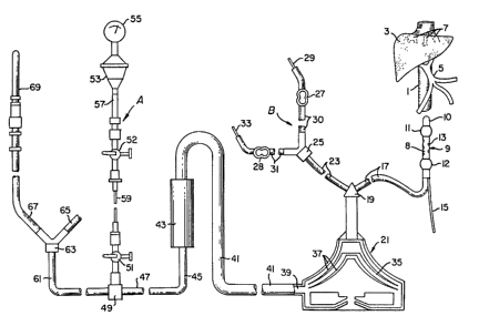
With respect to Figure 1, there is shown the featured components of
the apparatus assembly of the invention used to practice the process of the
5 invention in relation to a human body 2. Liver 3 is supplied with cancer
therapy drugs from syringe 4 through tubing leading to catheter 6 located in
hepatic artery 5. The hepatic venous blood cont~inin~ anti-cancer concentra-
tions of chemotherapuetic agent is passed via the hepatic veins 7 to the double
balloon catheter located in rVc 1. The balloons of the double balloon catheter
10 are positioned central and peripheral of the hepatic veins 7. The cont~min~qted
blood is passed through the double balloon catheter to tubing 17 to a point
exterior to the body 2, thence to a pump 21 such as a Bio Medicus BP-50
Bio-Pump having a priming volume of 48 ml, cont~inin~ two rotator cones and
providing a m~imum flow rate of 5 liters per minute. Pump 21 moves the
15 blood through the extracorporeal circuit at relatively constant low pressure,the object being to avoid raising or lowering the fluid pressure of the total
circuit r~n~in~ from the hepatic veins through the return to the body. The
cont~min~ted blood is transported through tubing 41 into detoxification zone
43, which in this case is a hemoperfusion cartridge cont~inin~ activated carbon.20 Suitable cartridge systems are obtainable from Clark Research and Develop-
ment, Inc., New Orleans, I~ 70121 and from Garnbro Dialysatoren KG, d-7450
Hechingen, Federal Republic of Germany AUT 224 (sold under the trademark
of ADSORBA~). The detoxified blood is passed through tube 44 to effect
infusion through the subclavian vein (not shown) by standard procedures in
25 the art.
With respect to Figure 2, there is shown the relationship of inferior
vena cava 1 to liver 3, hepatic veins 7 and portal veins 5. The hepatic artery is
not shown in the drawing Double balloon catheter 9 comprises central balloon
11 and peripheral balloon 12, each in juxtaposition to cylindrical fenestration
30 zone 8. Zone 8 contains fenestrations 13 sufficient in total area to allow the
complete removal of the hepatic venous flow into the catheter 9. The hollow
interior (main lumen) of catheter 9 is of sufficient size to completely remove
the blood from the hepatic veins without elevating hepatic back pressure.
- 29 -
133387~
BCHOOl
Catheter 9 is provided with channel 15 that is used to inject flu.l into the
balloons 11 and 12 for inflation or to ithdraw fluids for deflation. The ~enous
now is passed through catheter 9 nto openly connected lube 17. Tube 17 may
be interrupted by a pressure monitor the sarne as assembly A, discussed below,
5 that is later provided in the extracorporeal circuit. Tube 17 msy connect
directly with pump 21 or to Y-fitting 19, as shown. Also connected to Y-fitting
19 is ancillary feed s~stem B comprising tube 23, ~J-fitting 25, and multiple IVspikes 29 and 33 each connected to tubes 30 and 31 respectively, and each is
provided with a clarnp, 27 and 28, respectively. These lines can be used for the10 introduction of medications as required.
Pump 21 is a smooth rotator pump design and a particularly desirable
pump is a Bio Medicus BP-50 Bio-Pump having a priming volume of 48 ml,
cont~inir g two rotator cones and providing a m~ximum flow rate of 5 liters per
minute. The cont~min~ted blood is gently pu~hed between the smooth
15 rotators 37 in zones 35 and issued from the pump through port 39 into tube 41.
Tube 41 is connected to cartridge or canister 43 cont~inine a meshed sack of
activated carbon particles coated with an acrylic resin cont~ining heparin, see
Clark, supra. The outflow from cartridge 43 is fed to tube 45 and then to tube
47 that is connected to pressure monitoring assembly ~ Pressure monitoring
20 assembly A comprises a pressure monitor gauge 55 connected to fluid
membrane vessel 53 that contains a thin membrasre that separates the gauge
55 from the blood in vessel 53 and responds to the fluid pressure of the blood
in vessel 53. That response is read by the gauge. Vessel 53 is connected to
tubing 57, that is connected to stopcock 52. Stopcock 52 is connected to
25 flexible tubing 59 that in turn is connected to stopcock 51, the latter secured in
fitting 49.
Blood from tubing 47 is passed to Y-connector 63 via tubing 61, then to
tubings 65 and 67. Tubings 65 and 67 are each connected to catheter 69 and
another catheter (connected to tube 65) not shown. These catheters are
30 provided for returning the purified blood to the subclavian veins.
In Figure 3, there is shown a double balloon catheter design that can
have up to a 24 French (Fr) O.D. Zone 100 is provided with slotted fenestra-
- 30-
1333872
BGHOOl
tions 104 in the solid plastic tubing 102. The open end 118 termin~tes the
catheter. End 118 is tapered to the caliber of an angiographic guide wire that
will, under fluoroscope control, allow the catheter to be advanced from the
femorsl vein to the proper location in the inferior vena cava without risk of
5 injury to the interior of the vessels. Appropriate guide wires may be, for
example, 0.035, 0.038, or 0.045 inch in diameter. During treatment, the
catheter end hole is closed using a standard angiographic apparatus (tip-
occluding wire), that consists of a thin wire long enough to traver~e the lengthof the catheter at the end of which is a stainless steel bead just large enough to
10 obstruct the catheter'q end-hole when advanced into it (similPr to a metal
stopper that closes the outlet from a sink when advanced).
Alternatively, the end hole can be made 7-lZ Fr in diameter in order
to accommodate a return catheter. The return catheter can be used to return
treated blood to the systemic circulation. The return catheter is advanced
15 over a guide wire through the main lumen of the double balloon catheter and
through the end hole 118 into the right atrium or superior vena cava. The
return catheter can be made to graduslly taper its O.D. by decreasing its wall
thickness, leaving the I.D. constant, since the location of the tip of the return
catheter is not critical. The length over which the catheter tapers is arbitrary.
20 The taper is constructed so that the tip of the catheter is its narrowest O.D.
and the O.D. increases toward the femoral vein. As this return catheter is
advanced through the lumen of the main catheter the tip easily passes
through the end hole 118 of the double balloon catheter. The tapered end of
the return catheter is advanced until it obstructs the end hole 118, preventing
25 systemic blood from entering the double balloon catheter when the balloons
are inflated but lesving sn open lumen through the return catheter to return
blood beyond the isolated venous seg~nent without mi~ing with cont~min~ted
blood.
The catheter tubing (body) can be made of a variety of plastic
30 materials such as poly~roF~lene, polyethylene, polyvinylchloride, ethylene
vinylacetate copolymers, polytetrafluoroethylene, polyurethane, and the like.
A favorable plastic comhin~tion for catheters cont~inin~ a return lu~nen are a
homogeneous mixture of high density polyethylene and linear low density
- 31 -
1333872
BGHOOI
polyethylene. That combination gives favorable stiffness at arnbient condi-
tions and allows the use of especially thin wall thicknesses. When th~ surface
of the catheter is made of a p.astic that is dif~lcult to bond with a balloon, the
plastic may be treated first by one or more of a number of well known methods
5 that make bonding possible. The methods include plasma treatment, ozone
treatment, and the like. Balloons 110 and 114 may be made from a plurality of
elastomeric materials such as latex rubber, polyurethanes, spandex type
polyurethanes, EPDM rubber, and the like. The balloons are typically ad-
hesively bonded at sheath surfaces 108 and 112, respectively. A wide ariety of
10 a&esives may be employed. Polyacrylonitrile type adhesives, rubber latex
adhesives and the like may be used to secure the balloon to the sheath sur-
faces 108 and 112.
With respect to Figure 4, there is shown a cross section of a typical
catheter design such as that shown in Figure q. The interior of the catheter
15 contains main lumen 120 and 4 additional lumina 124 molded into the outer
wall 122. The additional lumina can be used for the various functions
described above.
With respect to Figure 5, there is shown a cross section of another
catheter design such as that shown in Figure 3 but cont~inin~ only three
20 ll-min~ The interior of the catheter contains main lumen 130 and two
supplementary lumina 131 molded into segrnent 133 of wall 135. The sup-
plementary lumina can be used for the various functions described above.
With respect to Figure 6, there is shown a double balloon catheter
design which can have an outside diameter of 24 French such as in the
25 fenestration zone 140 and an inside diameter of less than 22 Fr. Zone 140 is
provided with slotted fenestrations 141 in the plastic tubing 142. With respect
to Figure 7, it is a cross sectional view of another typical catheter design
showing a main lumen 150 and 3 supplemental lumina 151. Figure 8 provides
a more detailed schematic cross sectional side view of a typical double balloon
30 catheter 161. .In this depiction, the catheter sidewall 163 iS penetrated by a
plurality of fenestrations 165. The main lumen 169 contains at its periphery
supplemental lumina 170,171 and 173. Supplemental lumen 170 can be used
- 32 -
13338~2
saHool
to accommodate a guidewire, supplemental lumen 171 can be used to accom-
modate a pressure monitor, and supplemental lumen 173 is used to supply
fluid to the balloons 166 and 167 through openings 175 and 177.
Though this invention has been described with emphasis on the
5 treatment of cancer, it is quite apparent that the invention has broader
application. The invention is useful for the treatment of any organ in which
the treating agent would cause toxological ef~ects if it entered the body's
general circulation. For example, the invention could be applied to the
treatment of infectious deseases of organs such as fungal desea~es. A specific
10 illustration would be the treatment of hepatic fungal infections with Am-
photericen B. The procedures described above would be directly applicable to
extracorporeal recovery of this agent and its isolation from entering the
general circulation of the body during treatment of the liver with significant
concentrations of this drug.
Therefore, the breadth of the invention encompasses the perfusing of
a high concentration of an agent to treat an organ, such as anti-cancer agents
through a body organ cont~ining a tumor, without their entering the body's
general circulation, removing them from the organ with effluent blood and
transporting the cont~min~ted blood to an extracorporeal circuit where the
20 blood is treated to remove the cont~rnin~tion, and returning the treated blood
to the body. The process prevents toxic levels of the agents from entering the
body's general circulation while delivering lethal doses of the agents to the
tumor.
- 33 -