Note: Descriptions are shown in the official language in which they were submitted.
` 133~181
SYSTEM FOR SELECTIVE CELL
SEPARATION FROM CELL CONCENTRATE
TECHNICAL FIELD
The present invention concerns a novel system for
separating a specific cell population from a heterogeneous cell
mixture.
BACKGROUND OF THE INVENTION
In the field of cell separation, it is common to separate
cells from plasma in blood and also to separate by
centrifugation various types of cells such as red cells from
white cells and the like. However, there is often a need to
separate cells which are only slightly different from the other
cells in a suspension thereof. If the cells are of nearly equal
specific gravity, they may not be separated by centrifugation.
For example, it may be desirable to isolate various types
of leukocytes from a bone marrow concentrate or a peripheral
blood stem cell concentrate. It may be desirable to perform
selective separation of neuroblastoma cells from a bone marrow
concentrate. It may be desirable to selectively separate
specific T-lymphocyte subset populations (helper-inducer or
suppressor-cytotoxic T-lymphocytes) from a lymphocyte
concentrate that is prepared using a blood cell separator.
Additionally, it may be desirable to selectively separate
precursors of lymphokine activated killer (LAK) cells, tumor
infiltration lymphocyte (TIL) cells, or activated killer
monocytes, from lymphocyte or monocyte cell concentrates or from
a tissue cell preparation.
By current techniques of the prior art, such as Sauer, et
al, U.S. Patent No. 4,710,472, magnetic separations of
individual subsets of cells from larger populations in
- 2 - 1 33518 1
significant quantities become possible. This, in turn, opens up
new vistas of research and therapeutic techniques, making use of
the purified cell populations which may be obtained.
Another current practice in the field for cell separation
utilizes hollow fiber, flat sheet membrane or packed bed bead or
particle matrix materials with physically adsorbed or covalently
attached chemicals or biochemicals for the selective cell
separation from whole blood or the like. These devices are
designed to allow continuous whole blood or blood component
inflow and return. Since these devices operate at normal blood
flow rates under conditions in which the concentration of
desired cells can be very low compared with other cell types,
the separation process is often not efficient.
DESCRIPTION OF THE INVENTION
In this invention, a system is provided for improved
selective separation of a specific cell population from a
heterogeneous cell mixture. The heterogenous cell mixture may
first besubjected to a means for obtaining a selective cell
concentrate from the heterogeneous cell mixture by separating
the selective cell concentrate based upon the physical
properties of the concentrate. Preferably, the selective cell
concentrate is used with the system and method of the
invention. Specifically, centrifugation may be used as a first
separation step. There are many different types of cell
centrifuge systems for the separation of desired types of cells.
A flexible, collapsible, aseptically sealed container is
provided, having particle means inside, with the particle means
preferably having chemically covalently attached thereto a
substance capable of binding to the desired cells only, to the
exclusion of other cells. Examples of such a covalently bonded
substance may include antibodies, antigens, proteins,
glycoproteins, polysaccharides, or lipopolysaccharides. The
particle means to be in long term contact with the container
walls or the like.
1335181
Means are provided for intimately contacting either the
heterogenous cell mixture or the cell concentrate with the
particle means within the container. This may simply be a
flexible, aseptic conduit system which communicates between the
site of formation of the cell mixture orconcentrate and the
container which has the particle means. Incubation of the cell
mixture or concentrate with the particle means may then be
permitted, to cause selective binding of a specific cell
population from the cell concentrate to the particles, creating
a particle/cell conjugate.
Then, since the new particle/cell conjugate will have
significantly different properties from the remainder of the
cells, it becomes an easy matter to separate them from other
cells, by centrifugation, for example. Preferably, one may use
particles which are paramagnetic, then effecting separation
through a magnet system.
The initial cell fluid volume may remain within the
container during the incubation period, when binding to the
particles takes place. Also, means may be provided for
introducing additional volume of the cell mixture or concentrate
into the container during the incubation period.
After separation of the particle/cell conjugate from other
cells, as described above, one may separate the specific cell
population from the particles or vice versa, for example by
eliminating the bond between the particles and the cells in
known manner, so that a purified, selected population of cells
may be provided for further use. Alternatively, the unbound
cells may be the desired cells, being removed from the
particle/cell conjugates.
When the particles are paramagnetic, the particle/cell
conjugate may be retained in a fixed location by action of the
magnet means, as remain;ng, unbound portions of the cell
concentrate are removed from the location.
- 1 3 3 j l 8 1 Preferably, the magnet means which retains the
particletcell conjugate may define first and second spaced
magnet members, one being downstream of the other, so that the
second, downstream, spaced magnet member can pick up any bound
particle/cell conjugate that is lost by the first magnet member,
so that no particle/cell conjugate goes downstream with
remaining cells.
The magnet means may be positioned adjacent to means for
carrying and positioning the flexible, collapsible container
which carries the particle/cell conjugate, to permit at least
one inner wall portion of the container to be within the
magnetic field of the magnet means. Thus, this inner wall
portion serves as the fixed location at which the particletcell
conjugate is retained.
Flat-pressing means may also be provided, to press the
collapsible container flat while the inner wall portion is
within the magnetic field, to facilitate separation of the
particle/cell conjugate from the remainder of the cell
concentrate.
Specifically, the second magnet members possesses a
magnetic field that is shallower and less extensive in distance
than the magnetic field of the first magnet member, but stronger
adjacent the magnet surface for stronger paramagnetic particle
retention. Preferably, the magnetic reach of the first magnet
member is substantially equivalent to the width of the fluid
container being placed thereon. This ensures that the majority
of the paramagnetic particles in the container will fall within
the reach of the magnetic field and be drawn to the magnet
surface.
It is also preferred for the container which contains the
particle means to be aseptically connected to a flexible,
multiple-chamber insert member for a blood cell separation
centrifuge. Such an insert member may be the disposable,
- - 1335181
blood-carrying, inner, flexible portion typically used in such a
centrifuge. It may be integrally aseptically connected to the
container in accordance with this invention, so that freshly
collected blood cells may be aseptically transferred from the
insert member to the container having the particle means,
without any need of forming a sterile connection therebetween.
This greatly simplifies the use in accordance with this
invention, and also increases the likelihood that there is not
breach of aseptic conditions.
Also, a first container having the particle means may be
aseptically connected to further flexible, collapsible container
means, for receiving processed cells from the first container
which contains the paramagnetic particle means, or particle
means of another type, if desired. Typically, a second
flexible, collapsible container is sealingly, aseptically
connected to and positioned between the first container
described above and the further collapsible container means.
This may serve as a downstream catch area which is positioned
against the second magnet described above to catch any
particle/cell conjugate which escapes the first magnetic means
against which the first container is pressed during the cell
separation operation. The second flexible, collapsible
container may preferably be of hexagonal shape with inlet and
outlet ports at opposed corners, to cause generally slow flow of
cells through said second container relative to flow lines
connected thereto. The flow line diameter is typically no more
than one-fourth the width of the second container. Also the
flow path, particularly through the second container during
magnetic separation, should be of a very shallow depth,
typically 0.02 to 0.1 inch.
Further in accordance with this invention, one may
practice a method which includes the following steps. Blood
from the patient may either be collected in a first container or
the patient may be connected to a blood separation centrifuge,
- 6 - 133~ 181
the centrifuge being operated to form a cell concentrate which
is collected in the first container. The first container is
sealed. If the particle means are not already in the container,
they may be placed in the container in some aseptic manner, or
an inner container positioned within the container may be broken
from outside of the container to cause their release within the
container. Thus the cells and the particle means are mixed.
Then, the primary container is connected, if not already
integrally connected, to the inlet of a clamped separation set.
The primary container and a desired secondary chamber between
the primary container and the rest of the separation set are
both placed in magnetic separator means. A primary magnet
attracts the particles of the particle means, which by now are
bonded to the desired cells, to retain the particle means in the
primary bag as the remaining contents of the bag flow toward the
separation set. The same principle takes place in the secondary
chamber under the influence of a secondary magnet, to eliminate
or greatly reduce the possibility of any of the particle means
and attached cells flowing downstream with the rest of the
contents of the primary container.
A clamp is then opened and/or a pump actuated to initiate
flow from the primary container through the secondary container
and across the magnetic field of the secondary magnet, into
storage containers of the separation set. The storage
containers may then be sealed, followed by optional
disconnection of the storage containers.
Then the magnets may be removed and the adhering cells
flushed, so that one or more storage containers may contain pure
cell/particle conjugate, while other storage containers contain
the remaining contents of the primary container. After this, a
conventional process may be used to separate the particles of
the particle means from their attached cells, with the cells
then being separated by centrifugation or other desired means,
-- 7 --
1335181
such as filtration or magnetic separation, and sent to a
container for storage of the pure cells. The means used to
break the connection between the particles and the cells
depends, of course, upon the specific bonding agent. For
example, the particle means may be coated with a antibody for
the specific desired cells, with the result that the cells bond
to the particle means. Then, when the bond is to be broken, an
appropriate reagent may be used to break the bond.
An advantage of the method of this invention is use of
the magnetic separating system of this invention with a cell
concentrate in a batch process. In the cell concentrate the
specific cell population to be separated is present at higher
concentration, which tends to favor separation kinetics.
Another advantage is that numerous unwanted blood cell types, in
the situation where blood cells are being separated, may have
already been greatly reduced in number by the preliminary,
typically centrifugal cell separation process, to reduce
non-specific cell reactions. For example, the collection of a
lymphocyte cell concentration with minimal red blood cell,
platelet, and granulocyte contamination may be effected using a
blood cell separator.
Additionally, introduction of the particles of the
particle means to a cell concentrate under the conditions of
this invention allows incubation to take place at constant
volume conditions under storage within the container having the
particle means. Thus, the system solution composition can be
configured to more easily obtain favorable final incubation
conditions for formation of the particle/cell conjugate. These
conditions may then be optimized for a specific purpose in a way
which is far more versatile than otherwise.
The subsequent separation of the particle/cell conjugate
is also advantageous in the conditions of this invention, with
separation times being faster, and fewer cell types in the
original concentrate providing a final product with fewer
non-desired contaminating cells.
,i
1335181
As previously stated, the material which is covalently
attached to the particle means may be, for example, an antibody,
antigen, protein, glycoprotein, polysaccharide,
lipopolysaccharide. The material may also be a nucleic acid, a
lipid molecule, or a synthetic or chemically modified component
of such a substance which shows a selective binding affinity for
the cell population to be separated. The methods used for the
chemical covalent attachment of such are known and used in the
production of coupled matrix material for affinity
chromatography and other selective adsorption applications.
Examples of such techniques of covalent attachment to sepharose,
gelating, or other beads may be seen from the following
articles: Habeeb, "A Novel Preparation of Immunoadsorbents,"
Biochimica et Biophysica Acta, 673 (1981) 527-538; Cambier, et
al., "Isolated Phosphorylcholine Binding Lymphocytes. I. Use of
a Cleavable Crosslinking Reagent For Solid-Phase Adsorbent
Isolation of Functional Antigen Binding Cells," Journal of
Immunological Methods, 51 (1982) 209-221; and Bonnafous, et al.,
"Ligands Immobilized Through Cleavable Mercury-Sulfur Bonds.:
Journal of Immunological Methods, 58 (1983) 93-107.
The solution in which the particle means of this invention
may be suspended can be a buffered sale solution which may
contain a protein such as albumin, and is compatible with the
physiological requirements of the heterogeneous cell concentrate
and the biological binding material attached to the particle
means. This solution may be configured in its chemical
composition and properties to confer sterility to the substance
which is covalently attached to the bead or particle.
Furthermore, the solution may be configured in its chemical
composition and properties such that when a bead or particle
suspension is added to the heterogeneous cell concentrate, the
properties of the resulting mixture favor the formation of the
bead or particle conjugate.
- - 9 - 1335181
For example, the heterogeneous cell mixture or
concentrate may be a bone marrow preparation in which the
cells may be further concentrated in a cell concentrating
centrifuge or the like. Additionally, the heterogeneous
cell mixture can be a tissue derived cell suspension, or a
cell concentrate prepared from peripheral blood using such
centrifugal device. Examples of the latter are
concentrates of platelets, lymphocytes, granulocyte,
monocytes, or peripheral bone marrow stem cell preparations
prepared with a blood cell separator such as the previously
described CS3000 blood cell separator or Autopheresis-C
device.
The beads or particles used in the system may be
selected for particular size and specific gravity
properties so as to allow the subsequent separation of the
beads or particle/cell conjugate from the unconjugated
cells in the heterogeneous cell concentrate using the
centrifugal capabilities of a blood cell separator. A
solution such as Ficoll-Hypaque or Percoll might be used to
facilitate this separation.
The beads or particles of the particle means may be
composed of any number of different materials such as
polystyrene, latex, plastic copolymers, glass,
synthetically produced gel beads and the like. Preferably,
such materials will possess good mechanical properties to
prevent flaking or fracturing of the beads or particles,
and will allow chemical covalent attachment with ease.
It is further preferred for the beads or particles to
be formed around a magnetite particle, for example, to
allow separation of the bead or particle/cell conjugate
using magnets, as described above. For example, particles
may be produced in accordance with the methods as described
in International patent application of Chaeo-huei, J. Wang,
et al., published under W08004373 on May 18, 1989, entitled
"Process For Producing Magnetically Responsive Polymer
Particles and Applications Thereof."
- 10 - 1335181
The typical size of the particle means used in this
invention may be from about 2 to 10 microns, preferably about 3
to 5 microns. The particles may be added in a liquid
suspension, forming a typically dark sludge-like material.
On the order of 10 ml. of such liquid suspension may be
placed in the bag which is to receive the cells for separation
typically including one hundred thousand to 20 billion
particles. In the event it is undesirable for any reason for
the particles to remain in the bag for an excessively long time,
due, for example, to interaction with the bag wall, they may be
added separately to the bag by conventional means such as using
a sterile connector, or they may reside in a frangible container
within the bag, to be broken when their use is desired so that
they enter the bag interior from the frangible container.
As stated above, the various containers used in this
application are preferably integrally linked together in their
initial manufacture so as to avoid the need for sterile
connection during the processing in accordance with this
invention. However, they may also be connected together with
sterile connectors, numerous designs of which are well-known,
for example, those of U.S. Patent No. Re. 32,056.
The Dynal Company of Great Neck, New York manufactures
paramagnetic microbeads which may be used in accordance with
this invention.
For a typical blood cell separation, the number of
microbeads of the particle means used may number from about a
hundred thousand to one billion. It has been found, when making
use of a primary and secondary magnetic separator as described
above, that the removal of microbeads and the cells attached to
them from a cell suspension may be quantitative, with virtually
no microbeads found in downstream effluent after passage through
the magnetic separation system of this invention.
- - lOa - 1~35181
Other aspects of this invention are as follows:
An apparatus for the selective separation of a
specific cell population from a heterogeneous cell
mixture comprising;
first and second flexible, collapsible containers
designed to contain paramagnetic particles and the
heterogeneous cell mixture, each of the first and second
containers having inlets and outlets, with the outlet of
the first container being sealingly aseptically coupled
to the inlet of the second container by a length of
tubing, said first container having a width of about 0.5
inch to about 1 inch,
base means for receiving the first and second
containers in separate receptacles, with each of the
receptacles including a floor upon which the associated
containers rest, each of the floors being at least
partially formed by magnet members, the magnet member
forming the floor of the first receptacle having a first
surface and providing magnetic field local maxima of
about 7,000 to about 9,100 gauss at said first surface
and magnetic field local maxima of about 1,100 to about
1,700 gauss at a distance of lcm from said first
surface, and a magnet member forming the floor of the
second receptacle having a second surface and providing
magnetic field local maxima of about 7,300 to about
8,000 gauss at said second surface and magnetic field
local maxima of about 80 to about 500 gauss at a
distance of lcm from said second surface, and
a flat-pressing means associated with the second
receptacle for pressing said second flexible,
collapsible bag flat to provide a flow path through the
bag having a thickness of 0.02 to 0.1 inch.
_ - lOb - 1335181
A magnetic separator comprising:
a stand formed with at least first and second
container receiving portions, each of which first and
second container receiving portions includes at least a
floor area; and
a first magnet forming at least a portion of the
first container receiving portion floor area, said first
magnet having a first surface and providing magnetic
field local maxima of about 7,000 to about 9,100 gauss
at said first surface and magnetic field local maxima of
about 1,100 to about 1,700 gauss at a distance of lcm
from said first surface; and
a second magnet forming at least a portion of the
second container receiving portion floor area, said
second magnet having a second surface and providing
magnetic field local maxima of about 7,300 to about
8,000 gauss at said second surface and magnetic field
local maxima of about 80 to about 500 gauss at a
distance of lcm from said second surface, and
a first pressing means connected to the stand and
associated with the second receptacle, the first
pressing means being biased in a direction towards the
second receptacle floor area.
1335181
DESCRIPTION OF THE DRAWINGS
Figure 1 is a partially schematic plan view of apparatus
in accordance with this invention for selectively separating
cells.
Figure 2 is a perspective view of part of a magnetic
separation device used with the apparatus of Figure 1.
Figure 3 is a perspective view of another part of magnetic
separator used with the apparatus of Figure 1.
Figure 4 is a perspective view of the separation system in
operating position.
Figure 5 is a plan view of a compound magnet used in the
magnetic separator of Figures 2-4.
Figure 6 is an end view of the magnet of Figure 5.
Figure 7 is an end view of a second compound magnet used
in the magnetic separator of Figures 2-4.
DESCRIPTION OF THE SPECIFIC EMBODIMENTS
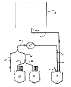
Referring to Figure 1, a d;sposable system 10 is disclosed
for separating an individual population or populations of cells
from a heterogeneous cell mixture in accordance with this
invention.
Cell concentrator portion 12 which may be used withthis
invention is schematically shown and may be of a design as shown
in U.S. Patent Nos. 4,379,452 or 4,410,026, or may be any cell
concentrator member usable in any known apparatus for the
concentrating of cells. For example, the CS3000T-M- separation
system sold by Baxter Healthcare Corporation may be used, or the
Autopheresis-CT M separation system, sold also by Baxter
Healthcare Corporation, or any other desired, similar device,
may be used.
At outlet port 14 of concentrator system 12, a desired
population of cells may be provided, for example lymphocytes,
separated in concentrator system 12 from whole blood. The
lymphocytes, perhaps mixed with other white blood cells, pass
- 12 - 1 33S 18 1
through flexible conduit 16 into flexible, collapsible container
18 of generally conventional design, which contains the particle
means of micron-sized particles including a paramagnetic
material such as ferric oxide, coated with plastic such as
polymethyl methacrylate, which, in turn, is further coated with
an antibody or other cell bonding agent to the specific marker
proteins in the cell wall of a given category of leukocytes, so
that the leukocytes and particles 20 selectively bond to each
other, to the exclusion of the remaining leukocytes and other
cells.
The apparatus of this invention also carries flexible
tubing 22 which communicates between flexible, collapsible
container 18 and a flexible collapsible container 24
communicating with tubing 22 at one end and communicating with
an outlet tubing 26 at this other end. Tubing 26 branches into
tubings 28, 30 each of which connects to another flexible,
collapsible container 32, 34.
While cell concentrator portion 12 is preferably
integrally connected to flexible container 18, the connection
may be made by a conventional sterile connector system, if
desired. Additionally, the particles 20 may be stored either
outside of container 18 and connected with a sterile connector
system, or they may be placed in a frangible container inside of
bag 28 with the container being breakable on use so that the
particles 20 do not have an excessive amount of time to interact
with the bag walls, in the event that some adhesion may take
place there. Of course, the particles 20 may reside freely
within the bag if the particles do not interact with the bag
wall.
After the cells have been separated and concentrated in
separation apparatus 12 and conveyed to bag 18, the primary,
cell-containing container 18 may be sealed by heat sealing of
line 16 and by operation of clamp 36 (although clamp 36 may be
closed at an earlier stage of the operation). If the microbeads
- 13 - 1335 181
or particles 20 are not already added to the container, they may
be so added at this time, followed by gentle mixing of the
contents of the primary container: specifically, the cells and
the microbeads or particles. At this time, bonding takes place
between the particular cells selected and the antibody coating
of the particles or microbeads 20.
Following this, if container 18 is not integrally
connected, as shown, with the downstream set portion of this
invention beginning with chamber 24 and including bags 32, 34,
such connection may be made in sterile, aseptic manner by
conventional means, for example, by use of a sterile connector
0 system of known design. In the preferred embodiment of Figure
1, the various components are integral to avoid the
inconvenience of connection and the risk of contamination of the
container contents.
Following this, containers 18 and 24 are placed into
magnetic separator 40, which is shown in various aspects in
Figures 2-7.
Separator 40 carries upper base member 42 which carries
magnet assemblies 44, 46. Upper rotatable, hinged presser
member 49 comprises a flat undersurface portion 50, and carried
by base 42, overlying in its closed position magnet 46. Upper
base member 42 thus provides two spaces, respectively against
magnet 44 and magnet 46, which are respectively proportioned to
receive bag 18 and bag 24. Channel 47 and space 48 provide room
for tubing 22, while channel 51 is provided to receive tubing 26.
Additionally, lower base member 52 is proportioned to
receive upper base member 42 in cradle area 54 so that tubing 26
can be connected to roller pump assembly 53 for controlled
pumping flow of cells out of bag 18, through intermediate bag
24, and into one or the other of containers 32 or 34.
Hinged cover 56 is positioned to be brought down on top of
bag 18 as installed in upper base member 42. Upper rotatable
hinged presser member 49 presses down in similar manner on
5 1 8 1
flexible chamber 24. The purpose of particularly presser member
49 is to provide precise definition of the thickness of the flow
path of chamber 24 during processing to cause the flow path
across the associated magnet 46 to be of very shallow depth, for
example, about 0.05 inch. Similarly, magnet 44 is positioned to
associate with chamber 18. As will be described more fully
herein, the magnets 44 and 46 are designed to provide suitably
configured magnet fields to capture and retain paramagnetic
particles passing through the containers positioned thereon,
respectively containers 18 and 24. In this regard magnetic 44
will possess a greatermagnetic field reach than magnetic 46 in
order to capture a larger percentage of particles. This places
the cells passing therethrough into a position to be strongly
influenced by the fields generated by magnets 44, 46. Thus,
those cells bonded to magnetic beads will be retained against
one or the other of the magnets.
One advantage for using a magnetic separator apparatus 40
having a separable upper base member 42 is that upper base
member 42 may be refrigerated to a temperature on the order of
4 C. prior to use. Thus, the cold magnets keep the cells cold
during operation, as well as providing some increase in magnetic
field strength. The cold cells are less active and better
preserved. Also, at low temperatures, non-specific cell
interactions with the paramagnetic beads can be reduced. For
example, phagocytes present are less active in ingesting
available beads when kept at low temperatures.
When containers 18, 24 are lying on magnets 44, 46, clamp
36 may be opened, and pivotable pressure member 56 may be gently
closed to press the container 18 flat and to press the cells and
their carrier liquids out of bag 18, which rests upon magnet
44. Presser 56 may also contain magnetizable metal strips that
are sized and positioned so that they are ~agnetically pulled to
the magnet 44 in a way such that the plastic bag 18 is squeezed
and the efficient flow from the bag is controlled and relatively
- 15 - 133S 181
constant. Alternatively, the roller pump 53 may be activated to
cause the cells and their carrier liquids to flow out of bag
18. As the cells and their carrier liquids flow through tubing
22, the magnetic field from magnet 44 attracts the particles or
microbeads 20, and the cells to which they are bonded, causing
such microbeads 20 to be generally firmly affixed against the
inner wall 57 (Fig. 6) of bag 18 that is closest to magnet 44,
holding the particles 20 and their attached cells as the
remaining cells and suspension liquid pass through tubing 22 out
of bag 18.
If any particles 20 and attached cells escape the first
magnet 44, they may be caught by the interaction between
container 24 and second magnet 46, upon which it lies, being
retained against inner wall 59 (Fig. 7). Even under significant
flow conditions, the enlarged container 24 exhibits relatively
slow flow conditions through it. Hinged presser member 49
contains four metal bolts 81 that are sized to hold the presser
member 49 in place with the proper spacing and mount of magnetic
force. The surface 50 of presser member 49 is precisely
machined to precisely define the thickness of the flow path
through container 24. The shallow depth of this flow path
causes any particles or microbeads present to drift into the
influence of the magnetic field of magnet 46, to be retained on
the inner wall 59 of container 24 as the remaining cells and
liquid flow by, out of container 24, through tubing 26, into bag
32, for example. In such a circumstance, tubing 30 may be
clamped off to keep bag 34 empty.
Then, when pressure member 56 has been used to squeeze all
possible liquid and cells out of bag 18, a small amount of
cell-compatible suspension liquid may be passed through the
system, via sterile connector port 61, priming line 33, or
another integrally connected container, to flush the remaining
cells which are unattached to paramagnetic particles through the
system into bag 32.
- - 1335181
Then, tubing 28 may be closed, and tubing 30 opened by
conventional clamp means. The system may be removed from the
field of influence of magnets 44, 46, and, more
suspension/solution may be passed into container 18, to flush
the cells which are bound to particles 20 through tubing 22,
through chamber 24, picking up any bound cells retained there,
and into container 34.
Then, containers 32, 34 may be separated from the system
by sealing and severing of tubings 28, 30 in conventional
manner, with the desired particular population of cells being
separated out from the main body of cells, and placed in
separate container 34 for separate use.
As stated above, magnet 44 is designed to possess a
greater magnetic field reach than magnet 46. Preferably, this
magnetic field reach is at least equal to about three-quarters
of the width of the container 18, and more preferably
substantially equivalent to such width. This ensures that a
majority of the paramagnetic particles within container 18 are
captured by the magnetic field and drawn to the surface of
magnet 44. Accordingly, magnet 44 will have a magnetic field
reach of from about one-half to one inch, preferably, one-half
to three-quaters of an inch.
Typically, magnets having greater magnetic field reaches
possess lower surface field strengths. One particle magnetic
assembly which provides the desired magnetic field reach, but
retains substantial surface field strength will be described
with reference to Figures 5 and 6. Magnet assembly 46 may be
made of similar construction. Magnet assembly 44 is shown to
comprise a stack of bar magnets 64 which are separated by, and
in contact with, steel pole pieces 66. As a particular
advantageous feature, the like poles of adjacent bar magnets 64
in the stack face each other and particular pole piece
separating them. This is demonstrated by the letters "N" and
"S", each of which indicate the combined north poles or south
1335181
poles of the respective magnets which are facing each other.
The bar magnets 64 define long sides 68 and ends 70, and north
and south poles of the bar magnets being defined along an
opposed pair of long sides as shown in Figure 5. Preferably,
the bar magnets are made of a high magnetism alloy of neodymium,
iron and boron. In Figure 5, the face 72 of the magnet assembly
shown rests in use against an outer wall of bag 18 so that the
magnetic field from magnet assembly 44 passes into container 18,
for retention of particles 20.
Turning to Figure 6, an end view of magnet assembly 44 is
shown, with face 72 being the face that is displayed in Figure
5. As shown, magnets 64, separated by pole pieces 66, rest upon
a non-magnetizable aluminum plate 74 or the like, to support the
respective magnets and pole pieces.
It can also be seen that pole pieces 66 each define an
angled groove 76 along parallel end faces which are spaced from
bar magnets 64, and which are opposed to face 72 of the assembly
44, which face is the fixed location for retaining the
particle/cell conjugate formed through the paramagnetic
particles as in this invention.
The bar magnets 64, pole pieces 66, and support plate 74
may be bonded together in any conventional manner with
non-magnetic cement, or making use of appropriate clamps or
retention straps. Appropriate magnets for use herein may be
obtained from the Crucible Magnetics Co. of Elizabethtown,
Kentucky. Magnet assembly 44 contains magnets 64 that are .50
inch thick and pole pieces 66 that are .25 inch thick. This
generates a magnetic field that has local maxima of 7000 to 9100
gauss (average 8300 gauss) at the surface of the magnet and
which decreases to 1100 to 1700 gauss (average 1400 gauss) at a
distance of 1 cm. from the magnet surface. This magnet assembly
gives both a relatively strong magnetic holding force at the
magnet surface and a relatively good magnetic "reach out" force
to capture beads some distance (up to 1") from the magnet
surface.
- 18 - 1~35 181
- Magnet assembly 46 contains magnets 64 that are .25 inch
thick and pole pieces 66 that are 0.1 inch thick. This
generates a magnetic field that has local maxima at the magnet
surface ranging from 7300 to 8000 gauss and which decreases to
80 to 500 gauss at a distance of 1 cm from the magnet surface.
Compared to magnet assembly 44, magnet assembly 46 has a
stronger magnetic holding force at the magnet surface, but has
less magnetic "reach out" force and thus has a shallower
magnetic field. The second flexible container 24 is designed to
take advantage of the particular magnetic field of magnet 46.
First container 18 is shown to rest on magnet assembly 44 for
cell separation.
Accordingly, a system is provided for isolating in an
aseptic, simplified manner a particular subclass of concentrated
cells, which may be separated as shown for any desired purpose.
The following examples and the other disclosure of this
application are provided for illustrative purposes only, and are
not intended to limit the scope of the invention of this
application, which is as described in the claims below.
EXAMPLE 1
This example illustrates a specific application of the
apparatus and method of this invention in the separation of
T-helper/inducer lymphocyte cells collected from whole blood.
Preparation of Mononuclear Cell Suspension:
Approximately 500 ml. of whole blood was collected into a
standard FenwalTM Sodium Citrate Double Blood Pack and divided
between the two packs to give 290 ml. in each pack. Hespan
Hetastarch (55 ml.) was added to each pack, and the contents
mixed by gently tilting of the pack in a back and forth motion.
The red cells were allowed to settle, and the plasma layer which
contains mononuclear cells was transferred to a standard Fenwal
600 ml. transfer pack using a Fenwal plasma extractor and
standard transfer tubing set. The transfer pack was centrifued
-- 19 --
13~5181
to pellet the cells from the plasma, and the plasma was
transferred to another transfer pack using the plasma
extractor. The cells were resuspended by adding 80 ml. of Hanks
Balanced Salt Solution (HBSS) which 10 percent Fetal Calf Serum
and gentle titling of the bag back and forth. An aliquot of the
cell suspension was counted in 2 percent acetic acid and used to
calculate a total cell harvest of 2.8 x 10⁹ mononuclear cells
for the cell suspension. After counting of the cells, an
additional 100 ml. of HBSS was added to the transfer pack, and
the cell suspension was mixed. One-half of the cell suspension
was transferred to a second transfer pack to give a total of
about 1.4 x 10⁹ mononuclear cells in 100 ml. HBSS in each bag.
Preparation of Paramagnetic Beads
Bonded ~ith Antibody
About 1 gram of paramagnetic beads (PandexTM beads with
amino surface functional groups) were washed five times with
saline solution and resuspended in 20 ml. of saline in a 50 ml.
glass test tube. Freshly prepared
N-succinimidyl-3-(2-pyridyldithio) propionate (SPDP) comprising
1.0 ml. of 20 millimolar solution of SPDP and absolute ethanol,
was added to the test tube. The tube was rotated end-over-end
for 30 minutes at 4 C. to form
3-(2-pyridyldithio)-propionyl-derivitized beads. The beads were
collected by magnet and washed five times with 20 ml. aliquots
of phosphate buffered saline. The beads were then suspended in
20 ml. of 50 millimolar dithiothreitol in sodium acetate (0.1 Ml
buffer, pH 4.5) and incubated with end-over-end rotation for 30
minutes at 4 C. to form the thiol derivative. The bead
preparation was then washed five times in phosphate buffered
saline, collecting the beads with a magnet after each wash.
Finally, the beads were resuspended in 20 ml. of phosphate
buffered saline.
- 20 - 1335181
Mouse anti-Leu 3a antibody (type CD4 antibody-2mg. in 2
ml. phosphate buffered saline) was added to 0.1 ml. of 20mM SPDP
in absolute ethanol. The mixture was dialyzed overnight against
500 ml. of phosphate buffered saline.
The resulting antibody solution was added to the
derivitized bead suspension in a 50 ml. glass centrifuge tube.
The tube was rotated end-over-end overnight at room temperature
to cause coupling of the antibody to the beads. Then, the beads
were washed five times with phosphate buffered saline and
resuspended in 30 ml. of phosphate buffered saline.
Thus, while the created antibody carries pyridyldithio
active groups, the beads, because of their dithiothreitol
treatment, carry bound sulfhydryl active groups. Accordingly,
upon being brought together, the antibody and the beads become
chemically bonded together through a disulfide linkage resulting
from a condensation reaction, with 2-thiolpyridine being split
off as a by-product.
Coupling of Mononuclear Cells to
CD4 Antibody Bonded To Beads
Approximately 300 mg. of the antibody-bonded beads
prepared as above (7 x 10⁹ beads) were added to each bag of cell
suspension prepared as previously described, using a 20 ml.
syringe with attached 21 gauge needle. The resulting suspension
had about a 5 to 1 bead to cell ration, and was gently mixed,
being then placed on a Cole-Parmer rotator at a setting of 4 for
30 minutes at 4 C. The bag 18 (Fig. 1) containing the bead and
cell suspension was connected to a 600 ml. Fenwal transfer pack
32 through tubing 22, 26 which communicates through an enlarge
chamber 24, as also illustrated in Fig. 1. Bag 18 was also
connected through the priming line 33 of the tubing set to a
1,000 ml. bag of physiological saline solution to allow priming
of the set between container 18 and the empty transfer pack 32.
- 21 - 133~181
After priming the resulting interconnected bags system was
installed into upper magnet tray 42 as shown particularly in
Fig. 2, with bag 28 being placed in area 44, and container 24
being placed in area 46. The appropriate tubing 22 was stored
in area 48, passing through channel 47, while tube 26 was
installed in channel 51, and threaded through roller pump 53.
The respective magnets 44, 46 were prechilled to about 4 C for
continued cooling of the cells. Hinged covers 49, 56 were then
closed, with the upper tray 42 installed on lower tray 52 (Fig.
4).
After about 5 minutes had elapsed, to allow complete
bonding of the appropriate cells to the beads through the bonded
CD4 antibodies, and to allow maximum capture of the resulting
bead-cell conjugates at the inner surface of bag 18 adjacent
magnet 44, roller pump 53 was actuated to pump at a rate of 10
ml. of fluid per minute into collection bag 32. Any bead-cell
conjugates that escaped from bag 18 were subjected to the
influence of secondary magnet 46 in bag 24, to be captured on
the inner surface of chamber 24 adjacent that magnet, while
cells which were not so bonded to the beads flowed out with
fluids to collection bag 32.
After substantial emptying of bag 18 and container 24 of
fluids, the bead-cell conjugate in bag 18 was resuspended in 100
ml. of physiological saline, while removing bag 18 and container
24 from their seating on the respective magnets, and closing
flow through tubing 26 while so doing. Following this, bag 18
and container 24 were reinstalled in their positions on upper
magnet tray 42, and the suspending saline solution withdrawn
from bag 18 by roller pump 53, as before, through tubing 26 into
collection bag 32, along with any remaining cells which were not
bonded to a paramagnetic bead.
Then, bag 18 and container 24 were once again removed from
the influence of the magnets, after closing off flow through
tubing 26. The bead-cell conjugate in bag 18 and container 24
- 22 - 1335181
was resuspended in 100 ml. of physiological saline containing
25mM dithiothreitol to break the bond between cells and beads.
Bag 18 and container 24 were placed on a rotator at 4 C for 20
minutes. Then, bag 18 and container 24 were connected to a 1
liter FenwalTM PL269 tissue culture flask (symbolized in Fig. 1 as
container 34). Following this, bag 18 and container 24 were
reinstalled in upper tray 42, the hinged presser plates were
closed, and the bag fluid contents were flushed into the tissue
culture flask 34 while the paramagnetic bead particles remain
trapped adjacent the magnets.
Thus, the free lymphocytes which are positive to the
action of CD4 antibody have been separated from other cells in
the mixture, and are provided in isolated form in the tissue
culture flask 34, or, if desired, a blood bag 34 as specifically
shown in Fig. 1.
Residual cells in the separation set downstream from bag
24 are also flushed into flask or bag 34 with more physiological
saline.
The cells in flask 34 may be collected by centrifugation
and resuspended in 100 ml. RPMI 1640 tissue culture medium
containing supplemental glutamine and placed in a carbon dioxide
incubator for subsequent further study.
EXAMPLE 2
By means of a process similar to that disclosed in Example
1, there may be removed from bone marrow cells generally, cells
of the following types: B-lymphoma, neuroblastoma, breast
cancer, or leukemia cells, when such cells express a tumor
associated antigen on their surface recognizable by a biological
attached to the bead particle surface. Thus, the invention may
be used as a process for purifying bone marrow cells prior to
autologous bone marrow transplant.
The bone marrow may be processed through a conventional
cell separator to extract a mononuclear cell preparation
- 23 - 1335 181
therefrom. This mononuclear cell preparation may be incubated
with a panel of mouse-derived monoclonal antibodies against the
tumor cells, washed to remove excess antibody, and then
incubated with paramagnetic beads as described in Example 1,
coated with goat antimouse antibody to selectively bind the
tumor cell to the bead. The tumor cell is then removed as in
Example 1, using a magnetic separator of the type disclosed.
EXAMPLE 3
As an alternative technique of the use of this invention,
T-lymphocytes may be removed from the mononuclear cell
preparation described in Example 2 before an allogenic
transplant to prevent Graft vs. Host Disease. This may be used
in the case where bone marrow grafts are provided for the
treatment of cancer, or for reconstitution of the bone marrow
after exposure to radiation apart from cancer treatment. In
this case, the Mononuclear cell preparation from the bone marrow
is treated with a mouse monoclonal antibody (for example, anti
CD3) to tag the T-lymphocyte cells for binding by the goat
antimouse antibody coated beads.
EXAMPLE 4
The mononuclear cell preparation previously prepared from
bone marrow as in Example 2 may also be treated in a manner
similar to that described above, making use of a mouse
monoclonal antibody bonded to the paramagnetic beads to allow
the selection of pluripotent stem cells from the mononuclear
cell preparation. The isolated stem cells can thus be used for
bone marrow transplant. This approach could be used to provide
for autologous bone marrow transplants which are substantially
free of tumor cells, while suppressing Graft vs. Host Disease.
Such isolated stem cells could also be used for gene therapy, in
which genes are inserted into the stem cells prior to their
implantation as a bone marrow transplant.
- 24 - 1335181
EXAMPLE 5
By a process substantially similar to the process of
Example 1, making use of an appropriate monoclonal antibody,
liver hepatocytes or insulin secreting pancreatic beta cells may
be isolated and thereafter cultured in an appropriate culture
medium to expand the cells for use in organ transplantation.
EXAMPLE 6
By a process similar to that of Example 1, making use of
an appropriate monoclonal antibody, specific populations of
cytotoxic T-lymphocytes may be selected, such as tetanus-toxoid
primed lymphocyte cells. These cells can be separated by an
analogous technique similar to that of Example 1, manipulated in
vitro and transfused to effect targeting of the cytotoxic cells
to specific B-lymphocytes which mediate autoimmune disease (for
example, Myasthenia Gravis). In this way, the disease mediating
cells could be destroyed in situ.