Note: Descriptions are shown in the official language in which they were submitted.
1 335963
AUTOMATED NUCLEIC ACID EXTRACTOR
Backqround of the Invention
This invention relates to the isolation and
purification of nucleic acids from cells, and
particularly to apparatus for automatically achieving
such isolation.
One of the first steps in the in vitro manipulation
of nucleic acids involves their isolation, for example
relatively pure samples of genomic DNA are required in
order to perform tests for genetic disease and
recombinant technology requires isolation of both the
vector DNA and the DNA to be cloned.
As a general rule, DNA does not exist as a free
molecule in a cell, but instead exists as a complex
association of DNA, RNA and proteins. This is a
consequence of the role of DNA as the genetic blueprint
of the cell. In order to express genetic information,
the DNA is used as a template for the production of
messenger RNA, which is translated by the ribosome into
protein. Proteins directly involved in the process of
gene expression, such as RNA polymerase and regulatory
proteins, interact with DNA in vivo to form nucleo-
protein complexes. DNA polymerase, DNA ligase, various
unwinding and supercoiling enzymes, recombination and
repair enzymes, and those proteins involved in the
initiation or maintenance of DNA
'`'X~ ~
~ ~ ~.
~ -2- 1 3 3 5 ~ 6 3
replication are aLso associated with DNA in vivo and
hence complicate the isolation of pure DNA. Because of
this complex association of DNA with these other
proteins and nucleic acids, the purification
(isolation) approach for obtaining DNA can generally be
thought o as a three step process: tl) releasing
soluble, high molecular weight DNA from disrupted cell
wall and membranes; ~2) dissociating DNA-protein
complexes by protein denaturation or proteolysis; and
(3) separating DNA from the other macromolecules.
Within this process, DNA of bacterial origin
(prokaryotic DNA) is typically purified by different
methods, depending on whether the DNA is chromosomal or
extrachromosomal, such as plasmids or bacteriophage.
In the purification of chromosomal DNA, the bacterial
cell wall is generally weakened by freeze-thawing or by
treatment with the enzyme lysozyme and the chelating
agent ethylenediaminetetraacetic acid (EDTA). Cell
lysis is accomplished by t~le addition of a detergent
such as sodium dodecyl sulfate (SDS) in a buffered
saline solution. Following lysis, the solution is
treated with pancreatic ribonuclease to hydrolyze RNA
and protease to degrade proteins. Residual proteins
and oligopeptides are extracted with an organic
solvent, such as phenol or an equal mixture of phenol
and chloroform. Most of the protein will denature and
enter the organic phase or precipitate at the interface
of the organic and aqueous phases, this phase
separation being accomplished by means of
centrifugation. The clear, viscous aqueous phase
containing the DNA is then removed. With the addition
of alcohol, the DNA precipitates out of the aqueou~
phase as a white fibrous material and can be spooled
onto a glass rod. Precipitation from alcohol serves to
concentrate the high molecular weight DNA while
removing the small oligonucleotides of DNA and RNA,
1 3~9~3
detergent, and the organic solvent used in the removal
of proteins. Residual detergent and salts can be
removed by dialysis of the resuspended DNA solution
against the desired buffer. In some instances, it may
be desirable to further purify the DNA by
centrifugation on isopycnic cesium chloride gradients,
or by hydroxylapatite chromatography. In the above
process for chromosomal DNA, typical protocols often
require at least two days for the DNA extraction and
purification process. (See Recombinant Techniques by
Raymond L. Rodrigues, and Robert C. Tact, 1983, p.
162).
During the purification of extrachromosomal
elements of prokaryotic DNA, including plasmids and
bacteriophage, it is desirable to minimize the amount
of chromosomal DNA contaminating the preparation. With
bacteriophage, this is often accomplished by first
purifying the phage particles from the infected
bacteria, then treating the purified phage particles
with protease and/or phenol to release the
bacteriophage DNA. Further purification of the DNA is
accomplished by means similar to those described for
chromosomal DNA. Due to its size, however,
precipitated bacteriophage and plasmid DNA cannot be
spooled out on a glass rod and is therefore generally
recovered by centrifugation. Again, three days is not
atypical for the entire isolation and puriication
process.
For eukaryotic cells, isolation and purification
of total cellular DNA is often achieved by a
modification of the detergent lysis procedure described
above for bacteria. The key difference is that
typically cell lysis and digestion of cellular proteins
are accomplished using proteinase K in the presence of
the detergent. (See M. Gross-Bellard, P. Oudet, and P.
.,
Chambon, Eur. J. Biochem., 36(1973) 32-38; N. Blin, and
~ 33~63
--4--
D. W. Stafford, Nuc. Acid. Res., 3(1976) 2303-2308; and
D. J. Law, P. M. Frossard and D. L. Rucknagel, Gene,
28(1984) 153-15~. The proteinase K is then removed by
extraction of the lysate with phenol or a
phenol/chloroform mixture. Typically, in the mixing
process as for the extraction of bacterial DNA, the
lysate/phenol or lysate/phenol-chloroform forms an
emulsion, the aqueous and organic phase~ of which are
separated by centrifugation. The upper, or aqueous,
phase containing the DNA is then poured off or removed
using a pipette, and this essentially protein-free
lysate is dialyzed to remove small molecular weight
cellular contaminants and residual phenol.
In the above approache~, a major limitation on the
extraction which critically limits the ability to
automate the process, i9 the need for centrifugation to
~eparate the aqueous and organic phases during the
phenol extraction. Often several extraction~ are
required to achieve the desired purity, each one
requiring centrifugation. Largely due to these various
centrifugations, the work is performed manually and is
therefore expen~ive. Al~o these confiqurations make
automating of the extraction process difficult and
expensive.
Swmmary of the Invention
In accordance with a preferred embodiment of the
invention, an automated apparatus is provided which
implements a new method of extracting and purifying
nucleic acids from cells without the use of
centrifugation. In the method, a lygate is created by
treating the cells with proteinage R in the presence of
a lysis buffer The lysate is mixed with a phenol-
based solve~t system, thereby creating an emulsion.The emulsion is heated to a temperature of a~ least
35C to promote phase separ~tion, and more preferably
to a temperature
1 335963
greater than 45C. For optimum results, a temperature
of about 55C is preferred. The rate of phase
separation is also enhanced by increasing the surface
surface area of the emulsion. Once the phase
separation is complete, the lower organic phase i8
removed and the upper a~ueous phase is repeatedly
extracted with the phenol-based solvent a preselected
number of times, and is finally extracted using
chloroform. The remaining aqueous phase is then
dialyzed to further purify and concentrate the nucleic
acid solution.
The apparatus for implementing this method
includes at least one extraction vessel for holding the
sample cells and a delivery system for delivering
reagents to the extraction vessel and for removing
fluids from the extraction vessel. ~ heating system is
provided for maintaining the desired temperature of the
extraction vessel during the phase separation process,
and a motor is used for gently oscillating the
extraction vessel to obtain mixing of the fluids
contained therein. The motor also rotates the
extraction vessel about a horizontal axis to achieve an
increase in surface area of the emulsion resulting from
treatment of the lysate with the phenol-based solvent
system. The combination of the high salt concentration
together with heating and increasing the surface area
of the emulsion results in phase separation in 2 to 8
minutes, and thus totally eliminates the need for
centrifugation.
A computer system is used for controlling the
heating system, the motor, and the delivery system, for
timing the flow of the reagents into the reaction
vessel to control volume, for monitoring the
temperature in the reaction vessel, and for monitoring
the flow of the organic phase out of the extraction
vessel. The computer also serves as the master timer,
1 335963
timing the mixing by the motor and the wait time during
phase separation, and operates according to a preselected
instruction set based on the above method. A pressurized
dialysis system is also included in the apparatus and is
coupled to the extraction vessel by the delivery system.
The dialysis system includes a pump for recirculating
dialysate from a bath vessel through a spectrophotometer
and a filter system back into the bath vessel in order to
reuse the dialysate and to avoid excessive replenishment.
Brief DescriPtion of the Drawings
Fig. 1 shows a schematic representation of a fluid
delivery system according to the invention.
Figs. 2A and 2B show two views of a chamber/rocker
system according to the invention.
Fig. 3 shows a pressurized dialysis system
according to the invention.
Fig. 4 shows a schematic representation of a
computer system according to the invention.
Fig. 5 illustrates a preferred design for an
extraction vessel and its attachment in the apparatus.
Fig. 6 illustrates the details of a suspension
system for dialysis bags in the dialysis system.
Detailed Description of the Invention Definitions
For the purpose of the description of the invention,
the following definitions will apply:
An "emulsion" is a mixture of two immiscible liquids
which are kept in suspension, one within the other. In
the context of the extraction of nucleic acids from
cells, after cell lysis and mixing of the lysate with a
phenol-based solvent system, an emulsion is formed, the
constituent fluids of which are an a~ueous phase
containing the nucleic acids, and an organic phase
containing the phenol, denatured proteins, and lipids.
In addition to nucleic acids,
~ 3359&3
6a
Detailed Description of the Invention Definitions
For the purpose of the description of the invention,
the following definitions will apply:
An "emulsion" is a mixture of two immiscible liquids
which are kept in suspension, one within the other. In
the context of the extraction of nucleic acids from
cells, after cell lysis and mixing of the lysate with a
phenol-based solvent system, an emulsion is formed, the
constituent fluids of which are an aqueous phase
containing the nucleic acids, and an organic phase
containing the phenol, denatured proteins, and lipids.
In addition to nucleic acids,
~ 7 ~ 33~9~3
the aqueous phase also typically contains impurities
which in many situations must be removed before further
in vitro manipulations can be performed. These
impurities include trace amounts of the phenol-based
solvent system, many small molecular weight cellular
constituents such as carbohydrates, amino acids, and
smaller nucleic acids such as nucleosides and nucleotides
(usually referred to as the cell sap).
"Dialysis" is a separation process that depends on
the differential transport of solutes of different sizes
across a porous barrier separating two iiquids where the
driving force is a concentration gradient only. In the
extraction of nucleic acids, dialysis is often used to
remove the impurities in the aqueous phase of the
emulsion.
"Restriction" is the selective cleaving or
endonucleitic cleaving of DNA at unique base sequence
sites. Generally, restrictability is considered a
stringent test for DNA purity.
Method
The approach of the invention relies on automation
of a new method to achieve separtion of the organic
(phenol) and aqueous (DNA and/or RNA) phases during a
nucleic acid extraction without the use of
centrifugation. The protocol used is adapted to
extraction of nucleic acids from mammalian cells and
particularly to high molecular weight DNA (about 108
daltons) from tissues such as peripheral blood
lymphocytes, liver, and amniocytes, although it can also
be used for other eukaryotic cells if the tissue is
properly prepared before the extraction is performed.
The protocol is as follows:
Step 1. The tisue from which the nucleic acid is to
be extracted is digested with the enzyme proteinase K in
the presence of a lysis buffer which has a high
concentration of a salt, the salt increasing the ionic
~r
~ 1 3359~3
--8--
strength. The preferred lysis buffer is the mixture 8M
urea; lM NaCl; 1% SDS, 50mM Tris-HCl, and 10mM EDTA, pH
8Ø The digestion is typically performed at about
55C for 3 to 6 hours, depending on the nature of cells
to be digested. The 8M urea concentration corresponds
approximately to saturation and so represents an upper
limit. Somewhat lower urea concentrations can be used,
although 8M is preferred for best efficiency. A lower
limit appears to be about 4M urea. Also, other
chaotropic agents, e.g., guanadine hydrochloride, may
... .
be substituted for urea. The concentration of the
detergent SDS can also be varied, typically from as low
as 0.5% to as much as 2%, but a concentration of about
1% appears to be more than adequate for most types of
mammalian cells. Other detergents such as Triton
X-100~, Nonedits~, and lauroyolsarcosine may also be
used. Similarly, the high salt concentration can be
varied from 0.5M, which considerably slows down phase
separation in Step 3 below, to as high as 2M, which
does not seem to appreciably increase the rate of phase
separation over that obtained with the preferred lM
NaCl solution. Other salts, e.g., KCl, may also be
used, the preferred concentration depending on the
ionic strength of the salt used. Also, chelating
agents other than EDTA may be used, for example
8-hydroxyquinoline, and in some instances, the
chelating agent may be omitted. The Tris-HCl serves as
a buffer.
Step 2. The lysate resulting from Step 1 is
gently mixed at room temperature for about 20 minutes
with an equal volume of a phenol-based solvent system,
preferrably phenol/chloroform/isoamyl alcohol at ratios
of about 50:48:2, the phenol for denaturing and
extracting the proteins, the chloroform to increase the
hydrophobicity, and to ensure that the DNA remains in
an aqueous phase (see Step 3), and the isoamyl alcohol
~ 3359~3
`-~
g
to serve primarily as an antisurfactant (antifoaming
agent). An emulsion is then formed as a result of the
mixing. Variations on this organic solvent system may
be used, such as replacing the
phenol/chloroform/isoamyl alcohol system with a phenol
saturated with Tris-HCl buffer, pH 8.0, but the
phenol/chloroform/isoamyl alcohol system is preferred
because of its eficiency in efecting the desired
phase separation in Step 3, and because the DNA does
not get lost in the interface between the organic and
aqueous phases, as can happen with the system using
phenol saturated with buffer. Similarly, the precise
volumetric ratios (i.e., 50:48:2) of this preferred
phenol based solvent system can be varied, but too low
of a concentration of chloroform often permits the DNA
to enter the organic phase rather than remain in the
aqueous phase. As a general rule, a ratio of phenol to
chloroorm of about 1:1 seems to provide optimum
performance. Similarly, too low of a concentration of
the antisurfactant can result in foaming which can clog
the tubes.
Step 3. Phase separation of the emulsion
resulting from Step 2 is then accomplished by
increasing the surface area of the emulsion while
heating to a temperature preferably between 35C and
55C, more preferably between 45C and 55C, and most
preferably to about 55C, that latter temperature being
close to the boiling point of chloroform. Increasing
the surface area is typically done by positioning the
reaction vessel containing the emulsion on its side
which increases the cross-sectional area of the
interface between the lysate and organic phase and
decreases the depth of the two phases. The combination
of the high concentration of salt, the large interface
area, and the high temperature during this step causes
the emulsion to separate into a two-phase system in 2
~ 3~59~
to 8 minutes, thus totally eliminating the need for
centrifugation.
Step 4. The reaction vessel is slowly returned to
its normal upright position to maintain the phase
separation.
Step 5. The lower phenol phase is withdrawn and
educted to waste.
Step 6. The extraction procedure, Steps 2 through
5 above are repeated until the upper phase containing
the nucleic acids is clear. Typically only one
additional extraction is required for the isolation of
nucleic acids from lymphocytes, whereas for some other
cell types, for example liver, as many as three
additional extractions may be required. Typically, the
final extraction is performed with chloroform alone
which removes most of the residual phenol, rather than
with the phenol/chloroform/isoamyl alcohol mixture.
Step 7. The aqueous phase remaining after Step 6
is removed.
Z0 Step 8. The solution removed in Step 7 is
dialyzed, typically under pressure, to concentrate the
nucleic acids and to remove residual organic molecules.
Step 9. As an optional step, after Step 8, the
aqueous phase can be treated with either RNase or
DNase, and the extraction Steps 2 through 8 repeated to
leave only DNA or RNA, respectively, in the aqueous
phase.
Step 10. Collect the purified sample.
Apparatus
In accordance with a preferred embodiment of the
invention, an apparatus for isolating and purifying
nucleic acids from cells is illustrated in Figs. 1, 2A
and 2B, 3, and 4, which show respectively, a fluid
delivery system 100 for routing the various reagents
and gases throughout the system; a chamber/rocker
system 200 for controlling the environment and the
1 335~3
physical orientation of extraction vessels during the
extraction process; a dialysis system 300; and a
computer system 400 for effecting automatic control.
The fluid delivery system 100 illustrated in Fig.
1 includes a series of reagent vessels, 1 through 9,
and a series of pairs of electrically operated gas
valves 21 through 38, with one pairl valves for each
reagent vessel. Each gas valve of the series 21
through 38 is connected either to a pressure manifold
40 or to a vent manifold 41, and to a particular
reagent vessel in the series 1 through 9, in order to
control the pressure in that reagent vessel. Such
reagent vessels are typically constructed of glass or
polyethylene, depending on the reagent contained
therein. For the particular protocol used in the
extraction process described above, the reagents
include the enzyme proteinase K; the lysis buffer made
up of 8M urea, lM NaCl, 1~ SDS, 50mM Tris-Hcl~ and 10mM
EDTA, pE~ 8.0; the mixture of phenol/chloroform/iSoamyl
alcohol (50:48:2); chloroform; and RNase and/or DNase.
For other protocols, such as for extractions from
bacteria and yeasts, other reagents which might be used
would include for example, lysozyme, SDS, ethanol,
Tris-HCl, EDTA, various saline solutions, and other
buffers.
Each of the reagent vessels is coupled to an
electrically operated valve block 50, which is an
assembly of zero dead volume valves similar to those
described in U.S. Patent 4,008,736, issued February 22,
1977, entitled VALVE A~RANGEMENT FOR DISTRIBUTING
FLUIDS, by Wittman-Liebold, et al., as are all other
valve blocks in the system. An example of such an
electrically operated valve block is manufactured by
Applied Biosystems, Inc., part number 600656. Another
example is described in copending application "Improved
Apparatus and Method for the Sequential Performance of
1 335963
-12-
Chemical Processes," filed November 10, 1982, Serial
Number 440,571,k by L. E. Hood, et al., assigned to the
same assignee. Fluid flow from the various reagent
vessels is controlled by opening the appropriate gas
s valve and closing the appropriate vent valve to
increase the pressure in the desired reagent vessel and
opening the appropriate valve in valve block 50. A
valve block 51 which is coupled to valve block 50 then
directs the flow to a particular extraction vessel, one
of vessels 11 through 15. Flow into these extraction
vessels is controlled via a series of pairs o~
electrically operated gas valves 61 through 70, each of
which is coupled to either a gas manifold 72 or to a
vent manifold 74. Once the extraction in the
extraction vessels is complete and organic and aqueous
phases have been separated, the organic phase (which is
on the bottom in the extraction vessels) is removed by
increasing the pressure in the desired extraction
vessel, and opening the corresponding valve in valve
block 51, thus forcing the fluid out of the vessel
through an educting tube, such as tube 17 on extraction
vessel 11. Valve block 51 directs the flow through a
conductivity detector 55 to another valve block 52 and
to waste. A large increase in conductivity is seen
when the organic phase has been educted, since the
aqueous phase has a high conductivity due to its high
salt content. This provides the signal necessary to
the computer system to indicate that the phase
interface has been detected and to stop any further
removal of fluid from the extraction vessel. Once that
signal has been received, the waste valve in valve
block 52 is closed and either the appropriate dialysis
ports of valve block 51, i.e., one or more of ports 82
through 86, are opened to educt the aqueous phase to
the dialysis system 300 or the dialysis ports are
closed forcing the fluid of the aqueous phase remaining
-
=
- 1 3~59~3
~ -13-
in the fluid delivery lines back into the appropriate
reaction vessel in preparation for another
phenol/chloroform/isoamyl alcohol extraction. A gas
valve 39 is used to force a buffer such as Tris-HCl
from an extraction vessel 10 into block valve 52 for
backflushing.
Tubing such as tube 16 in the above apparatus for
connecting the various vessels and valves is typically
constructed of Teflon~. Each tube for transferring
liquids in the system has a roughly calibrated flow
resistance and operates at a fixed known pressure
during transfers due to the pressure manifolds. Hence,
the length of time required Eor a transfer corresponds
directly to the volume of material transferred and is
controlled by the computer system. Gas flows
throughout the system are also controlled by the
computer system, the gas valves 21 through 39 and 61
through 70 typically being solenoid operated. An
example of such vaLves includes fluorocarbon valves
made by Angar, Inc. An example of an appropriate
conductivity detector 55 is a Wescon InstrumentS Model
219-200 conductivity flow cell coupled to a Model
212-100 conductivity meter.
Shown in Figs. 2A and 2B is the chamber/rocker
system 200 which includes a motor 201, a rocker arm
203, and an insulated chamber 205 containing band
heaters 221 through 225 (one for each extraction
vessel), and corresponding thermisters 231 through 235,
and the extraction vessels 11 through 15. In a typical
implementation, chamber 205 is a rectangular
parallelopiped, generally constructed of plexiglas for
ease of construction and because it is transparent, the
parallelopiped having a length Ll of about 20", a
height Hl of about 14", and a width Wl of about 12",
and having an insulating layer 211 located on the top
of the parallelopiped to assist in temperature control.
~' .
S -14- 1 335~63
Also, one or more fans (not shown) are generally used
to maintain ventilation through chamber 205 for cooling
the extraction vessels. A typical material for layer
211 is styrofoam about 1/2" thick. The above
dimensions for chamber 205 can vary considerably
depending on the desired number and size of extraction
vessels, and on the amount of space desired within the
chamber to facilitate manipulations of the extraction
vessels. Rocker arm 203 is typically a flat sheet of
insulating material such plexiglas about 1/4" to 1/2"
thick, about 16" long, and about 4" wide. Attached
firmly to the rocker arm 203, generally along the
length of the rocker arm, is a rocker arm shaft 213,
typically metal, which is essentially an extended drive
shaft for motor 201. Rocker arm 203 typically has
holes bored tllerethrough to accomodate threaded
fittings for holding each of the extraction vessels
firmly in place, the extraction vessels typically
having a threaded top and the fitting having three
holes therethrough to accomodate the teflon lines for
gas and liquid flow into the vessels. In the preferred
mode, motor 201 is a stepper motor, geared to provide a
slow oscillation of approximately one per second of the
extraction vessels during mixing operations to avoid
shearing of the DNA, that oscillation typically being
through an angle of 50 to 60. During phase
separation, the motor 201 is advanced to a position
corresponding to full horizontal for the extraction
vessels and held for several minutes, and is then
returned to normal position (i.e., upright for the
extraction vessels), generally over a period of about
10 seconds to ensure that phase separation is
maintained. An example of such a motor 201 is an
AIRPAX Model K82821-P2 geared down from 0.6 in pitch
diameter to 3.6.
7 3359~3
-15-
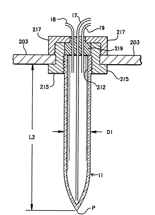
Fig. 5 illustrates a preferred design for the
extraction vessels 11 through 15. Each vessel is
constructed of glass and has a total length L2 of about
150mm, a maximum outside cross-sectional diameter Dl of
about 28mm, and tapers to a point P to acilitate the
removal of fluids. In the pre~erred mode, the vessel
is graduated and below a screw top 212 has a volume of
about 40ml. An example of such a vessel is a pyrex
conical screw cap centrifuge graduated tube available
as Corning stock number 8082. The fittings used to
hold the vessels in place in the rocker arm 203 are
typically constructed of three pieces: a threaded cap
217; a flanged coupling 215 having a inner threaded
opening to accept screw top 212, and an outer threaded
area to accept cap 217; and a teflon insert 219 which
is press fit into flanged coupling 215 and serves as an
inert stopper for the extraction vessel. Insert 219
generally has three holes therethrough to accomodate
the required liquids and gases. For example, for
extracton vessel 11, there is educting tube 17, a
pressure tube 18 which is coupled to gas manifold 72,
and a vent tube 19 coupled to vent manifold 74. A key
feature of this tube is the large change in surface
area of the fluid it contains which occurs when the
tube is reoriented from the vertical position to a
horizontal position, this change in surface area
enhancing the rate of phase separation of the emulsion.
Other tube shapes can also be used which can attain
this change in surface area, and in the general case,
the angle of rotation required to increase the surface
area of the fluid contained therein may not be 90.
Shown in Fig. 3 is a schematic representation of
the pressurized dialysis system 300 which is used for
further purifying and concentrating the nucleic acids
removed from the extraction vessels 11 through 15.
System 300 includes a manifold housing 351 and a bath
t 1 33~9~,~
-16-
vessel 350, both of which are typically rectangular in
cross-section, the bath vessel generally containing
about 8 liters of a dialysate 370. The bath vessel 350
and the manifold housing 351 both abut a cover 352
which is used to exclude foreign matter from the
dialysate, with vessel 350 sealed thereto via a seal
353, and manifold housing 351 connected thereto at one
side by a hinge 354 and by a clasp 355 at the other
side. Bath vessel 350 is generally vented to the
ambient atmosphere. In the preferred mode, the bath
vessel, the manifold housing and the cover are
constructed of plexiglas. Attached to the cover and
suspended through holes therein into the dialysate are
a plurality of dialysis bags 311 through 315 which are
typically in a corresponding relationship with
extraction vessels 11 through 15, coupled thereto via
valve block 51. Manifold housing 351 includes a bottom
piece 356 so that housing 351 is a closed container
except for holes through bottom piece 356 to accomodate
the dialysis bags, the holes for gas tubes 323 and 324
and for fluid lines 101 through 105 being sealed by
feedthroughs. Fig. 6 illustrates the details of the
suspension o the dialysis bags and the sealing system
so that pressure can be maintained in manifold housing
351 during dialysis. Generally, the dialysis bags are
first attached to a glass fitting independent of the
dialysis apparatus. For example, dialysis bag 311 i5
placed over a portion of a short piece of glass tubing
371 which is ground to a taper on one end. Then this
tubing 371 and the dialysis bag 311 are pushed firmly
into a mated piece of ground glass tubing 372 and given
a turn to effect a seal between the two pieces of
tubing thereby holding the dialysis bag firmly between
them.
The cover 352 is typically drilled to accomodate
the larger tubing 372 and the dialysis bag 311 and is
` -
1 335963
-17-
countersunk to accomodate an o-ring seal 374. The
dialysis bag and the glass fitting made up of tubing
371 and 372 is then placed through the hole. Bottom
piece 356 has a hole 375 which is aligned with the
glass fitting when the manifold housing is rotated to
the closed position about hinge 354. A grommet seal
376 is located in hole 375 and is used to affect a seal
between the glass fitting and the manifold.
The dialysis system 300 also includes a
recirculation system 330 having a recirculation tube
336 for extracting dialysate 370 from the bath vessel,
a peristaltic pump 337 for pumping the dialysate
through the recirculation system, a spectrophotometer
335 for monitoring the absorbance of the dialysate, and
a dual in-line filter 333 for filtering out phenol and
other organic materials. In a typical implementation,
filter 333 includes a carbonaceous filter 332 for
removing organic materials, and a mixed bed
ion-exchange resin filter 331 for removing inorganic
material. Also, spectrophotometer 335 typically
measures absorbance at 270nm to provide a measure of
the phenol remaining in the dialysate. ~he system
generally sets a flag when the absorbance (A270) drops
to 0.01 or below, indicating to the computer system
that the dialysis function is complete. During
dialysis operations, pressure in manifold housing 351
is maintained using an inert gas such as nitrogen and
is generally maintained at about 1200mm Hg (guage) when
using dialysis bags such as collodion bags from
Schleicher and Schuell having a volume of 2 to 8 ml.
Pump 337 typically provides a head of 15 psi and a flow
rate of about ll/min. Double distilled water i~
typically used as dialysate 370. The purpose of this
recirculation system is to allow the dialysate to be
reused instead of being replenished at regular
~ -18- 1 335963
intervals thereby further facilitating automatic
operation.
Fig. 4 shows a schematic representation of the
computer system 400 used for automatic control. The
system is made up of a microprocessor based computer
401 such as a Hewlett-Packard 85, which is coupled to a
converter system 403, for converting digital signals
from the computer 401 to analog signals to drive a
heater controller 409 for controlLing the heating of
the extraction vessels during extraction, and to
provide input signals to drivers 410, 411, and 412
which control the solenoids of the valve blocks, the
gas valves, and the peristaltic pump 436, respectively.
Converter 403 also serves as an analog to digital
converter for providing ~ignals to the computer 401
from spectrophotometer 33s and from conductivity meter
55, and from thermister 231 through 235. An example of
such a converter 403 is a Hewlett-Packard 3497
interface. The computer also provides signals to a
motor controller 405 for controlling a stepper motor
used to oscillate the extraction vessels during mixing
operations and to positon the extraction vessels
horizontally for phase separation. A typical example
of such a controller 405 is a Modulynx~ Motion Control
Interface Card, type lOD005A from Superior Electric.
Computer Software System
At the most basic level, software control of the
extraction apparatus is a matter of opening and closing
valves and turning switches on and off at the proper
times to achieve the desired flows of the various
materials from one vessel to another and to perform the
required operations. The fact that the method of the
invention is a sequence of steps lends itself
conveniently to software control. The following is an
- 35 example of a specific instruction set for each step of
the extraction metho~ which can be easily translated
~ 1 335963
--19--
into whatever programming language it is desired to
use. It is based on the assumption that reagent vessel
1 contains the lysis bufEer, reagent vessel 2 contains
the proteinase K, reagent vessel 3 contains the
phenol/chloroform/isoamyl alcohol, reagent vessel 4
contains chloroform, and reagent vessel 5 contains
RNase.
STEP 1: TISSUE DIGESTION
Command Number Command
Open valves 22; 50(port 91);
51(port 80, 81); 61.
Close all valves 4 minutes after
command 10.
Comment: Delivery of lysis buffer to
vessel 1 is now complete.
Open valves 24; 50(port 92);
51(ports 80,81); 61.
Close all valves 0.5 minutes
after command 30.
Comment: Delivery of proteinase K is now
complete.
Turn on heater 221 of vessel 1;
raise temperature to 55C and
maintain.
Turn on motor 201; angle of
rotation set for ~60 with
period of 1 second.
Turn off motor 201 and heater
221 3 hours after command 60.
Wait for cooling of digested
mixture.
STEP 2: EXTRACTION
90 Open valves 26; 50(port 93);
51(ports 80, 81); 61.
100 Close all valves except valve
61, 5 minutes after command 90.
-20- ~ 3 3 5 9 6 3
Comment: Delivery of the extraction mix-
ture (phenol/chloroform/isoamyl
alcohol) is now complete.
110 Turn on motor 201; angle of
rotation set for ~60; period 1
8 econd.
12U Turn off motor 201, 20 minutes
after command 110.
STEP 3: SEPARATE PHASES
1~ 140 Turn on motor 201, angle of
rotation set for 90.
150 Turn off motor 201 when angle of
rotation reaches 90.
Comment: Extraction vessel is now being
held in a horizontal position.
160 Turn on thermister 231; increa~e
temperature to 55C and
maintain.
170 Turn off thermister 231 12
minutes after temperature
reaches 55C under command 150.
STEP 4: RETURN EXTRACTION VESSSEL TO UPRIGHT
POSITON
180 Turn on motor 201, 12 minutes
after temperature-reaches 55C;
angle of rotation set for 0;
descent rate set for 9/second.
STEP 5: WITHDRAW PHENOL PHASE
190 Close valve 61.
200 Open valves 62; 51(port 81);
52(ports 71, 72).
210 Monitor conductivity with
conductivity meter 55.
220 1.0 seconds after conductivity
3 reaches 104 Mhos, close all
valves.
-21- 1 335963
Comment: The one second delay in command
220 after the conductivity goes
high is to ensure that residual
phenol left in the delivery
lines to valve block 52 has been
removed.
230 Open valves 52(port 74); 61.
240 Close all valves 30 seconds
after command 230.
250 Open valves 52(ports 71, 73);
39; 51(port 81); 61.
Comment: Command 250 backflushes valve
block 52 and forces aqueous
solution left in the delivery
lines back into extraction
vessel 11 for further extraction
or purification.
260 Close all valves.
STEP 6: REPEAT EXTRACTION PROCESS
270 Perform commands 90 through 250,
N times.
Comment: N, the number of extractions
performed, is chosen by the
programmer based on experience
and on the type of sample
tissue.
280 Open valves 28; 50(port 94);
51(port 80, 81); 61.
Comment: Command 280 adds chloroform to
extraction vessel 11 for the
final extraction.
290 Close all valves except valve
. 61.
300 Repeat steps 110 through 261 one
time.
~ -22- 1 33~963
STEP 7: REMOVE ~QUEOUS SOLUTION (To DiaLysis)
310 Open valves 51(port 81, 82); 62;
321.
320 Close all valves 5 minutes after
command 310.
Comment: The aqueous solution in
extraction vessel 11 is now in
dialysis bag 411.
STEP 8: DIALYZE AQUEOUS SOLUTION
330 Turn on pump 337.
340 Open valve 324.
350 Monitor A270 with
spectrophotometer 335.
15360 When A270 is less than or equal
to 0.01, close all valves and
turn off pump 337.
STEP 9: REMOVE RNA FROM AQUEOUS SOLUTION
(Optional)
20370 Open valve 324; open valve
51(ports 81, 82); 61.
380 Close all valves 5 minutes after
command 370.
Comment: The dialyzed aqueous solution in
dialysis bag 311 is in
extraction vessel 11.
39U Open valves 30; 50(port 95)7
51(ports 80, 81); 61.
400 Repeat Steps 90 through 360.
STEP 10: COL~ECT SAMPLE
410 Open valves 324; 51(port 82);
52(ports 71, 75).
420 Close all valves 5 minutes after
command 410.
Utility of the Invention
Examples. Preparation of DNA from Human LymphocyteS
S 1 335963
-23-
Lymphocytes are first washed from one unit of
whole blood and are resuspended in 4 ml balanced salt
solution. (Balanced salt solution is made up of 1
volume of a solution A and 9 volumes oE a solution B,
where solution A is 0.1% glucose, 5xlO-lOM CaC12,
9.8x10⁻⁴M MgC12, s.4Xlo~3M KCl, 0.145M Tris-HCl, pH
7.6; and Solution B is 0.14M NaCl.) Then 0.35 ml of
the lymphocyte suspension above is mixed with 4 ml
lysis buffer (lM NaCl, 1% SDS, 8M urea, 10 mM EDTA, 50
mM Tris-HCl, pE~ 8.0), and 1 mg of proteinase K in 0.65
ml lysis buffer to obtain a total volume of 5 ml. The
digestion is performed in a conical tube (extraction
vessel 11) as described earlier, at 55C for 3 hours.
About 5 ml of phenol/chloroform/isoamyl alcohol 50:48:2
is added to the tube and the two phases are mixed
according to the protocol for 20 minutes. The
extraction vessel is then rotated to the horizontal
position ~or 10 minutes at 55C to allow the phases to
separate. The e~traction vessel is then rotated slowly
back up to the vertical position over a period o~ about
10 sec~n~s, and the lower organic layer is removed to
waste. A second extrac~ioll is pe~Eo~ed ~ith the
pllenol/chloroform/isoamyl alcohol mixture and a third
extraction is per~orlned using 5 ml of chloroform. The
chloroEorm is relnoved to ~aste and the aqueous
DNA-containing layer is pressure transferred to a
dialysis bag, such as dialysis bag 311. Thi~ aqueous
solution is then pressure dialyzed according to the
protocol until A270 is below 0.01. The final DNA
solution is then educted from the bag and collected.
Following this process yields about 1 ml of DNA
solution containing about 250 micrograms o DNA The
resulting solution has an absorbance ratio A230:A260 of
0.52 and an absorbance ratio A260:A280 of 1.90,
demonstrating very high purity. (For absolutely pure
DNA, the absorbance ratio A230:A260 is 0.5+0.05 and
.~ _
S 1 335963
A260:~280 is 1.9+0.1.) Analysis of the sample using a
0.8% agarose gel and standard ethidium bromide staining
techniques shows a single band with a size greater than
50 kbase pairs (i.e., greater than 3.5~107 daltons).
Also most importantly, digestion with the enzyme Eco RI
is positive, indicating that the DNA is pure and
therefore restrictable, a very stringent test for DNA
purity.
Variations on the above example demonstrate the
importance of heating and increasing the surface area
to effect the phase separation step. For example, for
human lymphocytes, if the extraction vessel is not
heated, but is maintained at room temperature, and the
extraction vessel is not rotated to a horizontal
position, phase separation typically requires over 60
minutes. If instead the extraction vessel is heated to
55C but is not al~so rotated tv the horizontaL
position, the phase separation requires over 4 minutes.
~ith heating to 55C and rotation oE the extraction
vessel to the ~lorizontal position, the phase separation
typically re~uires only about 2.5 minutes. In each of
these variations, however, the high salt content is
enhancing the rate of phase separation by approximately
a factor of two.
While there has been shown and described a
preferred embodiment of the apparatus and method of the
present invention, it will be apparent to those skilled
in the art that many changes and modifications may be
made without departing from the invention in its
broader aspects. For example, it is apparent that the
extraction vessel need not be rotated to a horizontal
position to speed up phase separation, although it does
have a major influence. Similarly, the phase
separation can be performed at a temperature below the
preferred range of 45C to 55C, but it will proceed at
a slower rate, and the further below that range the
~`
1 335~63
-25-
slo~er the ra~e. In terms of apparatus it will be
apparent that automated devices according to the
invention can be constructed with either more or ~ewer
extraction vessels, reagent vessels, and dialysis bags.
~lso, some vaLves may be conveniently placed at
different locations in the apparatus, for example valve
bLock 52 may be placed ahead of conductivity meter 55.
However, this would result in some loss of the aqueous
phase when the flow is stopped. In addition, the
specific model numbers chosen for the various pieces of
apparatus included in the automated extraction system
are not meant to be restrictive as to the particular
models which can be used, but are offered by way of
example only. Also, it will be apparent that the
apparatus may be only partially automated, by providing
the fluid delivery system 100 and chamber/rocker system
200 independent o~ the dialysis system 300.