Note: Descriptions are shown in the official language in which they were submitted.
1 336064
METHOD FOR DETERMINATION OF A COMPONENT
OF A SAMPLE AND APPARATUS THEREFOR
FIELD OF THE INVENTION
This invention relates to an apparatus useful in
determining an analyte in a fluid sample. It also relates
to a method for determining an analyte in a sample, using
a four member, or "quaternary" complex involving the
analyte, a whole monoclonal antibody which binds to said
analyte, a labeled monoclonal antibody Fab fragment which
also binds to the analyte, where both of these are
obtained from the same animal species, and a solid phase
bound antibody which may or may not be monoclonal, which
binds to the Fc portion of a monoclonal antibody but not
to its Fab portion.
.
BACKGROUND AND PRIOR ART
The formation of sandwiches of antigen and antibody
and their use in immunoassays has been in use for over
fifteen years. The art has seen two distinct trends in
the field. The earliest trend was toward the formation of
ternary complexes, i.e., complexes of the form -
* *
Abl-Ag-Ab2 , where Ab2 carries some label. The later
trend is to multiple component systems, usually
quaternary, but sometimes involving five or more
components. The prior art discussion maintains this
distinction.
I. Ternary Complex Formation
The patent literature contains many examples of
inventions in this area. An early exampl~ of such an
assay may be found in Schuurs, et al., U.S. Patent No.
3,654,090 (1972), which is useful not only as historical
~"
-- 2 --
~ 33~064
background, but for an understanding of some of the key
facets of this field.
Schuurs, et al. teaches detection of an antigen using
a solid phase bound antibody against one epitope, or
binding site, of the antigen, as well as a soluble, enzyme
labeled antibody which binds with a second portion of the
antigen. The method disclosed in Schuurs, et al. involves
determination of the enzyme label after the sandwich
between bound antibody, antigen, and labeled antibody
forms. This is accomplished either in the solid phase, or
in the liquid phase, by addition of a substrate for the
enzyme label. Usually, the enzyme-substrate reaction
produces a color or change in color, which can be
recognized in "yes-no" tests, or quantitated where the
amount of substance present is to be determined.
Absent from Schuurs, et al. is an~7 discussion of
monoclonal antibodies or antibody fragments and this is
not surprising since Schuurs, et al. was filed in 1968,
and issued in 1972, i.e., much earlier than the
breakthroughs in hybridoma technology which occurred
following the development of the Kohler-Milstein method
for producing monoclonal antibodies.
Schuurs, et al. received another patent in 1974, U.S.
Patent No. 3,791,932, again directed to sandwich assays.
This patent describes a so-called "forward" sandwich
immunoassay. This type of assay calls for a specific
order of steps, i.e., the sample being tested is first
contacted with the insoluble binding partner and the
reaction between these two is allowed to proceed to
completion. The solid phase complexes are removed from
the solution, and the second binding partner, containing
an enzyme label, is then added to the solid phase.
Following binding to the complex, the enzyme level is
determined, following the standard techniques referred to,
supra. Again, thex~ is no mention of monoclonal
antibodies or antibody fragments.
1 336064
Ling, in U.S. Patent No. 3,867,517 (1975), taught
that enzymes were not the only label which could be used
in sandwich assays. This patent describes a forward
sandwich assaying as the label a radioactive antibody.
The radioactive label was 125I, a standard radioisotope.
Radiolabelling of antibodies is a standard technique, but
assumes the presence of the proper amino acids in the
antibody molecule for binding of the radioactive iodine.
Otherwise, the label does not hold.
Schuurs, et al., received yet another patent in 1977,
U.S. Patent No. 4,016,043. This patent claims to teach a
simpler version of rudimentary sandwich assays. It
teaches using an insoluble component of an antigen-
antibody reaction and a labeled sample of the same
component. This method assumes that the antigen being
detected has two identical epitopic sites. Further, the
use of two identical receptors precludes the use of
"simultaneous" assays, which are discussed infra. The
consequences of this is that the Schuurs ' 043 assay can
take as long as 60 hours to complete. In clinical or
diagnostic laboratory, the large amount of time requires
is unacceptable.
Piasio, et al., U.S. Patent No. 4,098,876 (1978)
taught a "reverse" sandwich assay. This patent is
important because it showed, first, that the component
being determined could be bound to the soluble, labeled
antibody first, and the immobilized antibody second. It
was also an improvement in that a washing step was
eliminated, which meant that time was saved in performing
30 the assay. Piasio, et al. teach that their assay could,
ideally, be completed in under one-half hour. This
paradigmatic system was not realized in their examples,
but the time was substantially less than the 60 hours for
Schuurs, et al., discussed supra. A significant drawback
of the method is that it requires enormous amounts of
immobilized antibody.
Niswender, U.S. Patent No. 4,048,298 (1977), is
actually not a sandwich assay, but shows an invention
where an immobilized antibody was used to bind another
antibody. This patent teaches an interesting variation on
older competitive immunoassays. Niswender contacts a
solid phase bound antibody with the sample being assayed
as well as a second, radiolabeled antibody which binds to
the first, but not to the component being determined. The
effect of this is to allow the investigator to determine
substance present by determining how much radiolabeled
antibody binds to the solid phase.
This patent shows that antibodies can bind to other
antibodies rather than just antigens. This property is
important in more recent assays, some of which are
discussed infra.
Schwarzberg, U.S. Patent No. 4,235,689 (1980)
recognized that antibodies possess two distinct portions,
the Fc portion, or "constant" region, and the Fab portion,
which is the part of the antibody which binds to an
epitopic site. Schwarzberg prepared complexes of labeled
Fab fragments bound to a ligand, such as a polypeptide.
This complex is then used in so-called "competitive"
assays. No solid phase binding, or sandwich assays, are
described.
Jeong, et al., U.S. Patent No. 4,244,940 (1981)
teaches a "simultaneous" sandwich immunoassay. Such an
assay requires an antigen with different epitopic sites,
because two different antibodies or receptors must be
used, for the reasons elaborated upon supra.
With Jeong, et al., it will be seen that by 1981 the
state of the art in this field did teach forward, reverse,
and simultaneous assay, always with ternary complexes
(i.e., complexes of three species) being formed. The art
had begun to see the use of Fab fragments as "linker"
molecules (Schwarzberg), but they had not been used as an
essential part of an immunoassay system, nor had
monoclonal antibodies been used.
6 ~
Both of these ideas were taught in patents which
issued in 1983. David, et al., U.S. Patent No. 4,376,110
(1983), overcame a prejudice in the art that monoclonal
antibodies were not "sticky" enough, i.e., possessed
insufficient affinity for use in sandwich assays. David,
et al., taught that all three forms of ternary sandwich
assays could be performed with monoclonal antibodies, as
long as they both had affinities of at least 108
liters/mole. Moussebois, et al., in U.S. Patent No.
4,397,060 (1983), taught an agglutination assay could be
performed using Fab fragments bound to a solid support.
This patent shows, yet again, that Fab fragments were not
being considered as partners of immunoassays, even though
monoclonal antibodies themselves were now being used.
Gallati, et al., U.S. Patent No. 4,467,031 (1984)
taught a specific sandwich assay, for determination of
carcinoembryonic antigen (CEA). The key feature of this
invention was the use of different salt concentrations to
improve complex formation. It is a "forward" sandwich
assay, as the term is defined herein, and discusses the
possibility of two monoclonal antibodies being used in the
assay. It will be seen that this, too, is a ternary
complex, and that an Fab fragment is not being used.
Woods, et al., U.S. Patent No. 4,469,787 (1984)
teaches a sandwich assay which requires the binding of a
label to the Fc portion of a second antibody. The label
is not directly attached to the second antibody, rather,
Woods et al. assert invention in that the label is bound
to the Fc portion of the antibody after the ternary
complex is formed. This is done so as to prevent
interference between the label and the immobilized first
antibody.
U.S. Patent No. 4,486,530 (1984), which issued to
David, et al., and is a continuation in part of U.S.
Patent No. 4,376,110, discussed supra, again teaches
ternary monoclonal antibody sandwiches and their
detection. This patent adds ~o the art by showing that
~ ~3~6~-64
sandwich assays can be performed in homogeneous phase,
i.e., without phase separation. This is performed by
labeling the monoclonal antibody components of the ternary
complexes with labels which do not react unless brought
together by the "glue" of a multiepitopic antigen.
Carro, et al., U.S. Patent No. 4,522,922 (1985)
combine sandwich assays with an older form of immunoassay,
the so-called "precipitation" test. This invention
teaches formation of a ternary sandwich, followed by
addition of a precipitating agent to precipitate the
complex out of solution. This is a radioimmunoassay,
which employs polyclonal antisera.
The most recent patents in the field show
modifications on the basic sandwich principle. Petska, in
U.S. Patent No. 4,623,621 (1986), teaches that an
oligomeric protein can be measured by using a solid phase
bound monoclonal antibody which is specific for an epitope
present once on the repeating protein portion of the
molecule. After solid phase binding, a second sample of
the same monoclonal antibody, only labeled, is bound.
Again, a ternary complex is formed, only with whole
antibodies, and simultaneous assaying is not possible.
II. Multiple Member Complex Formation
The earliest example of a quaternary system is
exhibited by U.S. Patent No. 4,343,896, which issued to
Wolters, et al. This patent which is based on a
disclosure filed in 1976, teaches the solid phase bound
complex Abl-Ab2-Ag-Ab3 . A crucial limitation in the
Wolters patent is that Ab2 and Ab3 come from different
animal species. The reason for this is because Abl has to
be directed against the constant region, i.e., "Fc"
portion of Ab2. All antibodies of a particular
immunological class which come from the same animal
species will have identical Fc portions. If Ab2 and Ab3
were from the same animal species, the art taught that not
1 336064
only would Abl-Ab2-Ag-Ab3 but one one would also obtain
Abl-Ab3 , both of which would bind to the solid phase,
causing interference and incorrect results.
Axen, et al., U.S. Patent No. 4,469,796 (1984)
teaches that more than three components may be involved in
an immune reaction, but the only four part complex taught
is a solid phase bound complex of Ag-Abl-Ab2-Ab3 . It is
noteworthy that in the description of reactants given at
column 1, lines 41-60, Axen, et al. never mentions Fab
fragments.
Tanswell, et al., U.S. Patent No. 4,624,930 (1986)
teaches four component complexes wherein a first and third
receptor in solution bind to the antigen while a second
solid phase antibody binds to the first antibody.
Tanswell's teaching is generic to the use of a double
antibody system and it does not specifically disclose
monoclonal antibodies.
Forrest, et al., U.S. Patent No. 4,659,678 (1987)
goes beyond the four part binding discussed supra, and
actually forms a pentavalent complex of antibody-hapten-
antibody-antigen-antibody. The tail end of the complex is
a radioactively labeled antibody. At least one antibody
must be a monoclonal antibody.
Forrest, et al. detail at some length the advantages
and disadvantages of multi-member complex forming assays.
The solution to the problems set forth at, e.g., column 2,
lines 1-5, is to use a solid phase bound mAb, to bind a
complex of Ab-Ag-Fab . The only time a solid phase bound
mAb is used to bind the complex mAb2-Ag-Fab , however,
Forrest requires that the mAb2 be found to another
antigen, so that the solid phase complex
mAbl-Ag2-mAb2-Agl-Fab is formed. It must be understood
in this context, however, that "Ag2" actually stands for a
linking agent, as mAb2 cannot possess two Ag binding
sites.
_ ~ 336064
SUMMARY OF THE INVENTION
This application is directed to a method for
determining an analyte in a fluid sample, involving
formation of a quarternary complex between a solid phase
bound antibody which binds to the Fc portion of a
monoclonal antibody but not to the Fab portion, a whole
monoclonal antibody which binds to the analyte, the
analyte itself, and a labeled Fab fragment of a monoclonal
antibody. It is also directed to apparatus which can be
used in such assays, but which are also useful in other
forms of assays including, but not limited to
immunoenzymometric assays, competitive assays, and
displacement assays.
How these and other aspects of the invention are
achieved will be seen upon review of the disclosure which
follows.
BRIEF DESCRIPTION OF THE FIGURES
Figure 1 depicts one embodiment of the invention, referred
to as the "1~ wick strip".
Figure 2 shows another embodiment of the invention,
referred to as the "double wick strip".
Figure 3 shows another embodiment of the invention
referred to as the "Delayed Physical Application System".
Figure 4 provides an embodiment of the invention referred
to as the "delayed diffusion application system".
Figure 5 is an embodiment known as the "loop strip".
Figure 6 shows an embodiment of the invention called the
"Integral Matrix Strip".
1 336064
Figure 7, shows a model of the invention known as the
"external pressure strip".
DETAILED DESCRIPTION OF PREFERRED EMBODIMENTS
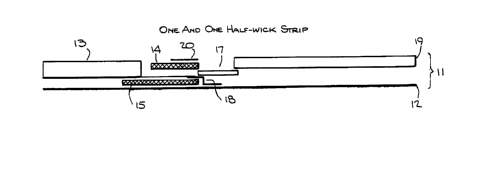
Referring now to Figure 1 a test strip 11 provided by
this invention is shown. A stable carrier foil 12 is
provided, which gives support to the entire apparatus.
Optional sponge 13 is shown, which may contain, e.g., a
buffer or other reagents useful in preparing the sample
for analysis. Sample may be applied to first zone 14, by,
e.g., pipette, and the sponge may be dipped directly into
a liquid, or liquid may be applied directly by, e.g., a
pipette.
The "first zone" is shown at 14, and contains at
least one of the analyte to be determined, an analogue of
this analyte, or a non-solid phase bound receptor which
binds to the analyte being determined. Various substances
are possible. Although it will frequently be the case
that the analyte being determined is an antigen, such as a
viral protein, a drug or drug residue, and so forth, other
substances may be determined, especially if the sample
being analyzed is not a biological fluid. The first zone
14 may also contain, in the case of a sandwich assay the
two monoclonal antibodies from the same species which bind
to the analyte being determined. When a sandwich assay is
being performed, one of the two monoclonal antibodies
contained in first zone 14 will carry a label, such as an
enzyme.
The "third zone" 15 contains a substrate for the
label carried by first zone 14. This substrate can ber
e.g., a substrate acted upon by the enzyme, such as a beta
galactoside when the enzyme is beta galactosidase. It may
be a substance which is necessary for the label to
function. For example, the substrate of the third zone
may be a substance which combines with the label of the
first zone to form a fluorescing moiety, or a functioning
- lo - 1 336064
-
molecule. For example, the label and substrate may be
halves of a complete enzyme which do not possess catalytic
activity until brought together.
The first and third zones must be kept separate from
each other, so that premature reaction between label and
substrate does not occur. This is achieved via the
blocking means 16, positioned between first and third
zones 14 and 15. This blocking means need not be made of
any particular material, as long as it prevents diffusion
between the zones in 14 and 15 until the sample has
entered the second zone.
The second zone 17 contains a solid phase bound
receptor which binds to any of the reagent in first zone
16 which does not react with analyte from the sample, or
when a sandwich assay is being performed, this second zone
contains a solid phase bound receptor which binds only to
the Fc portion of a monoclonal antibody.
An alternate construction divides this second zone
into two portions, one of which is Fc specific, and the
other which is not. In this case, formation of a signal,
of course, is related to whether the quarternary complex
formed, or did not. The non-specific matrix bound
antibody thus serves as a negative control.
It is to be noted that the first zone 16 and second
zone 17 must be in at least partial fluid contact with
each other. Second zone 17 and third zone 15 may be in
fluid contact, but need not be. The embodiment in Figure
1 actually shows no fluid contact between the second and
third zones, because of the presence of barrier foil 18.
This barrier foil serves to retard the passage of
substrate from the third zone into the second zone. This
permits whatever reactions are to occur between the solid
phase bound component and the unreacted reagent of the
first zone or sandwiches of mAb-Ag-Fab to occur without
premature formation of signal. As the substrate must,
eventually enter the second zone, barrier 18 is made of
fluid permeable material, preferably a polyvinyl alcohol,
1 336~
or material which, via contact with a surfactant or
surface active agent, is made fluid permeable.
Fourth zone 19 is in contact with second zone 17, and
receives excess sample and reagents therefrom. It acts as
a "waste receptacle" for the device as a whole.
An optional cover slip 20 is provided as well. This
gives additional stability to the device.
In Figure 2 a modification of the device of Figure 1
is shown. In device 21, all components are the same as in
device 11, except it will be noted that sponge 13 now
contacts first zone 14 directly, and does not contact
third zone 15 directly. Rather, there is partial fluid
contact between the first and third zones. Premature
contact of label and substrate is avoided by positioning
these at, e.g., the thatched positions 22 and 23, which
are separated from each other by barrier 16.
Figure 3 shows an embodiment where the optional
sponge 13 of Figures 1 and 2 is not used. Here, the
device 31 contains first zone 32, to which sample is added
directly. First zone 32 functions as does first zone 14
in Figures 1 and 2. It is in partial fluid contact with
second zone 33, which, of course, functions in the same
way as does the second zone 17 of Figures 1 and 2. A key
distinction between the device of Figure 3 and that of
Figures 1 and 2 is the placement and construction of the
third zone, which contains the substrate. As will be seen
by reference to Figure 3, the third zone, containing
portions 34 and 35, is below the second zone and is in
fluid contact therewith. The third zone contains
substrate in region 35, and is supported by layer 34.
When layer 34 receives fluid which has migrated
through component 43, it brings substrate 35 into contact
with the second zone. Blocking layer 44 prevents fluid
contact between zone 1 and component 43. Component 34 may
be, for instance, a compressed sponge which swells when
contacted by fluid.
- 12 -
1 3360f~
In this configuration, premature reaction of label
and substrate is not an issue, because by the time fluid
reaches the third zone and releases the substrate, any
reaction between the labeled reactant of the first zone
and the solid phase bound reactant of the third zone has
already taken place. The substrate diffuses into the
second zone, where the detectable moiety is formed.
Excess sample and reagents are carried into fourth zone
19, as in the embodiment of Figure 1, and the whole device
is again held together by carrier foil 12.
An optional feature presented by this device is the
covering means 36. The covering means allows for more
precise observation of the reaction going on in the test
strip. Generally, this covering means permits only
selective viewing by providing viewing means or "windows"
at various positions. Only one viewing means is actually
necessary, and this should be over the second zone 33, so
that formation of detectable moiety can be observed there.
If the covering means 36 contains additional viewing means
over fourth zone 19 one can Qbserve reaction between
substrate and labeled binding partner, e.g., or unreacted
labeled Fab fragment. Also, if covering means 36 is
adapted for use in, e.g., the device of Figures 1 and 2, a
viewing means can be provided at a point where zones 1 and
3 meet. This allows the investigator to determine if
premature mixing of label and substrate has occurred. The
covering means can be made of various materials, including
foils. It can also be an injection molded lid or cover
which is part of an injection molded case or container
means.
Figure 4 differs from the device of Figure 3, in that
protective layer 37 covers substrate 35 in the third zone,
and substrate diffusion into zone 2 is initiated by fluid
from zone 2 penetrating protective layer 37. This gives
greater assurance that prematuxe mixing of substrate will
not take place. The covering means 36, which, it has been
- 13 -
1 336064
pointed out, is optional, is not included in this
embodiment, although it could have been.
Figure 5, the "loop embodiment" depicts the
embodiment of the device where sponge 13 is used for
sample application, as in Figure 2, supra. The sample
passes into the first zone 14, where the reaction between,
e.g., analyte and binding partner or mAb, Fab , and Ag
takes place. The whole content of first zone 14 passes to
second zone 17, where either unreacted labeled substance
or sandwiches are picked up by the solid phase bound
reactant situated here. Anything not bound in second zone
17 is carried via means 38 through 39, which retains any
label. The excess portion of the fluid sample enters
waste 19 but, rather than being held here, the
configuration of the device is such that the sample is
forced into third zone 15, which contains the label
substrate. As the configuration forces passage into third
zone 15, it also precludes passage back to 19. Via means
52, the substrate containing material now passes through
barrier 53 back into second zone 17, where reaction of
solid phase bound labeled reactant and substrate takes
place. Barrier 53 is selected so that while sample can
pass through it from means 52, it cannot pass up from
second zone 17.
Figure 6 shows the "integral matrix" embodiment of
the device. In this embodiment, the second and third
zones essentially become one in matrix 42. The substrate
is incorporated into this matrix by means which may
include, but are not limited to, encapsulation. The
combining of the two zones in one matrix requires that the
substrate not be released until such time as the labeled
reactant from first zone 14 has reacted with solid phase
bound material contained herein.
The final pictured embodiment in Figure 7 shows
device 71. Here, all of the depicted elements are as in
Figures 1 and 2, except that in this embodiment the third
zone containing the substrate 15 is separated from second
- 14 - l 3 3 6 0 6 4
zone 17. Only by applying an external force to 15 can be
substrate be brought into contact with the solid phase
bound label.
Various assays may be performed in any and all of the
preferred embodiments shown in Figures 1-7. For purposes
of illustration, the mechanics of a different assay using
the device of Figures 1, 4, and 5 are set out, although it
will be clear to the skilled artisan that any and all of
these may be adapted for use in any of the devices.
In performing a test for the presence of thyroxin
(also called T4) in blood, e.g., a sample is applied to
first zone 14 of the device of Figure 1. Tap water is
applied to sponge 13 and migrates into third zone lS.i The
first zone contains T4 specific antibodies carrying the
enzyme label horseradish peroxidase, while zone 15
contains any of the standard horseradish peroxidase
substrates, such as orthophenylenediamine. The sample
begins moving toward second zone 17, which contains, in
solid phase bound and immobilized form, either T4 itself
or related molecule T3. In moving through zone 14, any T4
in the sample has reacted with the horseradish peroxidase
labeled T4 specific antibodies to form complexes. These,
together with uncomplexed antibodies wash into second zone
17 ahead of the fluid which travelled through zone 15.
The differential diffusion occurs because of the barrier
18.
While barrier 18 is dissolving, any uncomplexed
antibody reacts with the solid phase bound T3 or T4 in the
second zone 17, and the previously formed T4-antibody
complex passes into waste zone l9. Substrate for
horseradish peroxidase now passes into second zone 17,
where it reacts with the enzyme immobilized on the solid
phase. This produces a quantifiable signal, as will be
recognized by those skilled in the art. The amount of
enzyme caught by the solid phase is a measure of how much
T3 or T4 was in the sample.
- 1 336064
Similarly, one may perform a sandwich assay for,
e.g., carcinoembryonic antigen (CEA), a multiepitopic
substance, using the device of Figure 4. In such a test,
first zone 32 contains both mouse-anti-human CEA
monoclonal antibodies, and mouse-anti-human CEA monoclonal
antibody Fab fragments labeled with beta galactosidase.
Upon contact of first zone 32 with the sample, a sandwich
forms between the whole antibody (MAb), the CEA (Ag) and
the fragment (Fab ). This mAb-Ag-Fab sandwich, together
with unreacted mAb and Fab pass into second zone 33,
which contains, bound and immobilized to a solid phase, a
sheep-antimouse Fc specific antibody. This solid phase
binds both the sandwich described supra, as well as any
excess mAb. As Fab contains no Fc portion, however, this
is not bound, and passes into the waste zone. Meanwhile,
some of the sample has released the substrate resorufin
beta galactopyranoside, which moves into the second zone
33. This substrate reacts with the Fab fragments bound
in this region, giving an indication of the presence and
amount of CEA in the sample.
Using the device of Figure 5, one can perform a
competitive assay for determining if a subject has been
exposed to the HIV virus. This type of test assays for
antibody rather than antigen, so it shows that, for
purposes of this invention, these are equivalent.
Antibody to gpl20 of HIV which is conjugated to an
enzyme, such as a peroxidase, is incorporated into first
zone 14 of device 51. A serum sample which may contain
antibodies to HIV is introduced at sponge 13, and diffuses
into 14. The mixture of sample and conjugate passes into
second zone 17, which contains, immobilized in solid
phase, HIV gpl20 sufficient to bind all of the labeled IgG
if there is no other antibody present. Unbound conjugate
will pass via means 38 into trap 39, which removes any
free label from the sample. The remaining solution passes
through third zone 15, releasing substrate, which passes
via one way barrier into the matrix, where it reacts with
- 16 -
1 33606~
bound label. There is an indirect correlation - i.e., the
more label which bound, the less antibody there was in the
sample, and vice versa.
Different materials may be used in each facet of the
invention. As receptors, while antibodies are preferred,
additional materials such as protein A, and biotin-avidin
complexes, among others, can be used.
The immobilized receptor which forms the fourth part
of the quaternary complex may be any of the materials
listed supra, as long as it binds the first monoclonal
antibody and does not bind monoclonal antibody fragments.
Especially preferred are antibodies which bind to the Fc
portion of other antibodies, but do not bind fragments.
When an antibody is used as the solid phase, a
monoclonal antibody is preferred, although polyclonal
antisera can also be used. The species in which the solid
phase bound antibodies is generated is not important as
long as there is no cross reactivity between the first
receptor and the monoclonal antibody Fab fragment. The
monoclonal antibody which binds to the antigen and the
monoclonal antibody Fab fragment do derive from the same
species, however.
The label used on the Fab fragment may be any of the
conventional labels used in immunoassays, but especially
preferred are enzymes which react with their substrates to
form colored substrates. Examples of such enzymes are
beta galactosidase, horseradish peroxidase, alkaline
phosphatase urease and amylase, although it will be
recognized that these are only examples and are not to be
read as limits on what enzymes can be used. It will be
clear to the skilled artisan, that when avidin is the
matrix bound receptor, a biotinylated monoclonal antibody
can be used. When this is the case, the labeled component
need not be a Fab fragment, but can be a whole mAb.
The position~ng of the labeled Fab fragment or mAb
and first unlabeled or biotin/avidin labeled antibody in
the first zone is not a critical feature of the invention.
- 17 - 1 3 3 6 0 ~ 4
These can be positioned so that the sample reaches one
before the other, or so that there is simultaneous
contact.
The material of which the device is constructed can
include many different items. Of course, the various
zones must be absorptive of liquids and possess good
capillarity. Examples of such materials are bibulous
paper, nitrocellulose paper, sponges, and other absorptive
materials. These may be fibrous or not, and the different
zones can be composed of different materials possessing
different degrees of capillarity, absorption, and so
forth.
The receptor is immobilized via any of the standard
means known in the art for immobilizing such receptors,
e.g., to a solid support, such as by fixing with cyanogen
bromide.
As mentioned supra, when the barriers are used in the
apparatus, they must be chosen so that they permit fluid
passage. Inert polymers are preferred, and especially
preferred is polyvinyl alcohol (PVA). Other suitable
materials will be evident to the skilled artisan.
When the cover means is used, its openings must be
open, transparent or translucent. One preferred material
for this is transparent mylar, while the rest of the cover
can comprise suitably sturdy material, such as metallic
foil may or may not be covered with a transparent
material.
The additional features of the invention, such as the
support and the impermeable barrier between the first zone
and the substrate zone comprise conventional materials
known to the art.
The following example illustrates the operation of
the invention, but is not to be read as any limitative of
the preceding discussion.
- 18 -
7 3~0~4
Example
An apparatu~ for determining human chorionic
gonadotropin ~hCG) was prepared and teste~.
A piece of 4210 paper (Fi~ma Kalff) was cu~ into a
strip 2.6 cm long and .6 cm wide (first zone). One end
was impregnated with 10 ~l PBS buffer (pH 7.0, 1% BSA, .1%
Tween ~0), and its center portion was impregnated with 7.5
~l o~ a solution containing 20 ~/ml of a conjugate o~ an
Fab portion of a monoclonal antibody against hCG and beta
galactosidase. The monoclonal antibody fragment had no
cross reactivity against luteinizing hormone. This
; portion was also impregnated with 75 ~1 of a 100 ~g/ml ;~
solution of a monoclonal antibody against the beta chain
of hCG. The end of the strip opposite the buffer
impregnated end was impregnated with 10 ~l of an aqueous
solution of 5% polyvinyl alcohol. The resulting strip
overlapped .5 mm of a strip of 3512 paper from Schleicher
& Schull (second zone) which wa~ 1 cm long and .6 cm wide.
This paper had been activated using cyanogen bromide, and `
20 a sheep antibody against the Fc portion of mouse ;;
antibodies was fixed thereto. This strip overlapped .5 mm
of a 5 cm long and .6 cm wide strip of D28 paper
(Whatman), impregllated with 150 ~l o~ an aqueous soluti~n
of 18% polyvinyl alcohol (waste zone). The three ~trips,
overlapped as indicated to form a continuous strip, were
mounted on a 10 cm long, .6 cm wide strip of polystyrene
using adhesive tape. The strips thus produced were
dipped, one each, into urine samples calibrated as
containing 0, 100, 250, and 500 mIU/ml hCG. After 5
minutes, each strip was dipped into a solution of .8 mmol
resorufin beta-galactopyranoside in 100 mmol Hepes buffer
(pH 7.5), and allowed to develop for 5 minutes. All
strips dipped into hCG containing urine exhibited bright
fuchsia color at the second and waste zones, while the
strip dipped in the sample containing no hCG was yellow in
the second zone and fuschia in the third zone. The change
,~ ,
-- 19 --
- 1 336064
in color is indicative of the action of beta galactosidase
on the resorufin beta galactopyranoside in the second zone
and waste zone.
The foregoing example, it will be seen, could be
modified very easily by, e.g., having the resorufin beta
galactopyranoside impregnated into a separate zone in the
manner described supra, and the development of the color
change could be observed through a covering means as has
also already been described.
While there have been described what are at present
considered to be the preferred embodiments of this
invention, it will be obvious to one skilled in the art
that various changes and modifications may be made therein
without departing from the invention, and it is,
therefore, aimed to cover all such changes and
modifications as fall within the true spirit and scope of
the invention.