Note: Descriptions are shown in the official language in which they were submitted.
-1- 1 337495
M~TE~OD FOR TRF:P~TING E~A~ING DF:FICI~ICIF..':
This invention relates to the treatment of
hearing impairment in humans. More particularly, the
invention relates to an improved method and an improved
electronic hearing aid for effecting such treatment.
Two common types of hearing aids are tbe so-
called bone conduction devices and devices which
directly stimulate the tympanic membrane or ear drum.
A third type of device, utilized for direct neural
stimulation, is also sometimes employed. All three
devices have their strengths and weaknesses insofar as
their effectiveness in the treatment of hearing
impairment. Probably the most commonly used of these
devices is the air conduction type of device, which
uses a speaker to vibrate the air in the ear canal
adjacent the tympanic membrane.
The air conduction type of hearing aid
generally employs suitable electronics for amplifying
incoming sound waves, perhaps also with some processing
of the sound waves to change the shape of the response
curve. Reproduction of such sound waves by the speaker
stimulates the tympanic membrane.
Another type of hearing aid, which also
stimulates the tympanic membrane, employs a varying
magnetic field. The magnetic field displaces a piece
of magnetic material fixed to the lateral surface of
the membrane, thus displacing the membrane itself.
Many persons who could benefit from the use of
air conduction or magnetic hearing aids refuse to
utilize them. Typically, this refusal is based upon a
perceived stigma associated with hearing aids. Because
most air conduction or magnetic type hearing aids are
readily visible on the wearer, despite advances in
miniaturization, and because many people associate
hearing loss with aging and enfeeblement, the use of
such hearing aids is often avoided by persons who need
-2- 1 3374q5
them. In addition, where miniaturization of such
devices has been accomplished,such as to reduce
visibility, the performance of the device is often
compromised.
Many miniaturized air conduction hearing aids
are constructed with an outer housing which is molded
to conform to the contours of the auditory canal.
However, such devices are usually readily visible on
the wearer due to the inability to miniaturize such
devices to the extent that they may be recessed
substantially within the auditory canal as to be not
readily visible while still providing the degree of
performance necessary to effect a significant
improvement in hearing.
It is an object of the present invention to
provide an improved method and apparatus for treating
hearing impairment in a human.
Another object of the invention is to provide
an improved method and apparatus by which a hearing aid
of the air conduction or magnetic type may be made
essentially invisible on a wearer while still retaining
adequate performance to substantially assist hearing.
Another objection of the invention is to
provide an improved method and apparatus for
constructing and utilizing a miniaturized air
conduction or magnetic type hearing aid.
Other objects of the invention will become
apparent to those skilled in the art from the following
description, taken in connection with the accompanying
drawings wherein:
FIGURE 1 iS a view of the external portion of
the right ear of a human wearing the hearing device in
accordance with the invention;
FIGURE 2 is a schematic horizontal cross
section view of a human ear canal, taken along a plane
on the line 2-2 of FIGURE 1, illustrating the hearing
_3_ ~ 3374~5
aid device of the invention in position;
FIGURE 3 is a vertical cross sectional view of
the ear canal of a human, taken along a plane on the
line 3-3 of FIGURE 1, illustrating the hearing aid
device of the invention in position;
FIGURE 4 is an exploded perspectivç view of a
hearing device of the invention showing use of the
insertion and removal tool used therewith, and,
FIGURES 5 and 6 are schematic top and side
views, respectively, illustrating the locking
projection used on the hearing device in accordance
with the invention.
Very generally, in following the practices of
the present invention, the external auditory canal of a
human patient is substantially and surgically enlarged
in a region proximate the ear drum. In the enlarged
region, an electronic hearing aid is placed. The
hearing aid has an external housing which is molded to
conform with the shape of the enlarged region.
Accordingly, the hearing aid may be made sufficiently
large as to accommodate the electronics necessary for
satisfactory hearing assistance, while at the same time
remaining invisible to external view of the wearer.
The present invention makes it possible to use
a relatively larqe device, since, due to the surgical
enlargement of the auditory canal, more space volume is
available for the device, while at the same time
permitting the device to be positioned deeply within
the auditory canal and thus out of sight. The device
is not an implanted device in the sense of many prior
art surgically implanted hearing aids, since it is
positioned in the auditory canal. Such positioning
enableæ it to be readily inserted and removed for
cleaning, adjustment, servicing, etc.
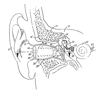
Referring now particularly to FIGURES 1 - 3,
the cross section of the human ear region is
- _4- 1 ~ 3 7 4 9 5
illustrated. Visible in the drawings, particularly
FIGURE 3, are the outer ear flap or pinna, the outer
skin and tissue 13, the mastoid area 15, the temporal
bone 17, the malleus 19, the incus 21, the stapes 23,
the cochlea 25, the cochlea nerve 27, the middle ear
promintory 29, the tympanic membrane 31, and the outer
auditory canal 33. As is well known, the ossicicular
chain comprises a malleus 19 which normally moves in
response to the tympanic membrane or ear drum 31. The
malleus is in turn connected to the incus 21, which is
connected to the stapes 23 which stimulates the cochlea
to produce neural transmission via the cochlea nerve
27.
The procedure by which, in accordance with the
invention, the outer ear canal is enlarged is designed
to be performed by an otolaryngologist trained in the
fundamentals of reconstructive ear surgery. The
procedure begins with an intraconchal incision and
separation of the canal skin from the underlying
fibrous and cartilaginous components to be removed. A
post auricular incision is made to facilitate
recontouring of the bony canal. Canal skin flaps are
developed and the bony canal is enlarged and
recontoured with suitable burrs and suction irrigation.
The canal skin flaps are returned to the new canal, and
a bolster is used to maintain adequacy of the meatal
opening during healing.
The preferred surgical procedure is as
follows:
The ear canal recontour procedure is performed
with the patient sedated but awake. The area in and
around the ear is cleansed, prepped, and prepared for
surgery. Pain is controlled with local injections of
analgesics.
The objective of the operation is to remove an
adequate amount of the meatal cartilage and
_5_ 1 337495
subcutaneous fibrous tissue, and bone of the bony
portion of the external auditory canal, while
maintaining all external auditory canal skin.
An initial crescent shaped incision is made in
the lateral surface of the auricular skin and is
carried down to the conchal cartilage. The plane
between the cartilage and the skin is dissected
medially until the medial extent of the posterior
meatal cartilage is reached. From that point, sharp
dissection is carried medially separating the
posterior, superior and inferior canal skin from the
deep subcutaneous and fibrous tissue that lies between
it and the bony external auditory canal. This
dissection is carried medially to the level where the
skin becomes more directly adherent to the bone of the
canal.
An incision is made through the cartilage
about three millimeters medial to the original skin
incision and carried superiorly along a line parallel
and immediately adjacent to the anterior edge of the
antihelix. This incision is then carried more deeply
through the subcutaneous meatal fibrous tissue to the
bony meatus defining the tissues to be removed later.
Three incisions are then made in the external
auditory canal skin. The first begins approximately
three millimeters lateral to the pars flaccid area of
the tympanic membrane, and is extended laterally into
the incisura area of the superior meatus. The second
begins about three millimeters from the tympanic
annulus at six o'clock and is brought laterally to
about 0.5 centimeters beyond the bony cartilaginous
junction. A third incision in the posterior canal skin
connects the medial extent of the first two incisions.
The posterior canal skin flap, thus
delineated, is elevated from medial to lateral with a
back angled elevator.
-6- 1 337495
Entering the anterior edge of the superior
canal incision, the superior meatal skin is separated
from the subcutaneous and fibrous tissue and the
anterior superior cartilage with sharp dissection.
These tissues are removed.
A post auricular incision is made
approximately one centimeter behind the post auricular
fold and the skin is elevated from the periostial and
fibrous tissue overlying the mastoid bone anteriorly
until the Spine of Henle and the posterior bony meatus
are encountered. About one centimeter posterior to the
Spine of ~enle a curvilinear incision is made into the
the investing fibrous tissue over the mastoid. The
fibrous tissue posterior to this inferior-superior
incision is elevated about three millimeters and the
fibrous tissue anterior to the incision is removed.
The elevated posterior tissue provides a stable
anchoring site for a bolster placed near the end of the
procedure.
The posterior canal skin flap developed
earlier is then lifted out of the canal and folded
laterally on its pedicle to reside temporarily within
the meatus. The postauricular incision is held open
and the posterior canal skin flap is held in place
within the meatus with a self retaining retractor, for
example, a Perkins' Tympanoplasty Retractor.
The pad of cut meatal cartilage, subcutaneous
and fibrous tissue earlier delineated is then removed.
Attention is then turned to widening of the
bony posterior canal wall. Using both cutting, and
diamond burrs, the bone of the posterior canal is
enlarged. A small amount of bone pate is saved for
later use.
An incision is made into the anterior canal
skin from the inferior to superior, about five
millimeters lateral to the tympanic annulus. The skin
_7_ 1 3374~5
lateral to the incision is elevated from the anterior
canal bony wall, to the point where it becomes adherent
to the cartilage of the anterior canal. Skin medial to
the incision is elevated several millimeters toward the
annulus to protect it from damage during drilling.
The bony anterior canal wall is then
recontoured with burrs and suction irrigation. At the
medial extent of the recontouring, a soft shoulder is
created, about five millimeters from the tympanic
membrane. Posterior and anterior canal wall
recontouring are merged resulting in a canal that is
enlarged and recontoured in all dimensions. The
recontoured canal is usually adequate when the lateral
diameter is about two centimeters and the mid canal's
diameter about one centimeter.
During the recontoùr procedure, caution is
exercised to avoid excessive widening of the ear canal
medially into the corda tympani nerve; anteriorly, the
temporal mandibular joint; and inferiorly, the facial
nerve.
! The anterior canal skin flap is replaced over
the recontoured canal bone. In order to maintain an
adequate meatal opening during healing, a specially
designed bolster is used. The bolster is made of a
low-resilliance foam covered with a thin layer of
Silicone rubber. To secure the bolster a 2-0 Tevdek (a
trademark) suture is passed through the superior
portion of the posterior canal skin flap and through
the æuperior portion of the previously created fibrous
anchor. It is then passed forward back through the
inferior portion of the anchor and back through the
inferior portion of the posterior canal skin flap.
Bone pate collected earlier is used to fill
any exposed mastoid cells.
The post auricular incision is closed with
subcuticular Vicryl (a trademark) sutures.
-8- 1 337495
The anterior and posterior canal skin flaps
are packed into place with chloramphenicol soaked in
Gelfoam PledgeS (a trademark). The bolster is
introduced into the meatus and tied in place with the
Tevdek Suture.
Half inch adhesive strips are placed over the
post auricular incision and a mastoid dressing is
applied. The dressing is removed by the patient at
home the following day.
The bolster remains in place two weeks and is
removed by the surgeon. One end of the suture emitting
from the posterior canal skin flap is cut flush with
the skin, the bolster is removed and the suture is
pulled out. The canal is then cleared of Gelfoam (a
trademark) and debris with a sterile suction tip.
Antibiotic ear drops are used for several days
and the patient is seen every week or two until healing
occurs.
Following surgical enlargement of the outer
ear canal as described above, an impression of the
recontoured canal is made. Typically this impression
is taken about 2 to 3 months after surgery, permitting
sufficient healing of the surgically modified region.
From this impression, the outer housing of the hearing
aid device itself is formed, as described below, so as
to fit the contours of the surgically enlarged ear
canal.
The volume of the surgically enlarged region
is of significance in practicing the invention. The
volume must be substantial enough to accommodate the
hearing aid as described below, and is preferable kept
substantially uniform in size and shape from patient to
patient to enable more uniformity in procedure and
manufacture. Too large a volume is undesirable in that
it involves a bulkier device and more extensive
surgery. A volume of two cubic centimeters is
9 1 3374q5
preferred:
The finished hearing device is moistened with
an antibiotic ointment and inserted. If the device is
comfortable, it is then worn with progressively longer
duration over the next few weeks.
FIGURES 1 - 3 show the hearing aid device 35
positioned in the surgically modified ear canal. It
will be seen that the device 35 is of sufficient size
to contain components adequate to provide superior
performance, while at the same time, due to the depth
which the devices recessed in the ear canal, the device
remains essentially invisible to outside observation.
The shoulder 37 (FIGURE 2) formed by the surgery
prevents the device from becoming dislodged and
engaging the ear drum, while the exterior contours of
the housing 38 of the device, since they are molded to
fit the surgically enlarged region, assist in retaining
the device firmly and comfortably in position.
As previously mentioned, once all healing of
the surgically modified ear canal has taken place, an
impression of the ear canal is taken. A general
procedure for making an earmold from an impression is
described in Chapter 21 the ~Basic Course for
Independent Study" published by the National Hearing
Aid Society. Unlike impressions made from prior art
hearing devices, where the impression is taken of the
pinna of the ear and continues to the external auditory
meatus opening, the impression taken in accordance with
the invention is from the ear drum itself out to and
beyond the external auditory meatus.
In order to prevent undue pressure on the ear
drum during taking of the impression, the material used
is of a low viscosity. The low viscosity also permits
the impression material to be inserted into the ear
canal while allowing the air therein to escape, thus
preventing the trapping of air bubbles which might lead
-lo- 1 337495
to an inaccurate impression. In addition to low
viscosity, the impression material should have a high
tear strength to prevent it from breaking or tearing
during removal, yet must have sufficient flexibility to
permit the impression to be readily removed from the
ear canal. It is preferred that the material have a
relatively short set up time, for example, 5 to lO
minutes, and be dimensionally stable so as to permit
the production of an accurate external shape for the
housing of the device. Due to the nature of the tissue
in the region where the device is positioned in a
patient, it is important to have accurate dimensional
stability and shape, since the tissue in this region is
not as compressible as the outer portions of the ear
where prior art devices are typically worn. It has
been found that a preferred form of impression material
is polyvinylsiloxane.
After the impression is made, the impression
itself is trimmed back approximately 2 or 3 millimeters
from the ear drum. This conforms to the point where
the shoulder is created by the physician during the
surgery. A lacquer coat is then brushed onto the
impression to provide a smooth finish. The impression
is then mounted to an investment casting base and an
2S investment mold is formed using a suitable investment
type process. Prior to this, the impression is
detailed in such a way as to account for any
imperfections and to provide a smooth surface and to
remove any rough or sharp edges.
Preferably, in making the investment mold, the
impression is mounted to an investment base with a
sticky wax, and the assembly is vibrated to insure that
the base of the sticky wax has adhered to the
impression. The impression is then coated with a
suitable separating oil and the investment container is
filled with an investment plaster. The plaster is
-11- 1 337495
permitted to harden, typically one-half hour, and the
impression is removed from the investment housing.
After curing of the plaster, such as heating in an oven
for approximately 15 to 20 minutes at approximately 92
to 100C, the mold is removed from the oven and allowed
to cool and dry.
Once the mold is completed, the housing for
the hearing aid device itself is molded. The housing
is formed using a polymer and monomer slurry to form
the biocompatible material in which the electronics of
the hearing aid are housed. A suitable material is
Audacryl, a trademark of Esschem of Essington, New
York. The formation of the housing or shell is such as
to provide a dimensionally accurate exterior surface,
and a thin continuous wall thickness with enough
strength to prevent the shell from being destroyed and
to protect the electronic contents. This is done by
subjecting the investment and the shell to a suitable
air pressure.
In a preferred procedure, the acrylic monomer
and polymer are mixed, slowly to avoid inducing air
into the mixture, and the mixture, at a temperature of
about 60F, is poured into the mold. The excess
material is poured out after approximately 3 to 5
seconds, with the mold being maintained at A
temperature of between 150 to 180F. This allows for a
thin, even coat of acrylic in the mold. Then the
polymerization process is begun. The mold container is
closed to form an air tight compartment and
approximately 30 pounds per square inch of air pressure
are injected into the container. This insures a
consistent even wall thickness throughout the shell.
After approximately one-half hour, the investment
container is opened and the shell is trimmed and
polished as necessary.
Once the shell or housing 38 iR formed, the
-12- ~ 33749~
electronics of the hearing aid device 35 are assembled
into the shell. The electronics include a microphone
39, a speaker 41, (or, in the case of a magnetic type
of device, a magnetic drive coil) and an
amplifier-signal processing section 43. The
microphone, speaker and amplifier are all mounted on a
ceramic support 46 inside the housing 38. In addition,
a face plate 45 with a battery door 47 and battery
contacts (not shown) is provided. It is preferred that
the device contain a suitable remote control (not
shown) to operate the volume and perhaps other aspects
of the device, since the device is inserted deeply into
the ear canal and is not readily accessible manually.
The microphone 39 is mounted in a microphone boot (not
shown) of a resilien-t type of material supported on the
face plate 45. The speaker 41, with a suitable shock
resistant type outer coating, not shown, is mounted
near the end of the device adjacent the ear drum.
Since the interior shape of the ear canal is
modified surgically, a more standardized cavity can be
developed, thus permitting a more standardized shape
and configuration for the hearing aid device 35. This
contributes to a more easily manufactured product.
The face plate 45 of the hearing aid has
provision for insertion and removal of the device from
the ear. The face plate, which is manufactured of
molded plastic, is bonded to the housing material and
is provided with a locking projection 49 which extends
outwardly from the external planar face of the face
plate. Referring to FIGURES 4 - 6 the locking
projection has a pair of opposed planar side 51 and a
pair of opposed partially cylindrical sides 53. The
cylindrical sides 53 are undercut at 55.
An insertion and removal tool 57 (FIGURE 4),
which is also cylindrical, is used by the wearer to
insert and remove the device. The tool 57 contains a
-13- 1 3374~5
pair of tabs 59 which project inwardly from the inner
surface of the cylindrical wall of the tool. The tabs
59, upon turning of the tool clockwise, slide under the
undercuts 55 formed in the cylindrical walls of the
locking projection.
Once the tabs 59 slide under the walls into
the undercuts, by turning the insertion and removal
tool one-quarter turn, the entire hearing device 35 may
be gently pulled outwardly to remove the device from
the ear canal. Conversely, when the device is inserted
into the ear canal, the insertion and removal tool may
be unlocked from the locking projection by turning in
the opposite direction, i.e. counterclockwise, until
the tabs slide out from the undercuts and clear the
planar sides of the projection. To provide a secure
lock, rubber pads or other cushioning, not shown, may
be provided on the interior wall of the insertion and
removal tool. When the tool is pressed into place, the
pads will then be compressed, giving a secure lock.
The insertion and removal tool may be provided with an
appropriate marker, not shown, to orient the tool in
the correct fashion in the wearer's hand. Thus, with
the tool properly oriented and inserted into the ear,
the tool may be readily seated on the locking
projection and then turned as appropriate.
The design of the electronics may be of any
suitable configuration to provide the desired hearing
assistance. Various hearing aid devices which operate
electronically, including devices which are adjustable
by remote control, are wetl known in the art and will
not be described in detail herein. Reference is made
to the description of an electronic hearing aid in the
book ~The Hearing Aid, Its Operation and Development~,
3rd Edition, 1984 authored by R.W. Berger and published
by the National Hearing Aid Society in Chapter 5,
entitled "Hearing Aids Today and Tomorrown.
-14- 1 337495
The hearing aid methodology and apparatus
described herein provide a significant improvement in
the treatment of hearing deficiency. The hearing aid
device may be conformed to reside completely within the
external auditory canal and can be removed and
reinserted easily by the patient. With the device
properly inserted, it is not readily visible by
external observation. Moreover, the device is capable
of accommodating sufficient electronics and power
supply as to provide a high quality device without
external obtrusiveness. A smaller air chamber may give
longer battery life because there iB Bmaller column of
air to vibrate. Sound reproduction may be better
because of fewer resonances off of soft tissue in the
ear canal.
Various modifications of the invention will
become apparent to those skilled in the art from the
foregoing description and accompanying drawings. Such
modifications are intended to fall within the scope of
the appended claims.