Note: Descriptions are shown in the official language in which they were submitted.
- 1 339005
Method of AssaY
This invention relates to assay techniques and to
means for putting such techniques into effect. In
particular it relates to an improved assay technique
which provides an enhanced signal to noise ratio and
enhanced sensitivity.
The assay techniques with which the present
application is concerned are based on the affinity
between the species which is to be assayed (hereinafter
called "ligand") and a specific binding material for the
ligand (hereinafter called "specific binding partner")
which is coated on a particular type of surface. Such
techniques are well known in the art, particularly in
relation to coated optical structures whereby binding of
the ligand to the specific binding partner results in a
detectable change in the optical characteristics of said
optical structure and have been described, for example,
in EP-0112721 (to Layton et al, published on July 4,
1984) and EP-0178083 (to North et al, published on April
16, 1986). The present invention provides an alternative
method of assay with considerable advantages over the
conventional assays.
In its broadest aspect, the invention is concerned
with improvements to a method of assaying for a ligand in
a sample which involves
a) incubating the sample in contact with a specific
binding partner for the ligand it is desired to
detect carried on one surface of an optical
structure;
b) irradiating another surface of the optical structure
at a suitable angle or range of angles to the normal
such that resonance and/or total internal reflection
of the radiation occurs within the optical structure
and/or the layer of specific binding partner; and
- 2 - 1 339 0 0 5
c) analysing the reflected and/or generated
radiation in order to determine whether,
and if desired the extent to which and/or
rate at which, the generated radiation and/or
optical characteristics of the optical structure
are altered by complex formation.
It is to be understood that the term generated
radiation as used herein includes fluorescence
and phosphorescence. Although the invention is
described hereinafter with particular reference
to fluorescence, it applies also to phosphorescence.
The optical structure comprises a substrate
and optionally one or more layers of material interposed
between said substrate and the layer of specific
binding partner carried on the surface of said
optical structure. Generally the layer of specific
binding partner may be either continuous or discontinuous.
However, in some embodiments of the invention as
hereinafter described and in particular where the
specific binding partner is carried directly on
the substrate surface of the optical structure
without any intervening layers of material therebetween,
a continuous layer of specific binding partner
may be preferred.
Unexpectedly, the method of the present invention
has been found to be of general applicability for
increasing the sensitivity of both direct and indirect
sensing methods of assay based on the optical properties
of certain surfaces. Direct sensing in the context
of the present invention involves monitoring the
modulation of a signal resulting from a biochemical
reaction (e.g. antigen/antibody binding~. Indirect
sensing involves monitoring a label (e.g. a fluorophore)
by a transducer in order to quantify a biochemical5 reaction.
The technique makes it possible to enhance
substantially the intensity of the electric field
t 339005
-- 3 --
at the surface of the optical structure thereby
enhancing the interaction between the exciting
radiation and the ligand/specific binding partner
complex, so maximising the response to complex
formation at the surface of the optical structure
and, in those embodiments employing indirect sensing
techniques, significantly reducing the background
signal levels.
Thus, where a direct sensing method is used
based on the change in refractive index of the surface
layer carried on an optical structure upon binding
of the ligand under assay, the method of the present
invention may be applied to enhance the surface field
intensity produced by the incident radiation source
and to sharpen the resonance(s) associated with the
coupling to modes which propagate in said surface
layer.
Thus, in one aspect, the invention provides
a method of assaying for a ligand in a sample which
method comprises the steps of
(a) incubating the sample in contact with a specific
binding partner for the ligand it is desired
to detect, the said specific binding partner
being carried on one surface of an optical
structure, said optical structure comprising
a substrate and one or more layers of material
interposed between said substrate and the said
specific binding partner;
(b) irradiating another surface of said optical
structure such that the radiation is totally
internally reflected and resonance occurs within
the optical structure, said resonance being
either long-rangé surface plasmon resonance
or resonant guided mode coupling; and
(c) analysing the reflected radiation in order
" 1 339~05
-- 4
to determine whether, and if desired the extent
to which and/or rate at which, the optical
characteristics of the sensor formed by the
optical structure and specific binding partner
carried thereon are altered by formation of
a complex between the ligand and the specific
binding partner.
For example, in one embodiment of the invention
the incident radiation may be coupled to a guided
mode which can be supported by an optical structure
of a particular geometry. Optical structures of
suitable geometry have been previously disclosed
in US4649280 in connection with a fluorescent immuno-
assay. However, it has not hitherto been appreciated
that similar techniques could be applied to a direct
sensing method of assay. Preferably, the optical
structure comprises a transparent (at least at the
wavelength of radiation used) substrate coated with
a thin metal layer such as silver or gold, which
metal layer is itself coated with a layer of dielectric
material such as silica. The layer of dielectric
material is of a thickness, for example of the order
of a wavelength of the incident radiation used, suffic-
ient to support one or more guided modes but it is
particularly preferred to employ a thickness of dielec-
tric material which will support only a single guided
TE or TM mode. For example, for a substrate of refractive
index 1.52 with a silver layer of 50 nm and an incident
radiation wavelength of 543 nm, the thickness of
a dielectric layer of silica for a single transverse
magnetic (TM) guided mode is from 350 nm to 750 nm.
A similar thickness is required to propagate a single
guided mode of transverse electric (TE) radiation.
In an alternative direct sensing embodiment
of the invention the incident radiat~on is coupled
to a long-range surface plasmon mode which results
from the interaction of surface plasmons on each
1 339005
-
-- 5
side of the metal layer. In this embodiment the
optical structure may, for example, comprise a glass
substrate and the layer of metal, such as silver
or gold, is displaced from the substrate by a layer
of dielectric having a refractive index lower than
that of the substrate, for example MgF2. Long-range
surface plasmon resonance (LRSPR) is conventionally
associated with a geometry in which the refractive
indices of the dielectric layers on each side of
a metal layer are identical. However, we have found
that LRSPR can still be achieved when there is a
modest index mis-match and that a range of sensitivities
are possible by using layers of differing thicknesses
and/or by selecting materials of appropriately mis-
matched refractive index. Thus, if the dielectric
layer thickness is decreased, the metal layer thickness
needs to be increased to optimise resonance coupling
between the incident radiation and the long-range
surface plasmon.
The invention therefore further provides a
sensor for detecting a ligand in a sample which comprises
an optical structure having a substrate, a layer
of metal and interposed therebetween a layer of dielectric
material having a refractive index lower than that
of said substrate, and a specific binding partner
for the ligand it is desired to detect adsorbed on
or bound to (directly or indirectly) the said metal
layer, said layers being such that, in use, long
range surface plasmon resonance may be propagated
therein.
Preferably the metal layer is of silver, 10-50 nm
thick, more particularly 15.5 nm thick and the dielectric
layer is of MgF2 of 10-2000 nm thick, more particularly
1500 nm thick.
The sensitivity of direct sens-ing assays can
be conveniently estimated from the resolution of
the sensor. The resolution may be defined as the
- 6 - 1 339 oo 5
ratio of the angular shift in the resonance peak
for a particular change in refractive index of the
medium adjacent to the metal surface to the angular
half width of the reflected resonance minimum. "Angular
S half width" as used herein means the angular range
between those angles of incidence or reflectance,
on either side of the angle of incidence or emergence
associated with the resonance reflectance minimum
at which the reflectance is at half its minimum value.
The greater the resolution the greater is the sensitivity
of the sensing system in resolving the changes in
resonance due to ligand binding, hence improving
the assay sensitivity. In comparable arrangements
and for the same change in refractive index, the
preferred materials and dimensions described in the
first embodiment above give an r value of greater
than 5 and those in the second embodiment above give
an r value of greater than 12 (with an angular half
width of about one sixth of a degree) compared to
an r value of 0.47-0.76 and an angular half width
of about 1.5 degrees using a bare silver film.
However, the method of the present invention
is particularly advantageous when applied to an indirect
sensing method of assay such as those techniques
based on surface-bound fluorophores. As already
described for the direct sensing techniques, the
method may be used to enhance the surface field intensity
produced by the incident radiation and to sharpen
the resonance peaks produced. This in itself produces
large improvements in the specificity and sensitivity
of fluorescence assays because the bound fluorophores
are excited by the evanescent field produced at the
outer surface of the optical structure. This minimises
excitation of unbound fluorophores and thus reduces
background signal. Iniaddition, the surface field
intensity is greatly enhanced compared to both direct
irradiation and evanescent irradiation via total
_ - 7 - 1339005
internal reflection and thus the available energy is
greater and the signal obtained from the bound
fluorophore very significantly enhanced.
However, still further advantages can be obtained by
coupling the emitted fluorescence to the detector via the
optical structure and the angular range of the detector
can be limited to ensure that substantially only that
radiation emitted by the bound fluorophore is detected.
Furthermore, by placing the detector outside the plane of
irradiation/reflectance, a further decrease in the
background signal may be achieved. Filtration of the
detected light will then be required only to remove
scattered, as opposed to reflected, excitation radiation.
Suitable arrangements for the optical detectors to
measure the fluorescence emission of bound fluorophores,
coupled via total internal reflection or surface plasmon
resonance into the optical structure, have been described
in, for example, EP-0170376 (to Shanks et al, published
on February 5, 1986) and the use of evanescent field
excitation using total internal reflection has also been
disclosed in US4608344, US4447546 and US4558014.
However, it has not previously been appreciated that the
methods could be generally applicable, in a modified
manner, alone or in combination, to provide substantial
advantages over the prior art.
In another aspect, the invention provides a method
of assaying for a ligand in a sample which method
comprises the steps of
(a) incubating the sample in contact with a specific
binding partner for the ligand it is desired to
detect, the said specific binding partner being
carried on one surface of an optical structure and
in the presence of a further reagent X being either
a fluorescently or phosphorescently labelled ligand
analogue specific for the same specific binding
partner or a
1 339005
-- 8
fluorescently or phosphorescently labelled further
specific binding partner for the ligand it is
desired to detect, said optical structure
comprising a substrate and optionally one or more
layers of material interposed between said
substrate and the said specific binding partner;
(b) irradiating another surface of said optical
structure in a plane perpendicular to the plane of
the said layers of material and at a suitable angle
or range of angles to the normal such that the
radiation is totally internally reflected and such
that fluorescence or phosphorescence is generated;
and
(c) monitoring the generated fluorescence or
phosphorescence which emerges from an edge of said
optical structure not in the path of the applied
radiation and analysing said fluorescence or
phosphorescence in order to determine whether, and
if desired the extent to which and or rate at
which, it is altered by complex formation.
For example, in one embodiment of the invention the
evanescent field associated with total internal
reflection can be used to excite fluorophores within
about one micron of the optical structure - sample
interface. The optical structure is irradiated with a
single reflection in a plane substantially at right
angles to the axis of the detection optics. This
provides an advantage over the arrangements disclosed in
the aforementioned US patents because the detected
radiation and the source radiation are in different
planes and thus resolution of incident and emitted
radiation is simplified. Filtration of the detected
light will be required only to remove scattered
radiation, although such scattering of radiation emitted
due to background solution fluorescence is substantially
eliminated using evanescent excitation.
Examples of fluorescent molecules which are
suitable for use as labels are rhodamine isothiocyanate,
dansyl chloride, FITC and XRITC.
~'
1 339005
g
In a further aspect, the invention provides
A method of assaying for a ligand in a sample which
method comprises the steps of
(a) incubating the sample in contact with a specific
binding partner for the ligand it is desired
to detect, the said specific binding partner
being carried on one surface of an optical
structure and in the presence of a further
reagent X being either a fluorescently or phosphor-
escently labelled ligand analogue specific
for the same specific binding partner or a
fluorescently or phosphorescently labelled
further specific binding partner for the ligand
it is desired to detect, said optical structure
comprising a substrate and one or more layers
of material interposed between said substrate
and the layer of specific binding partner carried
on the surface of said optical structure;
(b) irradiating another surface of said optical
structure at a suitable angle or range of angles
to the normal such that resonance occurs within
or at the surface of the optical structure,
said resonance being surface plasmon resonance
or long-range surface plasmon resonance and
such that fluorescence or phosphorescence is
generated; and
(c) analysing the fluorescence or phosphorescence
generated in order to determine whether, and
if desired the extent to which and/or rate
at which, it is altered by complex formation.
In a still further embodiment of the invention
the incident radiation is coupled into a surface
plasmon resonance (SPR) mode generated between a
thin metal layer, for example of silver or gold,
and a dielectric layer which may, for example, be
lo 1 339005
of silica, phosphate glasses or a silane (e.g. glycidoxy-
propyltrimethoxysilane). Silica can act as a passivating
layer to protect the metal from corrosion and to
provide a surface on which the specific binding partner
can be conveniently immobilised, for example, covalently.
However, it will be appreciated that the specific
binding partner may be directly adsorbed onto the
metal layer to itself form the layer of dielectric
material and in this particular embodiment it is
preferred that said specific binding partner forms
a continuous layer, at least over a discrete region
of the optical structure. It has been shown that
a surface plasmon's evanescent field intensity is
greatly enhanced, compared to that associated with
total internal reflection, due to the focussing effect
of coupling the incident radiation to a two-dimensional
surface wave. The surface field intensity attainable
using surface plasmon resonance is wavelength dependent,
being greater at longer wavelengths within the optimised
optical structure.
Fluorophores within the evanescent field will
be excited by a surface plasmon of the appropriate
wavelength and enhanced emission will occur. Thus,
the general advantages of enhanced surface field
intensity and specificity of excitation are attained
according to the method of the invention. However,
these advantages can again be magnified greatly using
a reciprocal optical arrangement whereby the excited
fluorophore is able to return to the ground state
by coupling its emission to a surface plasmon of
the Stoke's shifted wavelength. In this embodiment,
enhanced fluorescent emission will occur over a narrow
range of angles governed by the surface plasmon dispersion
and the fluorophore emission spectrum (see, for example,
Benner et al, Optics Communications-30, 145-149 (1979)).
The subsequent radiation of the surface plasmon energy
can then be detected by an optical arrangement similar
1 339005
to that described in EP-0170376 mentioned hereinbefore.
As with evanescent irradiation alone, unbound solution
fluorophore (i.e. fluorophore which is at a distance
from the surface which is substantially greater than
the wavelength of the incident radiation being used)
can only be excited by the scattering of incident
radiation but in view of the narrow angle of fluorescence
emission of fluorophores within the evanescent field
and the surface plasmon resonance properties of the
metal film, any such solution signal will be still
further attenuated by the metal film and hence the
background signal further reduced. The coupling
probability of the excited fluorophore to the surface
plasmons of the metal can be controlled by suitably
spacing the specific binding partner layer away from
the metal layer (see, for example, Weber and Eagan,
Optics Letters 4, 236 (1979)).
In a still further embodiment of the invention
the evanescent field associated with long range surface
plasmon resonance is employed to excite surface-bound
fluorophores. A sensor as described hereinbefore
in which LRSPR may be propagated is suitable for
use in such assays. In all respects the advantages
of using LRSPR are the same as those previously discussed
for surface plasmon resonance except that the surface
field enhancement is greater (x10) than for surface
plasmon resonance and the emission angles are narrower
which is of particular advantage where the emitted
light is itself coupled via LRSPR into the optical
structure.
The methods of the present invention have become
realistically attainable due to a number of modifications
of the instrumentation required, both for irradiating
the optical structure and for analysing the reflected,
transmitted and/or propagated radiation. Thus, the
present invention further provides apparatus suitable
for use in a method of assay hereinbefore described
1 339005
- 12 -
which comprises (a)a sensor, the said sensor comprising
a specific binding partner for a ligand it is desired
to assay carried on the surface of an optical structure
comprising a substrate and one or more layers of
material interposed between said substrate and said
specific binding partner; (b)a collimated source
of radiation which is capable of being arranged such
that, in use, the radiation enters said optical structure
at an angle suitable to produce total internal reflection
and optionally resonance (said resonance being surface
plasmon resonance, long-range surface plasmon resonance
or resonant guided mode coupling) within said optical
structure; and (c) means for in use analysing reflected
or generated radiation.
The radiation may be collimated, for example, to
within one or two degrees and may, in use, be introduced
into an optical structure positioned within said
apparatus, for example, through an edge of the substrate
of the optical structure or via a prism or a grating
coupler. Ideally the source radiation is polarised,
preferably transverse magnetic polarised, but an
unpolarised radiation source may also be used.
Where the method of assay involves coupling
the fluorescence of surface bound fluorophores into
the optical structure, for example by total internal
reflection, SPR or LRSPR, the sensitivity of the
method may be further enhanced using apparatus wherein
the angular range of view of the detector means is
restricted, for example to about 3, to correspond
to the coupled fluorescence emission angles. A theor-
etical analysis of this effect is given in EP-0170376.
It is particularly preferred to apply the method
of the invention to an immunoassay and in particular
to use a specifically reactive sample collecting
and testing device similar to that *escribed in EP-
0171148, together with the method of optical analysis
1 339005
- 13 -
disclosed in EP-0170376. Thus, the present invention
provides a specifically-reactive sample collecting
and testing device possessing a cavity or cavities
each having a dimension small enough to enable sample
liquid to be drawn into the cavity by capillary action,
and wherein at least one part of a wall of said cavity
comprises a sensor for detecting a ligand in a sample,
said sensor being of the type generally described
herein. In this particular embodiment of the
invention the optical structure comprises a planar
waveguide.
In a preferred embodiment of such a device,
the wall of the capillary cavity which is remote
from the wall comprising a sensor carries in dry
lS releasable form a fluorescently or phosphorescently
labelled ligand analogue or a further specific binding
partner.
However, it will be appreciated that the optical
structure used in the method of assay according to
the invention is not limited to planar waveguides
and includes within its scope other optical structures
such as gratings, prisms, optical fibres and slides,
provided that a suitable geometry can be chosen for
introducing the incident radiation other than via
the sample and at a suitable angle or range of angles
such that resonance and/or total internal reflection
can occur.
In a preferred embodiment of the invention
the ligand is an antigen and the specific binding
partner comprises an antibody to the said antigen.
However, the invention is not to be taken as limited
to assays of antibodies or antigens. Examples of
ligands which may be assayed by the method of the
invention are given in Table 1 below, together with
3S an indication of a suitable specific binding partner
in each instance.
- 14 - 1339005
Table 1
Ligand Specific Binding Partner
s
antigen specific antibody
antibody antigen
hormone hormone receptor
hormone receptor hormone
10 polynucleotide strand complementary polynucleotide
strand
avidin biotin
biotin avidin
protein A immunoglobulin
15 `immunoglobulin protein A
enzyme enzyme cofactor (substrate)
or inhibitor
enzyme cofactor enzyme
(substrate) or inhibitor
20 lectins specific carbohydrate
specific carbohydrate lectins
of lectins
- 15 - 1 339 005
The method of the invention has very broad
applicability but in particular may be used to assay:
hormones, including peptide hormones (e.g. thyroid
stimulating hormone (TSH), luteinising hormone (LH),
human chorionic gonadotrophin (hCG), follicle stimulating
hormone (FSH), insulin and prolactin) or non-peptide
hormones (e.g. steroid hormones such as cortisol,
estradiol, progesterone and testosterone, or thyroid
hormones such as thyroxine (T4~ and triiodothyronine),
proteins (e.g. carcinoembryonic antigen (CEA) and
alphafetoprotein (AFP)), drugs (e.g. digoxin), sugars,
toxins, vitamins, viruses such as influenza, para-
influenza, adeno-, hepatitis, respiratory and AIDS
viruses, or microorganisms.
It will be understood that the term "antibody"
used herein includes within its scope:
(a) any of the various classes or sub-classes of
immunoglobulin, e.g. IgG, IgA, IgM, or IgE
derived from any of the animals conventionally
used, e.g. sheep, rabbits, goats or mice,
(b) monoclonal antibodies,
(c) intact molecules or "fragments" of antibodies,
monoclonal or polyclonal, the fragments being
those which contain the binding region of the
antibody, i.e. fragments devoid of the Fc portion
(e.g. Fab, Fab', F(ab')2) or the so-called
"half-molecule" fragments obtained by reductive
cleavage of the disulphide bonds connecting
the heavy chain components in the intact antibody.
The method of preparation of fragments of anti-
bodies is well known in the art and will not be described
herein.
The term "antigen" as used herein will be under-
stood to include both permanently antigenic species
(for example, proteins, bacteria, bacterial fragments,
1 339005
- - 16 -
cells, cell fragments and viruses) and haptens which may
be rendered antigenic under suitable conditions.
The invention further provides a sensor for
detecting a ligand in a sample by a method described
hereinbefore which comprises an optical structure having
a substrate coated with a thin layer of metal, which
metal layer is itself coated with a layer of dielectric
material of a thickness suitable to support one or more
guided modes of radiation of wavelength employed when the
sensor is in use and which dielectric layer carries a
specific binding partner for the ligand it is desired to
detect.
Brief Description of the Drawings
Fig. 1 is a schematic depiction of an optical
structure used in Example 1 below;
Fig. 2 is a graph showing surface field intensity as
a function of angle illustrating results obtained in
Example 2 below;
Fig. 3 is a graph showing field intensity
penetration as a function of angle illustrating results
obtained in Example 2 below;
Fig. 4 is a graph showing reflectance and surface
field intensity as a function of angle, illustrating
results obtained in Example 3 below;
Fig. 5 is a graph showing intensity as a function of
angle, illustrating results obtained in Example 4 below;
Fig. 6 is a graph showing signals over an angular
emission range of 60-80, illustrating results obtained
in Example 5 below;
Fig. 7 is a graph showing signals over an angular
range of 60-80, illustrating other results obtained in
Example 6 below; and
Fig. 8 is a graph of reflectance versus angle of
incidence, illustrating results obtained in Example 6
below.
The following non-limiting Examples illustrate
particular aspects of the invention.
.~
17 1 ~39005
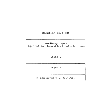
Example 1 Comparison of resonance excitation geometries
Table 2 below shows a comparison of the results
theoretically obtainable using an optical structure
as depicted schematically in Figure 1. The excitation
wavelength for all mechanisms was 543 nm and the
fluorophore was rhodamine B with a peak emission
at 570 nm and a half width of 35 nm (from 555 nm
to 590 nm).
Example 2 Total internal reflection
a) Figure 2 shows the surface field intensity
plotted as a function of angle for a number
of glass indices in water, with a wavelength
of 543 nm (green HeNe laser). For a glass
slide with a refractive index of 1.52 the field
intensity is 2.5 times that of the incident
radiation at 70.
20 b) Figure 3 shows the field intensity penetration
plotted as a function of angle. Away from
the critical angle this is a slowly varying
function of the angle of incidence. For the
example given in (a) above the field penetration
is about 90 nm at 70. It is not desirable
to work too close to the critical angle because
the penetration is too great and poor surface
discrimination would result. Thus, a balance
between field intensity and penetration depth
must be struck.
Example 3 Surface plasmon resonance
Figure 4 shows the reflectance and surface field
intensity of a 50 nm silver film in water irradiated
at 543 nm. A twenty-five-fold enhancement of the
field intensity results when compared with the intensity
1 339005
_ -- 18 --
of the incident radiation.
19 1 339005
o .,,
-~ 3
~n O O O
F~ S ~ o o
~ C~
O
,( ~ o o o
U~ ~ ~1 0
U~ Y 1-- ~ CO
E~ aJ o L~
~ O O O
o r
. .
X ~ ~I L'~
O C: C
O
O
O O
O O
O
U~
Q
- 1 339005
- 20 -
Example 4 Comparison of SPR and total internal reflection
Figure 5 shows a comparison of the excitation of
a rhodamine B solution by surface plasmon resonance
and total internal reflection. The emission intensity
enhancement and narrow emission range for the SPR
geometry can be clearly seen. The experimental details
follow.
(i) Fabrication of an Optical Structure
Glass slides measuring 25 mm x 75 mm were cleaned
ultrasonically in a solution of detergent. Using
vacuum deposition, chromium was deposited onto one
half of one surface of each slide through a mask,
to a thickness of 1 nm. In the same way, silver
was deposited onto the chromium layer to a thickness
of 54 nm. The mask was removed and a 10 nm coating
of silica was deposited onto the whole of said surface
by similar means, to ensure that both halves of the
surface of each slide had the same physico-chemical
properties. The slides were washed with ultra-pure
water. Each slide was scribed and cut into three
pieces (such that each piece was half-silvered).
Each piece was used to make a capillary cell using
another piece of glass of similar size and double-
sided adhesive tape.
(ii) Experimental Procedure
A sample solution was made up using an appropriate
concentration of a suitable protein labelled with
rhodamine B. The experimental set up was as described
in section (iii) of Example 5, below.
_ - 21 - 1339005
Example S Assay for human chorionic gonadotrophin
(hCG) using an indirect surface plasmon
resonance technique
(i) Fabrication of an Optical Structure
Glass slides measuring 25mm x 75mm were cleaned
ultrasonically in a solution of detergent. Using
vacuum deposition, aluminium was deposited onto one
half of one surface of each slide through a mask,
to a thickness of lnm. In the same way silver was
deposited onto the aluminium layer to a thickness
of 54nm. The mask was removed and a 10nm coating
of silica was deposited onto the whole of said surface
by similar means, to ensure that both halves of the
surface of each slide had the same surface chemistry.
The slides were suspended 5mm above a pool of GOPS
(glycidoxypropyltrimethoxysilane) for 2 hours at
20C in order to silanize the silica surface, following
which the slides were baked at 60C for one hour.
One 75ul drop of a 20ug/ml solution of ~12/17 anti-
hCG antibody in HEPES buffer was placed on each half
of each slide, and the slides were left to dry for
two hours. The slides were washed with ultra-pure
water and then a solution of sucrose with tris(hydroxy-
methyl)aminomethane and sodium azide was deposited
on the surface of the slide by means of spinning.
Each slide was scribed and cut into three pieces
(such that each piece was half-silvered). Each piece
was used to make a capillary cell using another piece
of glass of similar size and double-sided adhesive
tape.
(ii) Assay Methodology
A sandwich-type assay was performed using premixed
solutions in horse serum of hCG and XRITC-labelled
* TRADE-MARK
~,
1 339005
- 22 -
antibody immobilised in the capillary cell. The
concentration of XRITC-labelled antibody used was
2.5 ug/ml. Sample solutions were taken up by the
capillary cells prepared as in section (i) and allowed
to incubate for fifteen minutes before a reading
was taken.
(iii) Experimental set-up
The filled capillary cell under study was coupled
to a hemicylindrical lens using a fluid of suitable
refractive index. Light from a green helium-neon
laser was then directed at the slide (i.e. the plate
of the cell which carried the immobilised antibody)
through the planar wall of the lens at an angle suitable
for surface plasmon resonance to occur. Fluorescence
emission was monitored by rotating a photomultiplier
tube in a plane perpendicular to the plane of the
incident/reflected light. The light reaching the
PMT detector passed through a 610 nm bandpass filter
to remove any scattered excitation light. A slit
was placed behind the filter to give an angular resolution
of 1. Two lenses focussed the light passing through
the slit onto the detector. A comparison of the
fluorescence arising from surface plasmon resonance
with that arising from total internal reflection
was made by sliding the cell on the prism so that
the silvered and unsilvered halves of the cell were
interrogated in turn.
Results
Figure 6 shows a comparison of the signals obtained
over the angular emission range 60-80 from a capillary
cell containing 8105 mIU/ml hCG in sérum, as obtained
by SPR and TIR excitation. Particularly noticeable
is the strong peak in signal around 74 for SPR excitation.
1 339005
- 23 -
The integrated signal between 70D and 78D is 5.2
times greater using SPR excitation than using TIR
excitation. Optimisation of the metal layer and
improved protein immobilisation techniques should enable
greater enhancements to be achieved.
Figure 7 shows signals over the same angular range,
but this time from a cell filled with serum containing
no hCG.
The emission resulting from SPR excitation shows a
slight peak at 76 due to non-specific binding of the
XRITC-labelled antibody.
Example 6 - Demonstration of the sensitivity of a direct
guided mode sensor to changes in refractive index.
(i) Fabrication of the optical structure
Glass microscope slides were cleaned ultrasonically in a
detergent solution and extensively rinsed with ultrapure
water. Using vacuum deposition techniques, a layer of
aluminium, lnm thick, was deposited onto the surface of
the glass followed by a film of silver, 54nm thick. The
silver surface was then coated with a glass film by spin
coating a silica solgel (HT Products Inc., USA) onto the
device at 300 rpm. The optical structures were baked
overnight at 60DC. A capillary cell was fabricated from
the optical structure and another piece of glass of the
same dimensions using double sided adhesive tape.
(ii) Experimental set-up
The filled capillary cell under study was optically
coupled to a right angled crown glass prism using an
appropriate fluid. The cell was illuminated through the
glass substrate and onto the silver film with a TM
polarised HeNe laser, the reflected light intensity
1 339005
- 23a -
being measured with a photodiode device. The prism was
rotated through a range of angles during the
measurement. The cell was filled with ultrapure water
(refractive index 1.3316) and the position of the
reflectance minimum noted. The ultrapure water was
replaced by a 5% solution of sucrose (refractive index
of 1.3382) and then by a 10~ solution of sucrose
(refractive index of 1.3450), the position of the
minimum in reflected light intensity being measured in
the presence of the sucrose solutions.
(iii) Results
Figure 8 shows the positions of the minima in reflected
light intensity of the guided mode light in the optical
structure. As the refractive index of the medium in
contact with the optical structure increases there is a
shift in the angle at which the minium occurs,
demonstrating that the device is sensitive to refractive
index changes.
The binding of a ligand (eg an antigen) to an
appropriate biological molecule (eg an antibody)
immobilised on the device surface will result in a
refractive index change. This will allow the guided
mode sensor to be used as a direct optical immunosensor.