Note: Descriptions are shown in the official language in which they were submitted.
~ 339006
Dkt. 27934-A JPW
8E~F-CONTAIN~D ~ I-IMM~NOA88AY DIAGNO8TIC 8Y8TEM
Field of th~ Inventior.
This invention is in the field of immunodiagnostics.
More specifically, this invention provides test strips,
as well as kits containing, and methods employing,
these strips for use in the immunological detection of
analytes in aqueous liquids, particularly biological
samples such as blood, urine, and the like.
Backqround of the Invention
In recent years, the detection, or quantitative deter-
mination, or both, of analytes based upon reactionswith immunological reagents has gained considerable
importance, especially in the field of medical testing.
Commonly, these methods involve contacting a sample
su~pected of containing the analyte with a material
which exhibits specific immunologic reactivity with the
analyte, for example, an antibody directed to an
epitope present on the analyte. If the analyte is
present in the sample, it specifically conjugates with
the antibody to form a complex. A wide range of
~developer" or "reporter" mechanisms have been proposed
and are in use to indicate whether the con~ugation
reaction occurs. (See, for example, United States
Patent Nos. 4,366,242, issued December 28, 1982 to
.,'~ ~
1 339006
-2-
Neumann, et al; 4,278,653, issued July 14, 1981 to
- Harris, et al.; and 4,208,479, issued June 17, 1980 to
Zuk, et al.)
Such methods have become increasingly popular since the
introduction of monoclonal antibodies (MAbs), which may
be produced using the technology developed by Kohler &
Milstein, "Continuous Cultures of Fused Cells Secreting
Antibody of Preferred Specificity,H Nature, (1975) 256:
495-7, and which have unique specificity for the
analytes with which they conjugate. (See, for example,
United States Patent No. 4,376,110, issued March 8,
1983 to David, et al.).
As these methods have evolved, there has been a
parallel search for better ways to apply them on a day-
to-day basis. This has led to a range of test devices,
test kits, and the like. (See, for example, United
States Patent Nos. 4,623,461, issued November 18, 1986
to Hossom, et al.; 3,888,629, issued June 10, 1975 to
Bagshawe; 4,458,020, issued July 3, 1984 to Bohn, et
al.; 4,496,654, issued January 29, 1985 to Katz, et
al.; and 4,305,924, issued December 15, 1981 to Piasik,
et al.)
Desirable characteristics of a test kit or device
include the following:
1. The test device or kit should be easy to
store and to use.
2. It should give unambiguous results without
false positives or false negatives.
3. It should allow a multiplicity of samples
to be screened in a short period.
4. Ideally, it should provide the user with
positive indication that it has been used properly as
1 339006
_3-
confirmation that a false reading has not been
obtained.
5. Preferably, it should allow a plurality of
tests to be run simultaneously.
6. Preferably, the test can be configured to
use whole blood or its fractionated components.
It is a primary object of the present invention to
provide a test strip and a test kit based thereon which
provides these desirable features.
- 1 339006
Summary of the Invention
The present invention provides a test strip for
detecting, in a sample from a human subject, the
presence of an antigenic substance which
comprises a solid ~u~G~ ~, an antibody directed
against the antigenic substance bound to a first
discrete area on the solid support, an anti-human
antibody bound to a second discrete area on the solid
support as a positive control, and an antibody directed
against an antigen which does not naturally occur in
human subjects bound to a third discrete area on the
solid support as a negative control.
The invention additionally provides a test strip for
detecting, in a sample from a human subject, the
presence of an antigenic substance which comprises a
solid support, an antibody-based reagent directed
against the antigenic substance in an immune complex
and native human antibody thereto bound to a first
discrete area on the solid support, an anti-human
antibody bound to a second discrete area on the solid
support as a positive control, and an antibody directed
against an antigen which does not naturally occur in
human subjects bound to a third discrete area on the
solid support as a negative control.
The invention also provides a method for detecting in a
sample from a human subject the presence of an
antigenic substance using the aforementioned test
strips.
Thus, the present invention provides a test strip for
immunologically detecting the presence of one or more
analytes in aqueous specimens. The strip device
comprises a solid support which has a plurality of
,....t.
~` 5 1 3390~6
discrete areas on its surface. At least one of these
areas is a test area which carries a monoclonal
antibody (or a combination selected from polyclonal
antibody, mono- or divalent antibody fragment, hybrid
antibody, heterobifunctional antibody, or genetically
manipulated or cloned antibody) to an analyte to be
detected or quantitatively determined. This antibody
or combination is preferably conjugated to, or
immobilized on, the surface of the support. At least
one of the discrete areas on the test strip carries a
positive control, i.e., a control material which
produces a positive signal only when the test area ha~
been contacted with the specimen. In preferred
embodiments this positive control is an immunological
control, such that the control event includes an
immunological reaction between a species always present
in the specimen and its immunologically specific
partner immobilized in the positive control zone.
In other preferred embodiments, this test strip
includes at least one area carrying a negative control,
that is, a control material which does not give rise to
a detectable event in the presence of a normal specimen
but does give rise to a detectable event when the
sample is abnormal.
In another embodiment, this test strip contains a
plurality of separate test areas containing different
antibodies or combinations of antibodies so as to
detect more than one analyte in the specimen.
In another embodiment, this test strip contains a
plurality of separate test areas containing different
analytes or combinations of analytes to detect antibody
or other analytes.
,,
1 339006
In yet another embodiment, this invention provides a
test kit. This test kit includes the test strip just
described and a test plate comprising a plurality of
liquid holders, each shaped and sized to receive the
test strip and to permit the test areas and positive
control zone and negative control zone, if any, to
simultaneously contact each of a sequence of liquids
which are predeposited in the holders and at least one
of which includes the test specimen.
In a presently preferred embodiment of this kit, the
test plate comprises a plurality of liquid holders so
as to permit a plurality of specimens to be tested at
the same time, using a plurality of test strips.
. ~
"
--- i 339006
-7-
Description of the Figures
Figure 1 is a perspective view of a test strip in
accordance with the invention, showing its various
zones or areas in semischematic form;
Figure 2 is a perspective and semischematic view of
another embodiment of a test strip of this invention,
together with a sample test plate;
Figure 3 is a perspective view of a test plate adapted
to permit multiple specimens to be tested
simultaneously; and
Figure 4 is a block diagram of a typical test protocol
which may be employed according to the present
invention.
~' .
1 339006
Detailed Description of the Invention
This invention provides a test strip for detecting, in
a sample from a subject, particularly a human subject,
the presence of an antigenic substance. This test
~trip comprises a solid support, the antigenic
substance bound to a first discrete area on the solid
support, an anti-human antibody bound to a second
discrete area on the solid support as a positive
control, and an antibody directed against an antigen
which does not naturally occur in human subjects bound
to a third discrete area on the solid support as a
negative control. Such a test strip may also be
readily adapted so as to quantitatively determine the
amount or concentration of the antigenic substance
present in the sample.
Thi~ invention also provides a te~t strip which
comprises all of the elements of the aforementioned
test strip and additionally comprising an antibody-
based reagent directed against the antigenic substance
in an immune complex and native human antibody thereto,
bound to a fourth discrete area on the solid support.
In one embodiment of this invention, the antigenic
su~stance being detected is a virus or a viral protein.
For example, the antigenic substance may be HIV-l, HIV-l
GAG protein, HIV-l ENV glycoprotein, HIV-l POL protein,
HTLV-l GAG protein, HTLV-l ENV glycoprotein, HTLV-l POL
protein, or its epitope thereof.
In test strips according to this invention, the solid
support may comprise glass fiber filter paper,
nitrocellulose, scintered glass, plastic, synthetic
polymer, cellulose, cellulose acetate,
~ ."
9 1 339006
polytetrafluoroethylene, polyethylene, polypropylene,
or polyvinylidine fluoride.
In certain embodiments of this invention involving an
antibody-based reagent, this reagent comprises a
monoclonal antibody, polyclonal antibody, mono- or
divalent antibody fragment, hybrid antibody,
heterofunctional antibody, or genetically manipulated
or cloned antib~dy.
In a preferred embodiment of this invention the anti-
human antibody on the test strip is a monoclonal
antibody.
Similarly in a preferred embodiment of this invention,
the antigen which does not naturally occur in human
subjects and which is present on the test strip is a
synthetic organic molecule, e.g., dinitrophenol.
In preferred embodiments of this invention one or more
of the anti-human antibody, the antibody directed
against an antigen which does not naturally occur, or
the antibody-based reagent is labeled, for example,
labeled with a radioactive isotope, fluorophore,
chromophore, or an enzyme which catalyzes a chemical
reaction which produces a detectable product.
The invention also provides a test strip for detecting,
in a sample from a human subject, the presence of an
antigenic substance which comprises a solid support, an
antibody-based reagent directed against the antigenic
substance in an immune complex and native human
antibody thereto bound to a first discrete area on the
solid support, an anti-human antibody or other
appropriate anticellular structure bound to a second
-lO- 1 339006
discrete area on the solid support as a positive
control, and an antibody directed against an antigen
which does not naturally occur in human subjects bound
to a third discrete area on the solid support as a
negative control. As those skilled in the art will
appreciate, such a test strip may be readily adapted so
as to quantitatively determine the amount or
concentration of the antigenic substance present in the
sample.
The invention further provides a method for detecting
in a sample from a human subject the presence of an
antigenic substance which comprises pretreating the
sample to be tested so as to prevent non-specific and
specific binding of substances present in the sample to
proteins, including antibodies and recombinant
antigens, present on the test strip, and thus prevent
spurious results; contacting the resulting pretreated
sample to be tested with the aforementioned test strip
under conditions such that the antigenic substance
bound to the test strip forms a complex with any
antibody directed against it which is present in the
sample and in the case of test strips onto which both
the antigenic substance and the antibody-based reagent
are bound, so that antigenic substance present in the
sample binds to antibody thereto present on the test
strip; thereafter treating the test strip to remove
uncomplexed antibody directed against the antigenic
substance; contacting the resulting, treated test strip
with labeled antihuman antibody under conditions such
that the antihuman antibody forms a complex with any
human antibody bound to the test strip; detecting the
presence of labeled antihuman antibody complexed to the
test strip and thereby the presence of, or antibody to,
the antigenic substance and thus detecting the presence
,~
-11- 1 339Q06
of the antigenic substance in the sample; and verifying
the correctness of the detection so made by means of
the positive and negative controls on the test strip.
The invention additionally provides a method of
detecting in a sample from a human subject the presence
of an antigenic substance which comprises pretreating
the sample to be tested so as to prevent non-specific
binding of substances present in the sample to
proteins, including antibodies, present on the test
strip and specific binding of human antibodies to the
antigenic substance, and thus prevent spurious results;
contacting the resulting pretreated sample with the
aforementioned under conditions such that the antibody-
based reagent bound to the test strip forms a complex
with any antigenic substance which is present in the
sample; contacting the resulting, treated test stripwith labeled antibody directed to the antigenic
substance under conditions such that the labeled
antibody forms a complex with any antigenic substance
bound to the test strip; detecting the presence of
labeled antibody directed to the antigenic substance
bound to the test strip and thereby the presence of the
antigenic substance in the sample; and verifying the
correctness of the detection so made by means of the
positive and negative controls on the test strip.
In preferred embodiments of the invention the
hereinabove described method involves as the antigenic
substance, a virus or viral protein, and as the sample,
blood, serum, urine, or the like.
The invention also provides the aforementioned methods
wherein the verification comprises contacting the test
strip with labeled antihuman antibody under conditions
,
-12- l 339006
such that the labeled antihuman antibody binds to human
antibody bound to the test strip.
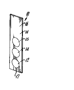
Thus, the present invention provides an improved test
strip device. One embodiment of this device is shown
as 10 in Figure 1. It includes a solid support 11
having a plurality of readable areas 12, 14 and 15.
While support 11 is shown as a solid "dip stick"
structure, it can be virtually any geometric shape,
including cube, block, rod, cylinder, prism, polygon,
spiral, sphere. or segmented combinations thereof. It
can ALso be a carrier device holding materials such as
plastic, metal, paper, or the like. It can be made of
a wide range of materials including plastic, such as
polystyrene or the like; metal; paper; glaæs fiber;
filter paper; nitrocellulose; scintered glass;
synthetic polymers; cellulose; cellulose acetate;
polytetrafluoroethylene; polyethylene; polypropylene;
polyvinylidine fluoride; or any other material
including the materials which it comes in contact with
2 during the test. Commonly, the materials deposited on
its surface are adsorbed, chemically coupled, or
otherwise bound to the surface of the test strip as
described hereinafter.
Readable areas 12, 14 and 15 are arrayed separately on
support 11 so as to give a plurality of potential
signals when the strip is processed in the analysis
method. At least one of the readable areas includes
antibody which is immunoreactive with a test analyte in
the test specimen. The readable areas may also include
an analyte reactive with test antibody or test analyte.
At least one of the readable areas includes a positive
control which reacts with a test specimen to verify via
a detectable signal when the test strip properly
-13- 1 339006
contacts a test specimen. Preferably, this positive
control is an immunologic positive control which
undergoes immunologic reaction with a nonanalyte
species in the specimen.
The readable areas can also include a negative control,
namely, a discrete area containing one or more reagents
that do not produce a reading with the specimen if the
specimen is properly handled but which do give a
reaction and produce a signal if the specimen is
defective or has been ~ishandled.
The relative position of the positive control area and
the antibody-containing areas may be important. Test
strip 10 is used as a "dip stick." In use, the
technician or automated test device grips or "holds"
the strip by top end 16 and dips it into the liquid
specimen and other reagents, lower end 17 first. This
means that area 12 will contact these liquids before
area 15 does. It is desirable, therefore, that the
positive control, which gives a positive reading only
when the specimen and reagents have properly contacted
it be positioned as the last to be contacted or
uppermost area, e.g., 15 in Figure 1. Conversely, the
negative control, if present, should be positioned as
the first to be contacted or bottommost, e.g., 12 in
Figure 1.
The positive control and the negative controls are
preferably immunologic. That is, they preferably
function by means of immunological reactions. Thus,
for example, when the test specimens are human serum-
based, a typical positive control could be a region
designed to be treated with a common immunoglobulin
always found in blood or its fractionated components.
.
,,
-14- 1 339006
The test reagent region (14 in Figure 1) is based on
and includes at least one antibody specific for the
analyte being tested for. A wide range of antibodies
against a wide range of potentially important analytes
have been described in the literature and may be used.
Turning to Figure 2, a variation on the test strip of
Figure 1 is shown as 21. Strip 21 is shown as part of
an overall analysis kit 20 which also includes a test
plate 22. Strip 21 includes support 11 and readable
areas 12, 14 and 15 as previously described and an
additional readable area 24. Area 24 may be an
additional control, an analyte, or an additional
antibody-containing test region. Area 24 may contain a
second antibody against the same antigen of area 14 so
as to provide additional confirmation or sophistication
to the test result or it may contain an antibody
against a second antigen-analyte. These various
reagents are generally immobilized in their various
regions to prevent cross-contamination and the like,
which will give ambiguous results. Antibody or most
analytes are commonly immobilized on plastics,
carbohydrate polymers, adsorbent fibers, polymer-coated
metal beads, silica gel, paper, glass filters, or the
like, by either direct adsorption tincubation for
limited times or until the reagent solution is
completely dry) or by chemical coupling to reactive
y~O~_ on the solid phase. The optimal conditions are
unique for each reagent.
In a typical application, the test strip 21 is employed
in concert with a test plate such as 22. Test plate 22
has several characteristics. For one, it contains a
plurality of liquid storage wells such as 25, 26, 27,
- 1 339006
-15-
28 and 29. The exact number is selected to accommodate
the various steps required to effect the desired assay
sequence. For another, the size of each of the wells
is set so as to permit the strip to be inserted. For
another, the wells are deep enough to accommodate a
depth of liquid "d" which is a depth adequate to
completely cover all the readable areas 12, 14 and 15;
or 12, 14, 24 and 15, present on the test strip.
In use, one or more of the wells of test plate 22 are
charged with predetermined quantities of reagents and
samples according to a predetermined p~otocol and then
the test strip is inserted into the wells, again
according to a predetermined protocol. This gives rise
to a readable detection event which is read together
with the results of the positive and negative controls.
On the basis of these readings, one may determine
whether a valid test has been conducted and whether the
analyte is present in the specimen.
For example, in a typical protocol a test strip might
be constructed with an antibody against a suspected
pathogen in human blood. Positive control 15 would
have an antibody against a common blood constituent,
and negative control 12 would have an antibody against
a material not commonly found in blood but of concern
as a contaminant, or the like.
Well 25 is charged with a premeasured quantity of
diluent, buffer or a like aqueous medium. A given
quantity of patient blood or other source is added to
the liquid of well 25 and mixed with it. The strip is
inserted into the liquid in well 25 for a preset
interval. This permits the test and control areas on
strip 21 to contact the liquid and the diluted blood
~ .
- 1 339006
-16-
constituents. This allows the immunological reactions
to occur between the antibodies in these areas and any
appropriate species in the blood.
The sample well 25 and any other well may contain
various reagents to prevent undesired reactions and the
like. These may include materials so as to alter the
properties of the sample, or materials to react with or
remove undesired cross-reactive materials in the
samples.
A current problem with most immunodiagnostic assays is
the undesirable interaction of sample constituents with
the coated solid phase (i.e. bac~Loul,d). These types
of interaction often cause negative samples to be
interpreted as false positives. Background may be
minimized by partitioning the undesirable reactants
(specific and nonspecific) onto a second solid phase
(usually be preincubating the sample before exposure to
the test solid phase). In the case of antibody capture
assays, the second solid phase would have bound to it
antibody specific for an analyte not found in the
sample or test (i.e., irrelevant specificity). This
type of second solid phase would remove both specific
and nonspecific background constituents for antibodies,
the solid phase, the blocking reagent, or a combination
of any of the three. The second solid phase may be
used in any or all parts of the assay although
preferably eliminated at the "developer" stage. In the
case of human derived viral components as analyte,
preinfected human components may be used as adsorbent
reagent for the second solid phase partitioning.
The configuration for the second phase should be
designed for the mechAnics of the reaction. Adsorbent
-17- l 339006
reagents would coat the chamber walls of the sample
tray for some solutions. For rapid immunoassay, a
critical step in controlling background is the initial
mixing of solid phase and sample. The second solid
phase should be free to move within the mixing solution
(a second strip of any geometric shape or particles).
A preferred method would employ an adsorbing means
comprising a suspension of particle beads (approximate
diameter of 1 to 10 micrometers) or any or all of the
chambers into which the test strip is immersed. The
advantage is a large total surface area for rapid-
reaction kinetics and maximum diffusion of reactants.
Preincubation of sample with adsorbent coated particlea
would quickly remove background components before
exposure to the test strip. Preparation of these types
of particles is described in the examples.
Next the test strip is removed from well 25 and
inserted into well 26. Well 26 may contain, for
example, a premeasured quantity of a wash solution to
remove interfering materials, or the like. Thereafter,
the test strip is passed to a well 27 containing a
detection system, for example, a labeled antigen, or a
labeled antibody to the bound antigen or the like.
Next, the strip may be moved to well 29 for washing and
finally to well 30 where a substrate (e.g., chromogenic
reagent) is added or already present. In some cases,
excess liquid on the strip may be removed and substrate
added directly to the strip. Other wells may be
present to contain other reagents as required or
desired.
Figure 3 illustrates an extension of the invention in
which test block 22 includes a plurality of sets (i.e.,
4 sets) of wells so that there are four sample wells
-
1 339006
-18-
25, four wash wells 26, etc. This permits multiple
samples to be tested at once with multiple test strips.
Figure 4 provides a ~lock diagram of the typical test
protocol just set forth and points out the sequential
nature of the test method.
It will be appreciated by those ~killed in the art that
this invention is not limited to particular antibodies,
reagents, particular samplefi, or particular utilization
chemistries and that reasona~le alternatives may b;~
employed without departing from its teachings.
It should be noted that suitable antibodies for use in
the test reagent region(s) may be monoclonal
antibodies, polyclonal antibodies, mono- or divalent
antibody fragments, hybrid antibodies, heterobi-
functional antibodies, or genetically manipulated or
cloned antibodies.
The test itself is, as noted above, a method of
immunologically detecting the presence of one or more
analytes in a specimen. Typically, the test is used
in either the detection or the quantitation of bound or
unbound label, wherein the amount of label detected
corresponds to the amount of analyte in the specimen.
As used herein, the "immunoassay procedure" of the
subject invention may be in the nature of immuno-
electron microscopy or fluorescence polarization, or it
may be an immuno-fluorescent assay, a radiometric assay,
an enzyme-linked immunoassay, or a photon-counting
bioluminescent or chemiluminescent assay. In the case
of the enzyme-linked immunoassay, the enzyme used is
normally selected from the group consisting of alkaline
, ~,
. . ~ ,
1 33900h
--19--
phosphatase, glucose oxidase, horseradish peroxidase,
urease, luciferase, and galactosidase. The label
itself may be virtually any detectable species, e.g., a
chromogenic compound, a radioactive isotope, an enzyme,
or a fluorescent, luminescent, bioluminescent,
chemilumine~^ent, phosphorescent, or ferromagnetic
material.
The analyte may be virtually any compound or organism
which is detectable using the aforementioned procedure,
i.e., a drug, hormone, vitamin, enzyme, ligand,
protein, including glycoprotein and lipoprotein,
antibody, polysaccharide, cell or tissue antigen, or
bacterium, protozoon, parasite, fungus, virus, blood
cell substance, blood fluid substance, or a component
of any of the foregoing. In a preferred embodiment, a
plurality of analytes is detected, which in a
particularly preferred embodiment includes HIV-l GAG
protein, HIV-l ENV glycoprotein, HIV-l POL protein,
HTLV-l GAG protein, HTLV-l ENV glycoprotein, and HTLV-l
POL protein purified from natural isolates, recombinant
genetic manipulations or chemical synthesis.
The aforementioned analytes may also be bound to the
test strip as the actual detecting reagents in
alternative assay procedures.
The specimen may be blood or its fractionated
components, urine, saliva, vaginal fluid, seminal
fluid, mucosa, birth fluids, tears, gastrointestinal
fluid and excrement, inflammatory fluids, pleural
effusion, pulmonary fluid, tissue extracts and the like
derived from mammalian, avian, reptilian, amphibian,
arthropod and the like.
-20- 1 339006
In alternative embodiments, the specimen or sample to
be tested may be an industrial sample, e.g. industrial
waste chemicals or water, and the analyte may be an
industrial pollutant or toxic chemical present therein.
The invention is further illustrated by the following
example~ which are not intended to and should not be
construed so as to limit in any way the scope of the
invention as defined by the claims which follow
thereafter.
-
-21- 1 339006
EXAMPLE 1
Detection of Human Anti-HIV-1 Antibody in
one Assay Minimization of False Positives
I. Preparation of the Test Stick
Test strip prototypes were constructed from the lids of
96-well tissue culture plates (Costar, catalog #3096).
Strips were cut from the lid using a sharp knife
attachment on a soldering iron so that the strip
contained 8 contiguous circles, each surrounded by a
raised peripheral ring. Strips can also be used as
support segments to contain highly adsorbent solid
phase (e.g., Nylon membrane or nitrocellulose paper).
The strip was washed in a buffer and allowed to air
dry.
Monoclonal antibodies, designated DNPl (CB6 mouse IgGl,
specific for dinitrophenol) and anti-HIgG (CB6 mouse
IgGl, specific for human immunoglobulin G) were gifts
from Dr. Pao-Min Loh's laboratory (University of Iowa).
Anti-HIV GAG (mouse IgGl, specific for Human
Immunodeficiency Virus-l) was purchased from Epitope,
Inc. (catalog #5001). Cocktails of MAbs with
specificity for each analyte can be used. Here, the
MAbs were made 100 mi~LG~rams/ml in Tris Buf~ered
Saline (TBS, 0.15 M NaCl, 0.05 M Tris, pH 7.2) and 10
microliters was applied directly to designated circular
segments. HIV-l (Litton Bionetics, HTLV-3, cat.
0 #37225, 1 milligram/ml, NP40 inactivated) was made 2
mi~GyLams/ml in TBS. 10 microliters were applied
directly to the designated circular segment. The
ordering of reagents was as follows:
3 Anti-human IgG (positive control) ~top]
- 1 339006
-22-
Anti-HIV GAG (detects virus antibody complex)
HIV-l (detects antibody)
Anti-DNP (negative control) [bottom]
The stick was placed horizontally (solution side up)
into a 45 C oven and the antibody and antigen allowed
to dry for about 30 minutes. The stick was placed in
blocking solution (TBS with 0.3% Tween; l mg/ml bovine
serum albumin, BSA, and 0.1% sodium azide) for a
minimum of 15 minutes.
II. Preparation of Adsorbent Beads
To minimize the contribution of certain samples'
affinity to bind to proteins denatured on solid
surfaces (or solid surfaces directly), latex
polystyrene particles were covalently coupled with
monoclonal anti-DNP antibody (purified human T-cell
plasma membrane can also be coupled to latex particles
when viral analyte is either directly adsorbed or
chemically coupled). In addition, the nonspecific
adsorbent can be coated on any other solid phase or
directly onto the walls of the chamber. Antibody is
made 200 micrograms/ml in PBS (O.l M phosphate-buffered
saline, pH 7.2, Pandex Laboratories published Research
Report, No. 4 (1984) "Coupling of Antigens to Latex
Particles by the Water-Solubl~ Carbodiimide Method," by
Michael E. Jolley, Ph.D.). The antibody solution was
added to washed and pelleted latex particle~ (Pandex,
Epicon carboxyl-polystyrene particles, catalog #31-OlO-
l). The solution was triturated and exposed to 20 sec
of an ultrasonic water bath. The fi~al concentration
of beads was 0.5%. Solid carbodiimide (Sigma, l-ethyl-
3-[3-dimethylaminopropyl]-carbodiimide, catalog #E-
7750) is added to make a final concentration of about 5
''':
Trademark
-23- 1 33~006
milligrams/ml. The solution is rotated gently for one
hour at room temperature and washed (with PBS) and
pelleted (12,000 x g, 3 min. room temperature) three
times. The final bead solution is made 0.01% to 0.5%
(w/v) in blocking buffer.
III. Sample Tray
A sample tray was configured by solvent welding plastic
cuvettes (Elkay Products, catalog #127-1010-400) with a
brand cyanocrylate glue. The cuvettes were 1 cm x 1 cm
x 4.5 cm. In this example, the tray contains 7
cuvettes (with lids). In this example, the first
compartment contained 2.5 ml of an approximately 1.0
mic~ G~L ams/ml, inactivated HIV-1 (Litton Bionetics,
HTLV-3, cat. #37225, 1 milligram/ml, NP40 inactivated)
in blocking buffer containing about 0.1% adsorbent
beads described above. The second compartment
contained 2.5 milliliters wash solution (TBS/0.3%
Tween). The third compartment contained a mixing
solution (2.5 milliliters of blocking solution with
about 0.1% adsorbent beads) for diluting the human
blood specimen. The fourth compartment contained 2.5
milliliters wash solution (TBS/0.3~ Tween). The fifth
compartment contained 2.5 milliliters of developing
conjugate solution; polyclonal goat anti-human
immunoglobulin coupled to alkaline phosphatase (Jackson
Immunoresearch Labs, catalog #109-5576) diluted 1:500
in blocking solution with about 0.1% adsorbent beads.
The sixth compartment contained 2.5 milliliters of wash
solution (TBS/0.3% Tween). The seventh compartment
contained 2.5 milliliters of presubstrate wash solution
(pH 10 alkaline buffer).
1 339006
-24-
IV. The Assay
The lid for chamber #3 was removed. A drop of blood
(obtained by a lancet puncture to a finger
alternatively, 1 to 50 microliters of serum may be
used) was allowed to drip into the third compartment.
With each chamber covered with its respective lid, the
entire tray was gently shaken several times to ensure
mixing of blood and bead solution. The tray was
returned to its original standing position and the lid
remove~ from chamber #1. While the bloo~ was
incuba~ing in chamber #3, a test strip was immersed in
chamber #1. The solution was gently stirred with the
test strip and allowed to incubate at 42C for about 20
minutes. Care was taken to ensure that the readable
was maximally -Yro~^~ to the solvent phase. The strip
was transferred to compartment #2 and washed for about
10 seconds. The strip was transferred to compartment
#3 containing the blood/adsorbent mixture. The
solution was gently mixed and allowed to incubate with
the strip for about 30 minutes at 42C. The strip was
transferred to compartment #4 and washed for about 10
seconds. The strip was transferred to compartment #S.
With about 5 seconds of gentle stirring, the strip was
allowed to incubate (stirring every 1 min.). After a
total of about 10 minutes incubation, the strip was
transferred to compartment #6 and gently stirred for
about 10 seconds. The strip was transferred to
compartment #7 and washed for about 10 seconds. The
strip was removed and tAppe~ lightly to remove excess
liquid. One drop (about 50 microliters) of substrate
(5-bromo-4-chloro-3-indolyl phosphate, Sigma catalog
#B-0766, in alkaline buffer, pH 10) was added to each
readable area. After approximately 10 minutes, the
- ~ 339006
-25-
color reaction of each readable area was recorded as
positive (blue color) or negative (no color).
V. Interpretation
The assay can only be valid if the segment containing
the anti-DNP MAb (negative control) remains colorless.
The adsorbent beads should remove all materials that
cause a false positive reaction and the negative
control readable area was used to confirm this
function. The anti-HIgG segment should bind human
immunoglobulin ~resent in blood samples. This reaction
must be positive or else the test reagents are not
working, thus invalidating the tect. A pocitive blue
color in the HIV-l segment indicates that there is
antibody to HIV-1 present in the blood sample. A
positive response in the anti-HIV segment suggests that
virus is present complexed with human anti-HIV
antibody.
.,
-26- l 339006
EXAMPLE 2
Detection of Human Anti-HIV-1 Antibody
Minimization of False Positives
I. Preparation of the Test Stick
Test strip prototypes were constructed from the lids of
96-well tissue culture plates (Costar, catalog #3096).
Strips were cut from the lid using a sharp knife
attachment on a soldering iron so that the strip
contained 8 contiguous circles, each surrounded by a
raised peripheral ring- Strips can also be used as
support segments to contain highly adsorbent solid
phase (e.g., Nylon membrane or nitrocellulose paper),
1 also, sticks can be constructed in such a way that can
contain reces6~ reactive areas. The strip was washed
in a buffer and allowed to air dry.
Monoclonal antibodies, designed DNP1 (CB6 mouse IgG1,
specific for dinitrophenol) and anti-HIgG (CB6 mouse
IgG1, specific for human immunoglobulin G) were used.
Cocktails of MAbs with specificity for each analyte can
be used. Here, the MAbs were made 100 mic~o~Lams/ml in
Tris Buffered Saline (TBS, 0.15 M NaCl, O.OS M Tris, pH
7.2) and 10 microliters applied directly to designated
circular segments. HIV-1 (Litton Bionetics, HTLV-3,
cat. #37225, 1 milligram/ml, NP40 inactivated) was made
2 micLo~Lams/ml in TBS. 10 microliters were applied
directly to the designated circular segment. The
ordering of reagents was as follows:
Anti-human IgG (positive control) ttop]
HIV-l (detects antibody) ~middle]
Anti-DNP (negative control) tbottom]
-27- 1 339~6
The stick was placed horizontally (solution side up)
into a 45C oven and the antibody allowed to dry for
about 30 minutes. The stick was placed in blocking
solution (TBS with 0.3% Tween, 1 mg/ml bovine serum
albumin, BSA, and 0.1% sodium azide) for a minimum of
15 minutes.
II. Preparation of Adsorbent 8eads
To minimize the contribution of certain samples'
affinity to bind to proteins denatured on solid
surfaces (or solid surfaces directly), latex
polystyrene particles were covalently coupled with
monoclonal anti-DNP antibody (purified human T-cell
plasma membrane or recombinant DNA related proteins
such as beta-galactosidase can also be coupled to latex
particles when viral analyte is either directly
adsorbed or chemically coupled). In addition, the non-
specific adsorbent can be coated on any other solid
phase or directly onto the walls of the chamber.
Antibody is made 200 mic~G~ams/ml in PBS (0.1 M
phosphate-buffered saline, pH 7.2, Pandex Laboratories
published ReceArch Report, No. 4 (1984) "Coupling of
Antigens to Latex Particles by the Water-Soluble
Carbodiimide Method," by Michael E. Jolley, Ph.D.).
The antibody solution was added to washed and pelleted
latex particles (Pandex, Epicon carboxyl-polystyrene
particles, catalog #31-010-1). The solution was
triturated and exposed to 20 sec of an ultrasonic water
bath. The final concentration of beads was 0.5%.
Solid carbodiimide (Sigma, l-ethyl-3-[3-
dimethylaminopropyl]-carbodiimide, catalog #E-7750) is
added to make a final concentration of about 5
milligrams/ml. The solution is rotated gently for one
hour at room temperature and washed (with PBS) and
-28- 1 339006
pelleted (12,000 x g, 3 min., room temperature) three
times. The final bead solution is made 0.01% to 0.5%
(w/v) in blocking buffer.
III. Sample Tray
A sample tray was configured by solvent welding plastic
cuvettes (Elkay Products, catalog #127-1010-400) with a
cyanoacrylate glue. The cuvettes were 1 cm x 1 cm x
4.5 cm. In this example, the tray contains 6 cuvettes
(with lids). The first chamber contained a mixing
solution (2.5 milliliters of blockinS solution with
about 0.1% adsorbent beads) for diluting the human
blood specimen. The second chamber contained 2.5
milliliters wash solution (TBS/0-3% Tween). The third
chamber contained 2.5 milliliters of developing
conjugate; polyclonal goat anti-human immunoglobulin
coupled to alkaline phosphatase (Jackson Immunoresearch
~abs, catalog #109-5576) diluted 1:500 in blocking
solution. The fourth chamber contained 2.5 milliliters
of wash solution (TBS/0.3% Tween). The fifth chamber
contained 2.5 milliliters of presubstrate wash solution
(pH 10 alkaline buffer). The sixth chamber contained 1
ml of substrate solution (5-bromo-4-chloro-3-indolyl
phosphate, Sigma catalog #B-0766, in alkaline buffer,
pH10).
IV. The Assay
The sample tray was eguilibrated in a 42OC water bath.
5 microliters (5-200 microliters can be used) of serum
or plasma was added into the first chamber. The sample
in chamber 1 was allowed to incubate for 3 minutes at
42 C. (40-45 C may be used). The solution was gently
stirred with the test strip and allowed to incubate at
-29- 1 339006
42 C for about 45 minutes. Care was taken to ensure
that the re~-dable area was maximally exposed to the
solvent phase. The strip was transferred to chamber #2
and washed for about 1-10 seconds. The strip was
transferred to chamber #3. With about 5 seconds of
gentle stirring, the strip was allowed to incubate at
42 C for about 45 minutes. The strip was transferred
to chamber #4 and gently stirred for about 1-10
seconds. The strip was transferred to chamber #5 and
washed for about 1-10 seconds. The entire sample tray
was removed from the 42-C water bath and placed on the
bench at room temperature. The strip was tra,sferred
to chamber #6 and allowed to incubate at room
temperature for 10-20 minutes. The color reaction of
each readable area was recorded as positive (blue
color) or negative (no color).
Three double-blind studies were performed to determine
the presence of antibodies to HIV-l in patient serum.
Test A: 25 out of 50 serum samples (Peralta Cancer
Center, Oakland, CA) were Western Blot (WB) positive
for HIV-l antibodies (predominantly gp41 only). The
other 25 were from a WB negative donor serum pool. The
test accurately identified 100% of the samples as
negative or positive as compared with the WB results.
Test B: 21 HIV-l WB positive blood samples, and 7
false positive blood samples (as originally screened on
the Abbott HIV-l antibody test at the Massachusetts
General Hospital Blood Bank, Boæton, MA) were tested
with the test. In the test, all 21 WB positive samples
were confirmed along with the 23 WB negative samples.
All 7 false positive samples were negative by the test.
- 1 339006
-30-
Test C: 9 HIV-l WB positive blood samples (NYU Medical
Center) and lO WB negative blood samples were correctly
identified by the test. Two HIV-l antibody positive
semen samples, two saliva samples and l urine sample
were also positive in the test.
`- 1 339006
-31-
EXAMPLE 3
Detection of HIV-l P24 Antiqen in Patient's Serum
I. Preparation of the Test Sticks
Test sticks are the same as those in Example 1. The
following monoclonal antibodies are adsorbed to the
stick: lF8 (Calypte Biomedical Company's anti HIV-l GAG
- monoclonal antibody, IgGl); anti-HIgG and anti-DNP.
Here, the MAbs were made 100 milligram/ml in Tris
Buffered S~line and 10 microliters applied directly to
a designat~d circular segment. The ordering of MAbs
was as follows:
Anti-Human IgG (positive control) [top]
Anti-HIV GAG (detects P24) [middle]
Anti-DNP (negative control) [bottom]
The stick is placed horizontally solution side up into
a 45 C oven and antibodies allowed to adsorb for about
60 minutes. The stick was placed in blocking solution
'TBS, 5% horse serum) for a minimum of 15 minutes.
II. Preparation of the Adsorbent Beads
To minimize the contribution of certain samples'
affinity to bind to proteins denatured on solid
surfaces (or solid surfaces directly) and to affect
removal of contaminating human anti-GAG (soluble or in
complexes), latex polystyrene particles were covalently
coupled with recombinant HIV-l p24 GAG protein. The
recombinant antigen is made 100-200 mi~o~ams/ml in
PBS. The antigen solution was added to washed and
pelleted latex particles. The solution was triturated
and eYros~ for 20 seconds of an ultrasonic bath. The
final concentration of beads was 0.5%. Solid
-32- l 339006
carbodiimide is added to make a final concentration of
about 5 milligrams per ml. The solution is rotated
gently for one hour at room temperature and washed
(with P8S) and pelleted (12,000 x g) three minutes,
room temperature 3 times. The final bead solution is
made 0.01% to 0.5% (w/v) in blocking buffer.
III. Sample Tray
The first compartment contained 2.5 ml of blocking
buffer containing about 0.1% recombinant antigen
adsorbent beads described above. The second
compartment contained 2.5 milliliters wash solution
(TBS). The third compartment contained 2.5 milliliters
of developing conjugate solution; polyclonal goat anti-
HIV-1 GAG coupled to alkaline phosphatase diluted 1:500
in blocking solution. The fourth compartment contained
2.5 milliliters of wash solution (TBS). The fifth
compartment contained 2.5 milliliters of presubstrate
wash solution (ph 10 alkaline buffer).
IV. The Assay
100 microliters of patient sample (blood, serum,
plasma) is added to a shallow well, containing twenty
microliters of dissociation buffer (1 M HCl-glycine
buffer). The solution is then transferred into chamber
#1. The solution was gently stirred with the test
strip and allowed to incubate at 42C for about 20
minutes. Care was taken to ensure that the readable
area was maximally eY~Q~e~ to the solvent phase. The
strip was transferred to compartment #2 and washed for
about 10 seconds. The strip was transferred to
compartment #3 with about five seconds of gentle
stirring. The strip was allowed to incubate for about
- 1 339006
-33-
minutes at 42C. The strip was transferred to
compartment #4 and gently stirred for about 10 seconds.
The strip was transferred to compartment #5 and washed
for about 10 seconds. The strip was removed and tapped
lightly to remove excess liquid. One drop (about 50
microliters) of substrate (5-bromo-4-chloro-3-indolyl
phosphate, Sigma catalog #B-0766, in alkaline buffer,
pH 10) was added to each readable area. After
approximately 10 minutes, the color reaction of each
readable area was recorded as positive (blue color) or
negative (no color).
SD 34
1 339006
SUPPLEMENTARY DISCLOSURE
Further embodiments of the present invention
provides a test strip for detecting, in a sample from a human
subject, the presence of an antigenic substance which comprises
a solid support, an antibody directed against the antigenic
substance bound to a first discrete area on the solid support,
an anti-human antibody bound to a second discrete area on the
solid support as a positive control, and an antibody directed
against an antigen which does not naturally occur in human
subjects bound to a third discrete area on the solid support as
a negative control.
The present invention also provides a test strip for
detecting, in a sample from a human subject, the presence of an
antibody which comprises a solid support, an antigenic substance
bound to a first discrete area on the solid support, an anti-
human antibody bound to a second discrete area on the solid
support as a positive control, and a negative control bound to
a third discrete area on the solid support.
The invention additionally provides a test strip for
detecting, in a sample from a human subject, the presence of an
antibody and an antigenic substance which comprises a solid
support, the antigenic substance bound to a first discrete area
on the solid support, the antibody directed against an antigenic
substance bound to a second discrete area on the solid support,
an anti-human antibody bound to a third discrete area on the
solid support as a positive control, an antibody directed against
3~ an antigen which does not naturally occur in human subjects bound
to a fourth discrete area on the solid support as a negative
control, and a second negative control bound to a fifth discrete
area on the solid support.
;. ~
SD 35 1 339006
- The invention further provides a test strip for
detecting the presence of an antibody and antigenic substance
additionally comprising an antibody-based reagent directed
against the antigenic substance in an immune complex and native
human antibody thereto, bound to the second discrete area on the
solid support. As a further embodiment, there may be other
control area on the solid support.
DESCRIPTION OF THE PREFERRED EMBODIMENT OF THE SUPPELEMENTARY
DISCLOSURE
This invention provides a test strip for detecting in
a sample from a subject, particularly a human subject, the
presence of an antigenic substance. This test strip comprises
a solid support, an antibody directed against the antigenic
substance bound to a first discrete area on the solid support,
an anti-human antibody bound to a second discrete area on the
solid support as a positive control, and an antibody directed
against an antigen which does not naturally occur in human
subjects bound to a third discrete area on the solid support as
a negative control. Such a test strip may also be readily
adapted so as to quantitatively determine the amount or
concentration of the antigenic substance present in the sample.
2~
It would be expected by one skilled in the art that
more than one antigenic substance could be detected using a
single test strip.
This invention also provides a test strip for
detecting, in a sample from a subject, particularly a human
subject, the presence of an antibody. This test strip comprises
a solid support, an antigenic substance bound to a first discrete
area on the solid support, an anti-human antibody bound to a
second discrete area on the solid support as a positive control,
and a negative control. Such a test strip may also be readily
adapted so as to quantitatively determine the amount or
concentration of the antibody present in the sample. The
negative control may be but is not limited to a blocking solution
SD 36 1 339006
- or a recombinant antigen which does not naturally occur in human
subjects.
It would be expected by one skilled in the art that
more than one antibody could be detected using a single test
strip.
This invention also provides a test strip for
detecting, in a sample from a subject, particularly a human
subject, the presence of an antibody and an antigenic substance.
This test strip comprises a solid support, the antigenic
substance bound to a first discrete area on the solid support,
an antibody directed against an antigenic substance bound a
second discrete area on the solid support, an anti-human antibody
bound to a third discrete area on the solid support as a positive
control, and an antibody directed against an antigen which does
not naturally occur in human subjects bound to a fourth discrete
area on the solid support as a negative control. Such a test
strip may also be readily adapted so as to quantitatively
determine the amount or concentration of the antigenic substance
and antibody present in the sample. As a further embodiment,
there may be other control areas on the solid support which may
or may not be blocking solution alone.
It would be expected by one skilled in the art that
more than one antigenic substance and antibody could be detected
using a single test strip.
This invention also provides a test strip which
comprises all of the elements of the aforementioned test strip
for detecting the presence of an antigenic substance and antibody
and additionally comprising an antibody-based reagent directed
against the antigenic substance in an immune complex and native
human antibody thereto, bound to the second discrete area on the
solid support.
In one embodiment of this invention, the antigenic
substance being detected is a virus or a viral protein. For
example, the antigenic substance may be HIV-1 GAG protein, HIV-1
SD 37 l 339006
ENV glycoprotein, HIV-1 POL protein, HIV-2 GAG protein, HIV-2 ENV
glycoprotein, HIV-2 POL protein, HTLV-1 GAG protein, HTLV-1 ENV
glycoprotein, and HTLV-1 POL protein and Hepatitis B surface
antigen or its epitope therewith.
In a preferred embodiment, a plurality of antibodies
to analytes is detected, which in a particularly preferred
embodiment includes antibodies to HIV-l, HIV-1 GAG protein, HIV-1
ENV glycoprotein, HIV-1 POL protein, HIV-2, HIV-2 GAG protein,
10HIV-2 ENV glycoprotein, HIV-2 POL protein, HTLV-1, HTLV-1 GAG
protein, HTLV-1 ENV glycoprotein, HTLV-1 POL protein, a Hepatitis
B core antigen, or an epitope thereof.
In another preferred embodiment a plurality of both
15analytes and antibodies to analytes is simultaneously detected,
which in a particularly preferred embodiment detects HIV-1 GAG
protein and antibodies to HIV-1 ENV glycoprotein or in another
particularly preferred embodiment the simultaneous detection of
Hepatitis B surface antigen and antibodies to Hepatitis B core
20antigen.
In test strips according to this invention, the solid
support may comprise glass fiber filter paper, nitro-cellulose,
scintered glass, plastic, synthetic polymer, cellulose, cellulose
25acetate, polystyrene, polytetrafluoroethylene, polyethylene,
polypropylene, or polyvinylidine fluoride.
In certain embodiments of this invention involving an
antibody-based reagent, this reagent comprises a monoclonal
30antibody, polyclonal antibody, mono- or divalent antibody
fragment, hybrid antibody, heterobifunctional antibody, or
genetically manipulated or cloned antibody. Heterobifunctional
antibodies are described by Urnovitz, et al. in "IgA:IgM and
IgA:IgA Hybrid HybridomasSecreteHeteropolymeric Immunoglobulins
35that are Polyvalent and Bi-Specific", J. Immunol., 140:558-563
(1988).
In a preferred embodiment of this invention the anti-
human antibody on the test strip is, but is not limited to, a
SD 38 l 339006
monoclonal antibody.
Similarly in a preferred embodiment of this invention,
the antigen which does not naturally occur in human subjects and
which is present on the test strip is, but is not limited to, a
synthetic organic molecule, e.g., dinitrophenol.
In preferred embodiments of this invention one or more
of the anti-human antibody, the antibody directed against an
antigen, or the antibody-based reagent is labeled, for example,
with a radioactive isotope, fluorophore, chromophore, or a ligand
which can be used with an enzyme that catalyzes a chemical
reaction which produces a detectable product that can be further
amplified in a secondary reaction.
The invention further provides a method for detecting
in a sample from a human subject the presence of an antigenic
substance which comprises pretreating the sample to be treated
so as to prevent simultaneously non-specific and specific binding
of substances present in the sample to proteins, including
antibodies, present on the test strip, and thus prevent spurious
results, contacting the resulting pretreated sample with the test
strip for detecting an antigenic substance under conditions such
that the antibody directed against the antigenic substance bound
to the test strip forms a complex with antigenic substance
thereafter treating the test strip to remove uncomplexed
antibody, contacting the resulting, treated test strip with
labeled antibody to the antigenic substance under conditions such
that the labeled antibody forms a complex with the antigenic
substance complexed to the antibody bound to the test strip,
detecting the presence of labeled antibody complexed to the test
strip and thereby the presence of the antigenic substance in the
sample, and verifying the correctness of the detection so made
by means of the positive and negative controls on the test strip.
The invention additionally provides a method for
detecting in a sample from a human subject the presence of a
human antibody directed to an antigenic substance which comprises
pretreating the sample to be tested so as to prevent
.
r .~ii
SD 39 l 339006
simultaneously non-specific and specific binding of substances
present in the sample to proteins, including antigenic
substances, present on the test strip, and thus prevent spurious
results, contacting the resulting pretreated sample with the test
strip for detecting an antibody under conditions such that the
antigenic substance bound to the test strip forms a complex with
any human antibody directed against it which is present in the
sample, thereafter treating the test strip to remove uncomplexed
antibody, contacting the resulting, treated test strip with
lo labeled antihuman antibody under conditions such that the
antihuman antibody forms a complex with any human antibody
complexed to the test strip, detecting the presence of labeled
antihuman antibody complexed to the test strip, and thereby the
presence of human antibody in the sample, and verifying the
correctness of the detection so made by means of the positive and
negative controls on the test strip.
The invention additionally provides a method for
detecting in a sample from a human subject the presence of an
antigenic substance and a human antibody directed to an antigenic
substance which comprises pretreating the sample to be tested so
as to prevent simultaneously non-specific and specific binding
of substances present in the sample to proteins, including
antibodies and antigenic substances, present on the test strip,
and thus prevent spurious results, contacting the resulting
pretreated sample with the test strip for detecting the presence
of an antigenic substance and antibody under conditions such that
the antibody directed against the antigenic substance bound to
the test strip forms a complex with the antigenic substance and
the antigenic substance bound to the test strip forms a complex
with any human antibody directed against it which is present in
the sample, thereafter treating the test strip to remove
uncomplexed antibody, contacting the resulting, treated test
strip with labeled antibody to the antigenic substance and
labeled antihuman antibody under conditions such that the labeled
antibody forms a complex with the antigenic substance complexed
to the antibody bound to the test strip and the antihuman
antibody forms a complex with any human antibody complexed to
the test strip, detecting the presence of labeled antibody and
"
1 339006
SD 40
~~- labeled antihuman antibody complexed to the test strip, and
thereby the presence of the antigenic substance and human
antibody in the sample, and verifying the correctness of the
detection so made by means of the positive and negative controls
on the test strip.
In preferred embodiments of the invention the
hereinabove described method involves as the antigenic substance,
a virus or viral protein, and as the sample, blood, serum, or the
like. In a particularly preferred embodiment, the sample is
urlne .
A preferred method would employ an adsorbing means
comprising a suspension of particle beads (approximate diameter
of 0.5 to 10 micrometers) in any or all of the chambers into
which the test strip is immersed.
The invention as described in this Supplementary
Disclosure is further illustrated by the following examples which
are not intended to and should not limit in any way the scope of
the invention as defined by the claims which follow thereafter.
EXAMPLE 4
HIV-l GAG Protein Antigen Test Format
I. The DiPstick
Test strip (dipstick) prototypes were constructed from
injection molding of polystyrene or polystyrene treated to give
a white opaque stick to have the shape of 5 wells in a row with
the same spacing as that of a 96 well microliter plate (see
Figure 1).
All proteins added at 50 microliters per well were in
0.1 M sodium bicarbonate buffer pH 9.6.
Stick Well #1 - anti-human IgG (5 micrograms per well)
[Top]
Stick Well #2 - anti-HIV-l GAG protein (5 micrograms per
well)
SD 41 t 339006
- Stick Well #3 - negative control monoclonal (5 micrograms
tBottom] per well)
Sticks were placed in a humidified incubator/oven for
1 hour at 45C. The sticks were removed, solutions were
aspirated and 100 microliters of 5% non-fat milk/TBS were added
to each well. Sticks were incubated for 60 minutes at 37C in
a humidified incubator/oven and then solutions were aspirated.
100 microliters of a 10% sucrose/4% PVP (polyvinylpyrrolidone),
0.1% sodium azide solution was added to each well, incubated for
15 minutes at room temperature and then aspirated. After air
drying for about 10 minutes, the sticks were then stored
desiccated at 2-8C.
II. The Assay
Tube 1
Tube 1 was pre-coated with 3.0 ml of 50 micrograms/ml
each of equine IgG, goat IgG, and non-reactive mouse IgG in TBS
azide by incubation for 30 minutes in a 37C water bath followed
by decanting of the coating solution. The tubes were allowed to
dry in an inverted position and then used.
Tube 1 contained beads coated with either 1) goat
immunoglobulin (IgG) or 2) bovine IgG or 3) horse IgG or 4) non-
reactive mouse monoclonal antibody 0.01% v/v for each of the fourtypes of coated beads (0.04% v/v total coated beads), 9% serum
(3% horse, 3% bovine, 3% goat) in 0.05N Tris-HC1 buffer pH 7.2
and 0.15 M sodium chloride (TBS). The final volume was 1.5 ml.
30150-300 microliters of patient whole blood or 50-150
microliters of fresh serum or plasma (preferably not frozen) was
used.
The sample was allowed to incubate at room temperature
35with the beads/tube for 5 minutes before the dipstick was added.
The tube with dipstick was incubated at 37C in a water
bath for 60 minutes.
4~
1 339006
SD 42
The dipstick was removed, tapped gently on an absorbent
pad, and then placed in Tube 2.
Tube 2
The tube contained TBS was solution. The final volume
was 5-40 ml depending on specific application.
The dipstick was swirled for 5-10 seconds, tapped
gently on an absorbent pad, and then placed in Tube 3.
Tube 3
Tube 3 contained biotinylated rabbit anti-HIV-l GAG
protein and 5% horse serum in TBS. The final volume equaled 1.5
ml.
The dipstick was incubated for 60 minutes at 37C. It
was then gently tapped on absorbent pad and added to Tube #4.
Tube 4
Tube 4 contained a TBS wash. The final volume was 5-40
ml depending on the specific application.
The dipstick was swirled for 5-10 seconds, then gently
tapped on an absorbent pad and added to Tube 5.
Tube 5
Tube 5 contained a 1.5 ml solution of strepavidin
horseradish peroxidase 5~ horse serum and TBS. The dipstick was
incubated for 30 minutes at 37C. It was gently tapped on an
absorbent pad and then added to Tube 6.
Tube 6
The tube contained a TBS wash. The final volume was
5-40 ml depending on the specific application.
The stick was swirled for 5-10 seconds, gently tapped
on an absorbent pad and then added to Tube 7.
Tube 7
SD 43 1 339006
~ The tube contained a TBS wash. The final volume was
5-40 ml depending on the specific application.
The stick was swirled for 5-10 seconds, gently tapped
on an absorbent. The stick was then placed horizontally on a
flat surface.
III. Color Development
One drop (50 microliters) of 3,3', 5,5'
Tetramethylbenzidine (TMB) substrate was added to each well.
After 10 minutes, the reaction was stopped with a drop (15-25
microliters) of 2N HC1. The wells were then read as either
negative, weak, or positive according to the presence or absence
of a yellow color.
IV. Results
The optical density is given in Table 1.
Table 1
Serum Serum + Antiqen
Negative Control MAb 0.045 0.051
Anti-GAG MAb 0.065 0.142
EXAMPLE 5
HIV-l Antibody Test Format
I. The Dipstick
Test strip (dipstick) prototypes were constructed from
injection molding of polystyrene or polystyrene treated to give
a white opaque stick to have the shape of 5 wells in a row with
the same spacing as that of a 96 well microliter plate (see
Figure 2).
All proteins added at 50 microliters per well were in
TBS/0.1% sodium azide.
Stick Well #1 - positive control - anti-human IgG
[Top] monoclonal antibody (5 micrograms per well)
Stick Well #2 - HIV-1 recombinant ENV protein (0.5
~ ~ ..3
SD 44 l 339no6
~~ micrograms per well)
Stick Well #3 - negative control (no coat)
Stick Well #4 - HIV-1 recombinant GAG protein (0.5
~Bottom] micrograms per well)
Sticks were placed in a humidified incubator/oven for
1 hour at 45C. The sticks were removed, solutions were
aspirated and 100 microliters of 5% horse serum/TBS azide were
added to each well. Sticks were incubated for 40 minutes at room
temperature, then solutions were aspirated. 100 microliters of
a 10% sucrose/4% PVP (polyvinylpyrrolidone), 0.1% sodium azide
solution was added to each well, incubated for 15 minutes at room
temperature and then aspirated. After air drying for about 10
minutes, the sticks were then stored desiccated at 2-8C.
II. The Assay
All steps of this procedure are carried out at room
temperature unless otherwise noted.
Tube 1
Tube l was pre-coated by adding 3.0 ml. of 50
microgram/ml of each of equine, horse and goat IgG in 0.05 M
Tris-HCl buffer pH 7.2 with 0.15 M sodium chloride (TBS) and 0.1%
sodium azide (azide). The solution is incubated for 30 minutes
in a 37C water bath and then decanted. The tubes are used after
drying in an inverted position on an absorbent pad. Tube l
contained beads coated with either 1) goat immunoglobulin (IgG)
or 2) bovine IgG or 3) horse IgG 0.01% v/v for each of the three
types of coated beads (0.03% v/v total coated beads), 9% serum
(3% horse, 3% bovine, 3% goat) in 0.05M Tris-HC1 buffer pH 7.2,
0.15 M sodium chloride (TBS), and 0.1% sodium azide. The final
volume was 1.9 ml.
300 microliters of patient whole blood or 100-150
microliters of fresh serum or plasma (preferably not frozen) was
used.
~,
SD 45 1 339006
--~ The sample was allowed to incubate for 5 minutes at
room temperature with the beads/tube before the dipstick was
added.
The tube with dipstick was incubated for 30 minutes at
room temperature (22-25C).
The dipstick was removed, tapped gently on an absorbent
pad, and then placed in Tube 2.
Tube 2
The tube contained TBS wash solution with 0.1% sodium
azide. The final volume was 5-40 ml depending on the specific
application.
The dipstick was swirled for 5-10 seconds, tapped
gently on an absorbent pad, and then placed in Tube 3.
Tube 3
Tube 3 contained goat anti-human IgG - alkaline
phosphatase, 5% horse serum in TBS, and 0.1% sodium azide. The
final volume equaled 2 ml.
The dipstick was incubated for 10 minutes at room
temperature (22-25). It was then gently tapped on absorbent pad
and added to Tube #4.
Tube 4
Tube 4 contained a TBS wash with 0.1% sodium azide.
The final volume was 5-40 ml. depending on the specific
application.
The dipstick was swirled for 5-10 seconds, then gently
tapped on an absorbent pad and added to Tube 5.
Tube 5
Tube 5 contained a TBS wash with 0.1% sodium azide.
The stick was swirled for 5-10 seconds and then gently
, .,
SD 46 l 339006
tapped on an absorbent pad. The stick was then laid horizontally
on a flat surface.
III. Color Development
One drop (50microliters) of 5-bromo-4-chloro-3-indolyl
phosphate (BCIP) substrate (0.1 M glycine buffer, pH 10.4, 0.1%
sodium azide) was added to each well. After 10 minutes, the
reaction was stopped with a drop (15-25 microliters) of EDTA
(25mM). The wells were then read as either negative, weak, or
positive according to the presence or absence of a blue color.
IV. Results
TEST A
Using the HIV-1 recombinant ENV protein well as an
indicator of an HIV-l exposure, the subject invention's test
results agreed with an FDA licensed anti-HIV ELISA test in 51 out
of 53 double blind samples (Table 2). The two samples that
tested differently, wherein the subject invention tested negative
and the FDA licensed anti-HIV ELISA tested positive, were Western
Blot indeterminate.
A second FDA licensed HIV-1 ELISA antibody test results
differed from the first FDA licensed anti-HIV ELISA test in 14
out of 53 samples. In all 14 samples, the second FDA licensed
HIV-1 ELISA test was positive and the first FDA licensed HIV-1
ELISA test as well as the subject invention's were negative.
- 1 339006
SD 47
TABLE 2
Sample# l* Difference Difference
2* 3* 2vs3* 3vsl* Comments
-- _ +
2 + + +
3 + + +
4 + + +
-- _ _
6 + + +
7 _ _ +
8 + + +
g _ _ + +
11 + + +
12 + +
13 - + + + Western Blot
Indeterminate
14 + + +
+ + +
16 - - +
17 + + +
18 + + +
19 + + +
+ + +
21 - - +
22 + + +
23 - _ +
24 - _ +
26
27 + + +
1 339006
SD 47a
28 - - + +
29 + + +
+ + +
31
32 - _ + +
33 - _ _
34
36 - _ +
37 - _ +
38 + + +
39
+ + +
41 + + +
42 - _ + +
43 _ + + + Western Blot
Indeterminate
44 _ _ + +
+ + +
46 + + +
47 _ _ + +
48
49
51 + + +
52 + +
53
l* - Subject Invention Test
2* - First FDA Licensed anti-HIV ELISA Test
3* - Second FDA Licensed anti-HIV ELISA Test
~.,
1 339006
SD 48
These data suggest that there is agreement between the
results of the subject invention's test and the first FDA
licensed test, except in the case where the first FDA licensed
test is positive and Western Blot results are indeterminate. The
two samples that fall into this exception are tested negative
with the subject invention test. In 14 out of 53 samples, the
subject invention test and first FDA licensed test results were
negative whereas the second FDA licensed test was positive.
These data collectively suggest that the subject invention test
has no false positives unlike the second FDA licensed test and,
in addition, does not register a response when the Western Blot
is indeterminate.
TEST B
Twenty double blind samples (serum or plasma) were
utilized. Using the ENV wells of the subject invention as an
indicator of an HIV-l exposure, 14 samples were identified as
positive and 6 as negative. These results agreed with the second
FDA licensed HIV-l ELISA test in all but two samples. Two
samples were known to be false positives according to the second
FDA licensed HIV-l ELISA test (tested positive by the second FDA
licensed HIV-l ELISA test, negative by Western Blot). One of
these two samples contained icteric blood and the other contained
lipemic blood. These data suggest that the subject invention
test had no false positives unlike the second FDA licensed HIV-l
ELISA test and could correctly identify that test's true
positives. (See Table 3.)
, ~
1 339006
SD 49
TABLE 3
l* ~*
SA~PLE # GAG ÇNV
2 - -f~ise positive
3 + + +
4 - + +
S +
7 - + +
9 + + +
0 - + t
; 1 - .
12 + + +
13 + + +
1 ~ . .
+ + +
16 + + +
17 - -~alse Dositive
1 8 + +
19 + +
+ + +
l* - Subject Invention Test
2* - Second FDA Licensed anti-HIV ELISA
Test
.
,,
~ 33900f~
SD 50
TEST C
Patient 1 was followed over the course of a two month
period from Day 1 to Day 73. Eighteen different sample points
were taken. On blood samples drawn on Day 1, Day 8 and Day 10,
seven FDA licensed HIV-1 antibody tests all registered negative
for the presence of HIV-1 antibody (Tests 1-7 in Table 4). The
subject invention test is shown as Test 8 in Table 4. All
observers (5) scored a negative for samples drawn on Day 1 and
Day 8. However, all observers scored a positive reaction for the
subject invention for the Day 10 sample. All observers (5)
scored a negative for samples drawn on Day 1 and Day 8. However,
all observers scored a positive reaction for the subject
invention for the Day lO sample. All samples from Day 15
thereafter were positive with the subject invention test. On the
Day 15 sample, Day 17 and Day 22 samples, only the seventh test
picked up a positive response. On Day 24, the second and seventh
samples now registered positive and HIV-1 antibody. From Day 29
on, all seven licensed tests detected the presence of HIV-1
antibody. The Western Blot test had been negative up to Day 15
and became GAG positive (ENV negative) from Day 17 to Day 24.
Only at Day 29 when all the other license~ test kits were
positive was the Western Blot positive for both ENV and GAG.
These results suggest the subject invention test is
more sensitive than all seven commercially licensed HIV-1
antibody tests. Both the subject invention and the seventh test
appeared to be more sensitive than the Western Blot.
1 33900h
SD 51
TABLE 4
DETECTION OF anti-HIV AhD HIV ANTIGEN(S) IN
LONGITUDINALLY COLLECTED SAMPLES FROM A PLASMA DONOR (Donor C)
Test Run Referencel
DAY NO. (1) (2) (3) (4) (5) (6) (7) (8) (9) (10) (11~ (12)
-- -- _ _ _ _ _ _ _
8 - _ _ _ _ _ _ _ +
_ _ _ _ _ _ + _ _ _ +
- - - - _ + +
17 - - - - _ + + _ +/_
P22 + + +/ +
A 24 - + - - - - + + _ +/_ +
N 29 + + + + + + + + - + +
E 31 + + + + + + + + - + +
L 36 + + + + + + + + - + +
43 + + + + + + + + _ + +
C 45 + + + + + + + + _ + +
57 + + + + + + + + _ + +
59 + + + + + + + + _ + +
64 + + + + + + + + _ + +
66 + + + + + + + + ~ + +
71 + + + + + + + + _ + +
73 + + + + + + + + _ + +
(I~ = anti-HIV ELISA
(2) = anti-HIV ELISA
(3) = anti-HIV ELISA
(4) = anti-HIV ELISA
(5) = anti-HIV ELISA
(6) = anti-HIV ELISA
(7) = anti-HIV ELISA
(8) = subject invention
(9) = anti-H9 ELISA
(10) = anti-HIV WESTERN BLOT
(11) = anti-HIV IFA
(12) = HIV Antigen(s) ELISA
ELISA results were generated using commercially available
FDA licensed anti-HIV screening tests. The IFA, WB and HIV
Antigen data were generated with unlicensed products (re-
search use only) whose test characteristics have not yet
been fully determined.
All samples were assayed under code by individuals who
routinely use these test procedures.
-
- SD 52 1 339006
TABLE 5
- DETECTION OF anti-HIV AND HIV ANTIGEN(S) IN
LONGITUDINALLY COLLECTED SAMPLES FROM FIVE PLASMAPHERESIS DONORS
Test Run Referencel
DAY NO.(1) (2) (3) (4) (5) (6) (7) (8) (9) (10) (11) (12)
-- -- _ _ +/ _
8- - _ _ _ +/_
P 22 +/
A
N 36- - - - - - - + - +/-
E
L 43- - - - - _ _ + _ +/_
E 50- - - - - - - _ _ +/_
64- - - - _ _ _ +/_
85- - - _ _ _ _ +/_ _ +
92- - - _ _ _ _ +/_ _ +
96+ + + + + + + + _ + +
(1) = anti-HIV ELISA
(2) = anti-HIV ELISA
(3) = anti-HIV ELISA
(4) = anti-HIV ELISA
(5) = anti-HIV ELISA
(6) = anti-HIV ELISA
(7) = anti-HIV ELISA
(8) = subject invention
(9) = anti-H9 ELISA
(10) = anti-HIV WESTERN BLOT
(11) = anti-HIV IFA
(12) = HIV Antigen(s) ELISA
- SD 53 1 339006
TEST D
A second patient was followed longitudinally beginning
at Day 1 and ending Day 96. (See Table 5) The Western Blot had
been equivocal (pp 53/55 +/-, GAG negative, ENV negative) for the
first nine samples and not until the tenth sample (Day 96) was
the Western Blot positive. In addition, all seven licensed kits
were negative for the first nine samples and all were positive
by Day 96.
Although there were discrepancies among the five
independent observers, weak reactions had been observed in the
subject invention test as early as the first sample (Day 1).
Four out of five observers observed weak to positive reactions
on samples drawn on Day 36 and Day 43. Three out of five
lS observers noticed weak to positive reaction on ENV for the Day
43 sample. The ENV reaction was not observed by any readers on
the Day 50 sample.
These data suggest that for this patient, the subject
invention test is more sensitive than the seven licensed test
kits and the Western Blot. Over a 4 month period, there was a
waxing and waning of a response to both GAG and ENV until Day 96
when there was a clear response to ENV and GAG.
TEST E
Twenty-five patient samples, all positive for the
presence of antibody to HTLV-1, were used for this test. Five
of these samples were reportedly reactive in a licensed HIV-1
antibody test. The subject invention test recorded a positive
reaction for both GAG and ENG in the noted HIV-1 antibody
positive serum. However, three sera were noted with reactivity
to ENV, where one sample (#R) was clearly ENV positive according
to the subject invention test and shown to be Western Blot
positive for ENV. These data would suggest that a tube was
possibly mislabeled as HIV-l antibody negative. The other two
positive samples were both weakly reactive, suggesting that these
patients may have a weak response to HIV-l ENV. With respect to
those samples that were ENV negative by the test of the subject
invention, 1 out of 17 sera were positive for GAG according to
~,
1 339006
SD 54
the test of the subject invention. These data suggest that the
GAG antigen used in the subject invention test is highly specific
for HIV-1 with minimal cross reactivity to HTLV-1. However, one
should be cautious in interpreting GAG reactivity alone (no ENV
reactivity) in the subject invention test. It would be advised
that GAG reactivity alone would be a suspicious exposure to a
human retrovirus but could only be confirmed as HIV-1 infection
at a later date if ENV reactivity appears.
TEST F
The plasma of twelve HIV-1 infected patients were
tested for HIV-1 antigen in a commercially-available antigen GAG
antigen HIV-1 ELISA test system. Three out of twelve patients
showed significant levels of HIV-1 antigen (GAG antigen) in their
blood.
In the subject invention test measuring antibodies
against GAG and ENV, all twelve patients were strongly reactive
in the ENV well. However, only eight patients were weakly to
strongly reactive in the GAG well. These eight patients were
shown to be antigen negative by the commercially-available
antigen test. Three negative subject invention GAG patients were
all reactive in the commercially-available antigen test. One
subject invention GAG negative sample was negative on the
commercially-available antigen test.
From this study, it would appear that the loss of
subject invention test GAG reactivity in HIV-1 infected
individuals correlates with the rise of antigenemia.
TEST G
Ten normal volunteers provided blood drawn in tubes
containing the anti-coagulant EDTA. 3 00 microliters of whole
blood was added to the subject invention tube #1 and the test
processed. The identity of all 10 volunteers remain blinded.
In ten out of ten cases there were no positives in the negative
control, GAG or ENV wells for any of the bloods. In the case of
normal whole blood spiked with patient serum, the subject
invention test result was not inhibited by the presence of whole
, ., ,i j
~''AO';t
SD 55 l 339006
blood.
TEST H - URINE SAMPLES
Ten concentrated urine samples (lOOX) were used. All
S ten samples were coded. The test procedure was different from
the format described previously for blood. The sample was
diluted 1/20 and allowed to incubate with the beads and dipstick
for a total of two hours at room temperature. The stick was
washed in tube #2 and then allowed to react in tube #3 for a
total of 30 minutes at room temperature. The rest of the
reaction was carried out as described in the original format.
Seven urines were reactive for the ENV. Of those seven, two were
reactive also for GAG. Three urines were unreactive for ENV and
GAG. All ten urines were reactive with the positive control
(anti-human immunoglobulin). The seven ENV positives identified
by the subject invention test were all Western Blot positive HIV-
1 infected individuals. The three ENV, GAG subject invention
negative urines were from normal volunteers (non HIV-1 infected
individuals).
The results are shown in Table 6. These data show that
the subject invention antibody test for blood can be designed to
detect HIV-l antibodies in concentrated urines.
TEST I
Fifty unconcentrated urine samples were tested with a
recombinant HIV-1 envelope antigen (the carboxyterminus of gpl20
and an immunoreactive part of ENV). Twenty-seven samples were
from asymptomatic HIV-1 seropositive individuals. Three samples
were from AIDS/ARC patients. Twenty samples were from HIV-1
seronegative individuals. Undiluted samples were incubated with
the beads and dipstick for a total of two hours at room
temperature. The stick was washed in tube #2 and then allowed
to react in tube #3 for a total of 30 minutes at room
temperature. The rest of the reaction was carried out as
previously described. All 30 urine samples from HIV-1
seropositive individuals were positive for HIV-l recombinant
envelope. All 20 urines from HIV-l seronegative were negative
for HIV-1 recombinant envelope. All 50 samples were reactive
.
1 339no6
SD 56
_
with the positive control (anti-human immunoglobulin). These
data suggest that the subject invention antibody test for blood
can be designed to detect HIV-l antibodies in unconcentrated
urines.
~,
1* 2~ Western RIPA
Blot
SAMPLE# GAG E~rV ELISA gpl60gpl20 p66 pSS pSl gp41 p31 p24 pl9
+ + + .~
2 + + + ~ gpl60,p66,gp44,p24
3 - + + ~ - - gpl60,gp41
4 - + +
S - + + ~ - gpl60,p66,p24
6 + + ~ ~
7 - + + ~ ~ - - gpl60,pSl,p24
g
11
1~ - Subject Invention Test
2~- Commercial anti-HIV ELISA Test ~O
o
1 339006
SD 58
SU~ARY
From two longitudinal studies, the subject invention
antibody test appears to be more sensitive than all seven
commercially licensed FDA HIV-1 antibody tests and the Western
Blot.
From the Test A, the subject invention test appears to
be more sensitive for ENV than the Western Blot.
The subject invention test appears to have no false
positives as judged in Test A nor is it reactive in the case of
Western Blot indeterminates.
The subject invention HIV-1 antibody test seems to be
highly specific for HIV-1 exposure with minimal cross reactivity
in patients infected solely with HTLV-1.
The subject invention test may be used to follow HIV-1
infected individuals that develop antigenemia by observing the
levels of anti-GAG protein reactivity.
The subject invention test appears to work on whole
blood. This means a physician can draw blood in an anti-
coagulant tube, and directly add the whole blood to the subject
invention test kit and know the results within 1 hour at room
temperature.
The subject invention test kit also detects HIV-1
antibodies in urine. The fact that human immunoglobulin could
be found in all ten urine samples (both normal and HIV-1 infected
individuals) suggests that the located beads and coated tubes now
used in the subject invention test kit for blood will be required
for urine.
Selecting the correct ENV protein in this equilibrium
assay allows the subject invention to pick up HIV-1 antibodies
in unconcentrated urine. Urine is a preferred choice of sample
material for two reasons: 1) unlike blood, urine contains none
to little HIV-1 infectious material; and 2) no venipuncture is
.~
1 339006
SD 59
required, minimizing infection due to accidental needle sticks.
EXAMPLE 6
Simultaneous HIV-1 GAG antigen and HIV-1 ENV antibodv test format
I. The Dipstick
Test strip (dipstick) prototypes were constructed from
injection molding of polystyrene or polystyrene treated to give
a white opaque stick to have the shape of 5 wells in a row with
the same spacing as that of a 96 well microliter plate (see
Figure 2).
All proteins added at 50 microliters per well.
Stick Well #1 - positive control - anti-human IgG
[Top] monoclonal antibody (5 micrograms per well)
Stick Well #2 - anti-HIV-1 GAG protein monoclonal antibody
(5 micrograms per well)
Stick Well #3 - negative control monoclonal antibody (5
micrograms per well)
Stick Well #4 - HIV-1 ENV antigen (0.5 micrograms per well)
Stick Well #5 - negative control (no coat)
tBottom]
The stick was placed horizontally solution side up into
a humidified 45 oven and reagents were allowed to adsorb for
about 60 minutes. The stick was placed in blocking solution (TBS
5% horse serum for negative control and ENV; TBS 5% nonfat milk
for antibodies) for 60 minutes at 37C.
II. Assay
Tube 1
Tube 1 was coated before use with 3.0 ml of 50
micrograms/ml in TBS/azide of each of equine IgG, bovine IgG and
goat IgG by incubation for 30 minutes in a 37C water bath
followed by decanting of the coating solution. The tubes were
_j ~
.,
,, ,
SD 60 l 339006
allowed to dry in an inverted position and then used.
Tube 1 contained beads coated with either 1) goat
immunoglobuli (IgG) or 2) bovine IgG or 3) horse IgG or 4) non-
reactive mouse monoclonal antibody 0.01% v/v for each of the fourtypes of coated beads (0.04% v/v total coated beads), 9% serum
(3% horse, 3% bovine, 3% goat) in 0.05M Tris-HCl buffer pH 7.2
and 0.15 M sodium chloride (TBS). The final volume was 2.5 ml.
300 microliters of patient whole blood or 100-150
microliters of fresh serum or plasma (preferably not frozen) was
used.
The sample was allowed to incubate at room temperature
with the beads/tube for 5 minutes before the dipstick was added.
The tube with dipstick was incubated at 37C in a water
bath for 60 minutes.
The dipstick was removed, tapped gently on an absorbent
pad, and then placed in Tube 2.
Tube 2
The tube contained TBS wash solution. The final volume
was 5-40 ml depending on the specific application.
The dipstick was swirled for 5-10 seconds, tapped
gently on an absorbent pad, and then placed in Tube 3.
Tube 3
Tube 3 contained both biotinylated rabbit anti-HIV-1
GAG protein and biotinylated rabbit anti-human in 5% horse serum
and TBS. The final volume equaled 2.5 ml.
The dipstick was incubated for 60 minutes at 37C. It
was then gently tapped on absorbent pad and added to Tube #4.
Tube 4
Tube 4 contained a TBS wash. The final volume was 5-40
SD 61 l 339006
ml depending on the specific application.
The dipstick was swirled for 5-10 seconds, then gently
tapped on an absorbent pad and added to Tube 5.
Tube 5
Tube 5 contained a solution of strepavidin-horseradish
peroxidase in 5% horse serum and TBS. The dipstick was incubated
for 30 minutes at 37C. It was gently tapped on an absorbent pad
and then added to Tube 6.
Tube 6
The tube contained a TBS wash. The final volume was
5-40 ml depending on the specific application.
The stick was swirled for 5-10 seconds, gently tapped
on an absorbent pad and then added to tube 7.
Tube 7
The tube contained a TBS wash. The final volume was
5-40 ml depending on the specific application.
The stick was swirled for 5-10 seconds, gently tapped
on an absorbent pad. The stick was then placed horizontally on
a flat surface.
III. Color Development
One drop (50 microliters) of 3,3', 5,5' - Tetramethyl-
benzidine (TMB) substrate was added to each well. After 10
minutes, the reaction was stopped with a drop (15-25 microliters)
of 2N HC1. The wells were then read as either negative, weak,
or positive according to the presence or absence of a yellow
color.
IV. Results
Table 7 shows the optical density for the following
serum samples:
1. normal human;
2. normal human plus GAG antigen;
SD 62 1 339006
3. HIV-1 seropositive, GAG antigen negative; and
4. HIV-1 seropositive, GAG antigen negative plus GAG
antigen.
SD 63 1 339006
TABLE 7
Simultaneous Testinq for HIVl AntibodY and Antiqen
Serum Sample
Well Contents NHSNHS+Aq Patient Patient+Aq
Negative Control 0.010.01 0.01 0.01
HIVl ENV 0.010.02 0.22 0.19
Negative MAb Control 0.06 0.07 0.06 0.06
Anti-GAG MAb 0.06O.lS 0.06 0.19
Anti-Human IgG 1.020.90 0.85 0.98
NHS=Normal Human Serum
Patient=HIVl seropositive, GAG antigen negative
Ag=GAG Antigen, 300 pg/ml
MAb=Monoclonal Antibody
HIV-l GAG Antigen free serum was spiked with 300 pg per ml. of
GAG protein. Serum was then added to the El format and test-
ed. The results show that the E1 dipstick can simultaneously
detect the presence of antibody in the same serum sample. In
addition, the stick can be used either for antibody capture or
antigen capture.
~,...