Note: Descriptions are shown in the official language in which they were submitted.
20~016~
S P E C I F I C A T I O N
BACKGROUND OF TME INVENTION
Field of the Invention
The invention relates generally to surgical instruments
and procedures and more particularly to apparatus and a
procedure for verifying proper femoral intramedullary channel
preparation and seating therein of a prosthetic femoral hip
implant.
~rior Art
In a hip replacement surgical procedure where the head
and neck of the posterior femur are removed and replaced with
a prosthetic implant it is required that, once installed, this
prosthetic device remain stationary for proper healing and
prosthesis functioning. In practice, if a prosthetic implant
is loose such that rotational micromovement of the implant
within the bone will occur, particularly for a prosthesis that
is secured by means of friction or porous ingrowth coatings,
that rotational movement will loosen the fit, shearing away
the ingrowth, and prohibiting healing.
Accordingly, the present invention is directed to a
procedure and apparatus for verifying mechanical fixation of a
prosthetic femoral implant during a hip joint replacement
surgical procedure.
The present in~ention involv~s a system and apparatus
for torsionally testing a prosthetic hip implant to verify
proper seating. In this procedure, proper seating is assumed
where it is determined the implant will maintain stability
21~ 9
when subjected to application of a certain torsional force in
inch points, as has been determined experimentally. While
torsional testing apparatus and procedure have heretofore been
practiced in other surgical disciplines, such have not
involved prosthetic hip implants. For example, a patent to
Boland, U.S. Patent No. 4,576,158, shows a torsional testing
device for testing bone stability; with a patent to Cordey,
U.S. Patent No. 4,359,906, showing a device for tightening a
screw into a bone material to a pre-set force; and a patent to
Daniel, et al., U.S. Patent No. 4,712,542, that shows a device
and procedure for verifying ligament isometric positioning and
tensioning. Where tooling for placing and positioning of
certain hip prosthesis are shown in patents to McKee, U.S.
Patent No. 3,801,989; Amstutz, U.S. Patent No. 3,857,389; and
Kaufer, et al., U.S. Patent No. 3,868,730; these patents do
not consider torsional testing of a seated hip femoral
prosthesis.
SUMMARY OF THE INVENTION
It is a principal object of the present invention to
provide apparatus and a process for torsionally testing to a
certain force applied for a period of time to an installed
prosthetic hip femoral implant to verify proper seating.
Another object of the present invention is to provide a
mechanical system for precisely verifying both proper femoral
preparation and torsional stability of a seating prosthetic
hip implant.
Still another object of the present invention is to
provide apparatus and a process for imparting a certain
torsional force for a period of time to a prosthetic hip
2~
femoral implant, which force application, provided the implant
does not experience rotational micromovement, has been
determined in practice will verify proper implant seating.
The present invention is in a procedure and apparatus
for use by a surgeon conducting a hip replacement surgical
procedure. The procedure is practiced to verify both proper
preparation of the femoral intramedullary channel to receive a
prosthetic hip femoral implant, and to verify that a seated
prosthetic hip femoral implant will not experience rotational
micromovement. The apparatus includes a torque wrench to
apply, through an adapter, a measured torsional force on a
rasp used in preparing the exposed femoral intramedullary
channel for testing the seating of prosthetic hip femoral
implant. This same torque wrench and adapter is then utilized
to verify proper seating of a prosthetic hip femoral implant
by applying a determined torsional force to the friction
seating prosthetic. For this force application held for a
certain time the implant is judged to be properly
mechanically fixed in place where it does not experience
rotational micromovement as would disrupt porous ingrowth to
the implant.
BRIEF DESCRIPTION OF THE DRAWINGS
These and other objects and features of the present
invention will become more apparent from the following
description in which the invention is described in detail in
conjunction with the accompanying drawings.
Fig. 1 shows a profile perspective view of the proximal
femur wherefrom the head and neck areas, above the lesser
trochanter, have been removed and the intramedullary channel
2~o~_6~
prepared to receive a prosthetic implant, which preparation is
shown being tested by application of a torsional force through
an adapter to a rasp that is shown inserted in that prepared
intramedullary channel.
Fig. 2 is an enlarged profile perspective view of the
rasp of Fig. 1, removed from the intramedullary channel;
Fig. 3 is an enlarged profile perspective view of the
adapter of Fig. 1 rotated to the vertical;
Fig. 4 is a profile perspective view showing the
proximal femur of Fig. 1 with a prosthetic hip femoral implant
aligned for installation in the prepared intramedullary
channel, the adapter of Fig. 3 shown straddling the prosthesis
neck with a torque wrench aligned for attachment to that
adapter; and
Fig. 5 is the assembled view of the components of
Fig. 4.
DETAILED DESCRIPTION
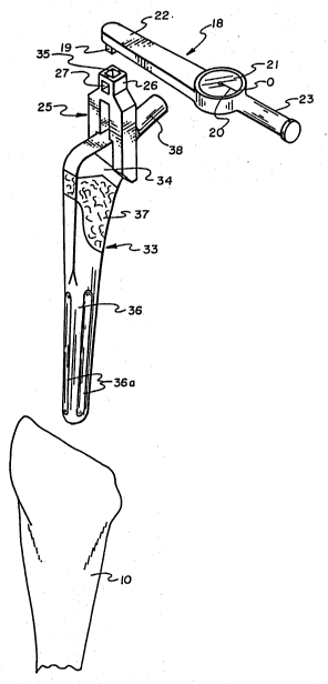
In hip replacement surgery the proximal femur 10, as
shown in Fig. 1, is prepared to receive a prosthetic femoral
hip implant by cutting the bone along a diagonal across the
femur neck, above the lesser and greater trochanter, as shown
at 11. The proximal end of the femur intramedullary channel
is thereby exposed for enlargement to receive a prosthetic hip
femoral implant utilizing a rasp 12. Rasp 12 is shown in Fig.
2 as including, below a flat head end 14 and narrow
rectangular portion a body 13, a round cross-section that is
tapered inwardly to a blunt lower end. Below the head end 14,
2~ 9
in the rectangular portion, the rasp body is holed laterally
at 15 to receive a rod or like tool, not shown, that is fitted
therethrough for applying a torsional force to the rasp.
Fig. 1 shows the rasp 12 seated in the prepared
intramedullary channel end. In that intramedullary channel
preparation the rasp is moved up, down and turned therein such
that cutting ridges 16, as shown in Fig. 2, will file away the
channel wall, appropriately enlarging it to a suitable
diameter and depth to accommodate a prosthetic implant like
the prosthetic hip implant 33, that is shown in Figs. 4 and 5.
In this filing process, as shown in Figs. 1 and 2, a force may
be applied to the rasp as by tapping it with a hammer, on the
rasp head end 14. Which rasp 12, as set out above, can be
turned by fitting a rod, not shown, through rasp hole 15, and
manually turning it. For preparing the intramedullary
channel, the rasp 12 provides, as an arrangement for sizing
the channel to a certain opening that will fit a particular
size of prosthetic hip implant as determined by the surgeon,
lines, shown as A, B and C that are scribed around the rasp
mid-portion, as shown in Figs. 1 and 2. The lines A, B and C
represent different sizes of prosthetic hip femoral implants.
In practice, the rasp 12 is urged into the intramedullary
channel until a select line A, B or C is aligned with the
lowest edge of the intramedullary channel, which positioning
indicates that the intramedullary channel is appropriately
prepared for the particular size of prosthesis.
With the rasp 12 fitted in the intramedullary channel,
as set out above, the seating thereof is then torsionally
tested. This testing is preferably accomplished utilizing a
6 2000I 69
torque wrench 18 that, as shown in Figs. 1, 4 and-5, includes
a dial 21 for indicating force in points that is applied
through a square drive 19. The square drive 19 is operated
through an arm, not shown, that is connected to turn a pointer
20 that is pivoted over scale graduations formed around the
face of dial 21. The square drive 19, that is journaled in an
under surface of housing 22, is arranged to transmit a torque
therethrough as applied at a handle end 23 of the torque
wrench, which force is displayed as pointer 20 travel over the
dial 21 scale graduations. So arranged, the pointer
positioning over a scale graduation is indicative of a
torquing force being applied through square drive 19. In
practice, a dial indicating torque wrench, model ~"DA",
manufactured by Utica Toll Company, Inc., has been used
successfully as the torque wrench 18.
Shown in Fig. 1, the torque wrench square drive 19 is
aligned to fit into a square opening 28 that is formed in a
neck end 26 of an adapter 25. Shown best in Fig. 3, the
adapter 25 is preferably formed to have a U-shape with co-
planar parallel legs 29 that extend from the ends of a web
portion 28. The parallel legs 29 are shown stepped apart from
a first narrow opening 30 adjacent to the web portion 28, to
second opening 31.
Fig. 1 shows the square drive 19 aligned to enter the
adapter square opening 27, which adapter 25 straddles the
rectangular cross-section end of the rasp 12. The rasp end is
shown seated between the parallel legs 29, and have traveled
therein to the first opening 30. So arranged, after the rasp
12 has been used to prepare the bone intramedullary channel
Trademark
2aQo~s
for seating a prosthetic implant, a torsional force is applied
thereto to verify proper intramedullary channel preparation.
In practice, when the rasp 12 does not experience rotational
micromovement at an applied torque of approximately sixty (60)
inch pounds applied for approximately fifteen (15) seconds it
can be assumed that the intramedullary channel is properly
prepared to receive the prosthetic hip femoral implant 33
seated therein.
Shown in Fig. 4, the prosthetic hip femoral implant 33,
hereinafter referred to as implant, is aligned for
installation in the prepared intramedullary channel and has
the adapter 25 fitted over a neck 34 thereof. The preferred
implant neck 34 is of a thickness to just fit between the
parallel legs 29 at the second opening, the area between the
second and first openings to butt against a top surface of
that neck. As shown in Fig. 1, the adapter parallel legs 29
has sloped ends 32 that butt against an upper edge of a
compressed metal shavings matt 37 that is arranged as a mid-
section of the implant, below a dogleg bend, hereinafter
referred to as matt 37. Matt 37 is to provide an area of
multiple ridges and depressions for encouraging bone growth
into the matt as will occur in the natural healing process.
Shown best in Fig. 4, the prosthetic implant edges,
below matt 37 are curved to essentially a round cross-section,
of a bottom portion 36. The implant bottom portion includes
elongate depressions 36a that are formed in opposite surfaces
thereof that are for receiving bone growth therein to further
lock the implant in place.
20(~0169
Fig. 4 shows the wrench square drive 19 aligned for
fitting in a second square opening 35 of the adapter 25, which
square opening 35 is longitudinally formed into the adapter
neck 26, at a right angle to the square opening 27. In Fig. 5
the torque wrench 18 is shown connected through adapter 25 to
apply a torsional force to the implant 33, after which implant
has been seated in the prepared intramedullary channel.
Whereafter a ball, not shown, of a ball and socket hip joint
prosthesis can be secured to the implant head end shaft 38.
Fig. 5 shows the torque wrench 18 with its square drive
19 connected to the adapter 25 at the second square opening
35. So arranged, the adapter parallel legs 29 straddle the
implant 33 to impart a torsional force thereto as reflected by
the positioning of pointer 20 over one of the scale
graduations of dial 21. In practice, the implant 33 is
secured by its friction engagement in the prepared
intramedullary channel. With bone growth to the implant as
occurs in the healing process to further secure the implant in
place. Should, however, that implant, after seating, be
subject to rotational movement, that movement will tend to
shear away the porous bone ingrowth, tending to loosen the
friction fit, destabilizing the appliance. Accordingly, it is
highly desirable to test implant seating prior to olosure.
The present invention provides for such testing by the
application of a torsional force of approximately sixty (60)
inch pounds for approximately fifteen (15) seconds thereto.
At such force application, if the appliance does not
experience rotational micromovement, the implant friction fit
can be judged to be secure. Providing, of course, the implant
9 2000169
33 is otherwise stable. The applied force can vary for
different sizes of implants and accordingly, for a full
range of sizes of a preferred prosthetic implant identified
as an "Anatomic Hip", manufactured by Zimmer, Inc., the
torsional force to be applied to confirm an acceptable
friction fit is approximately sixty (60) inch pounds of
torque, plus or minus ten (10) pounds for fifteen (15)
seconds, plus or minus five (5) seconds. Of course, a greater
force application for a longer period of time can obviously be
used within the scope of this disclosure.
Hereinabove has been set out a preferred system and
apparatus of the present invention for practicing a torsional
testing process to verify a proper friction mounting of a hip
prosthetic implant. It should, however, be understood that
the present disclosure is made by way of example~only and that
the apparatus and process set out herein may be varied without
departing from the subject matter coming within the scope of
the following claims, and any reasonable equivalency thereof,
which claims I regard as my invention.
* Trademark