Note: Descriptions are shown in the official language in which they were submitted.
~o~o~
l BACKGROUND OF THE INVENTION
FIELD OF T~E INVENTION:
The present invention relates to an immuno-
assay method and more particularly, to a method for
immunoassay which is suitable for analyzing and deter-
mining the immune component in a sample utilizing a
photoacoustic spectroscopy.
STATEMENT OF THE RELATED ART:
For determination of an antigen or antibody
contained in body fluids with high sensitivity,
attention has recently been brought to a method for
immunoassay using a photoacoustic spectroscopy. This
method is described, for example, in Japanese Patent
Application KOKAI (Laid-Open) No. 63-44149. According
to the prior arti a particulate antigen-antibody complex
is formed in liquid and the resulting dispersion of the '~
complex is introduced into a photoacoustic cell, where
photoacoustic measurement is made therein. In this
case, utili~ing particle diameter~dependent sensitivity
in photoacoustic spectroscopy, the sensitivity of a
photoacoustic spectrometer is rendered highly sensitive
to the particulate substance to be analyzed, using an
exited light having a wavelength either identical with
or similar to the siæe of the particulate substance to
be analyzed, whereby the particulate substance is
1 -- i
:
,, ~ . ,
,; ~ . . . . .. .
~V~)8~L7
1 ~selectively detected and quantitatively determined.
`~ An invention of U.S. Serial No. 283,B14 filed
December 13, 1988 also relates to immunoassay using
photoacoustic spectroscopy. In the prior invention, an
immune reaction is caused within a reactor to form an
immune complex labeled with ine particles on the solid
phase. Then, the fine particles are separated from the
solid phase in the reactor and the dispersion of the
fine particles is introduced into a measuring cell for
photoacoustic spectroscopy. The fine particles in the
dispersion are measured.
According to Japanese Patent Application KOKAI
No. 63-44149 supra, the particulate antigen-antibody
complex can be detected in such a state that influence
by othex particles is minimized. ~owever, the
concentration of rheumatoid factor, cancer specific
antigen, etc. is extremely low and it is thus desired to
develop a method for measurement with much hi~her
sensitivity. The prior invention described above is
also desired to achieve measurement with much higher
sensitivity.
SUMMARY OF THE INVEN~ION
An object of the present invention is to
provide a method for immunoassay which permits to
measure an antigen or antibody contained even in a trace
amount in body fluids with high sensitivity, utilizing a
photoacoustic spectroscopy.
-- 2 --
'' ~ ~ ' ' ': , . '
. ..
. . . . . .
; ~ , .
: .
8~7
1 According to the present invention, the
antigen-antibody complex is labeled with a color
material and photoacoustic properties of the label are
determined by photoacoustic spectroscopy. As the color
material, a fluorescent substance, a dye or colored
particles can be used. ?
The immune reaction of the labeled antigen or
- antibody is carried out in liquid under the condition
that the solid phase is present. As the result of
immune reaction, the labeled antigen-antibody complex is
immobilized on the surface of the solid phase. Then,
the solid phase is separated from the liquid and moved
to the position for exposure to light and intermittently
irradiated by the light. An acoustic signal given ~ -
thereby is detected. According to the present inven-
tion, a pressure medium for photoacoustic determination
~ is a gas.
; In a photoacoustic spectroscopy, it is
essential that a substance absorbs light. By absorbing
20 light~ the suhstance takes in light energy, and atoms or ;
molecules constituting the substance are excited. Owing
to the light energy which the atoms or the molecules
have taken in, the atoms or the molecules release heat
energy when they return to the ground state. In this
25 process, an acoustic wave is generated by the heat ~;
energy. According to the photoacoustic spectroscopy,
thus generated acoustic wave is detected.
A photoacoustic spectrometer comprises as main
-- 3 --
,, .... . ~ . , .: ;. . ,. .. ;.. - . . . .
;,: : ,
, . - ~ , : .
,.. ' ~:, , ,.. : ': . .. : , - ~. . :
,: ~-, : . , , :
. . . -
~)0817
1 constitutional elements a la~er light source, a chopper
for intermitting a light, a photoacoustic cell, an
acoustic sensor, an amplifier and a signal processor
system. After light from the light source has passed
through a photometer, the light is converted to inter-
mittent light by means of the chopper to be casted on
the photoacoustic cell. In the cell, a sample absorbs
the intermittent light, whereby a photoacoustic signal
is generated. The photoacoustic signal is detected by
the acoustic sensor such as a microphone.
In a preferred embodiment of the present
invention, a solid phase to be determined is placed in
the photoacoustic cell which is sealed in such a state
that gas is present, and a leak into the gas of heat
generated as a result of the light absorption by a
sample is detected as a per70dic pressure change by
means of a highly sensitive microphone.
A pressure change (photoacoustic signal)
generated when a sample is exposed to an incident light
is converted into an electric signal ~generation of
voltage) by means of a microphone or a piezoelectric
element. By previously preparing a calibration curve
between the concentration of antigen and the photo-
acoustic signal converted into electric signal using an
antigen having known concentrations, the antigen-
antibody reaction can be quantitatively analyzed by a
photoacoustic spectroscopy, whereby the desired antigen
or antibody can be quantitatively determined.
. .. ; . , . ~ . ,
i, . . .
~0(~817
1 According to a preferred embodiment of the
present invention, trace components which could only be
determined from a practical viewpoint by radioimmuno-
assay (RIA) heretofore can be quantitatively determined
rapidly with high sensitivity in a simple manner.
Further by using a plurality of species in the
standard substance, a plurality of analyses can also be
determined concurrently.
BRIEF DESCRIPTION OF THE DRAWINGS
Fig. 1 shows an outline of the whole construc-
tion of a photoacoustic apparatus for practicing an
embodiment of the present invention. Fig. 2 shows an
example of calibration curve for determination of HCG.
Fig. 3 shows a calibration curve for concurrent deter-
mination of AFP and CEA. Fig. 4 shows an example of a
calibration curve for determination of TSH. Fig. 5
shows a pretreatment equipment within the analytical
apparatus shown in Fig. 1. Fig. 6 shows a cross
sectional construction of measuring cell used in the
apparatus of Fig. 1.
`, ~''~ ' .
DE~AILED DESCRIPTION OF THE PREFERRED EMBODIMENTS
In a preferred embodiment of the present
invention, a first antibody immobilized to the solid
phase is reacted with a labeled second antibody capable
o~ specifically reacting with the first antibody. By
this reaction, the immune complex labeled with a color
: .. : . . . .
.: ~ . :
,; . . . .
.,
.: ., . : . :.
8~7
l material is formed on the surface of the solid phase
together with the antibody-bound antigen contained in
the sample. The solid phase is separated from the
liquid, introduced into a measuring cell and measured by
a photoacoustic spectroscopy. In this case, the second
antibody labeled with a label is not required to be
specifically reactive with the antigen to be analyzed
but is sufficient to specifically react with the first
antibody. For example, when the analyte is a-feto-
protein ~AFP) and the first antibody is rabbit-anti-APP,
sheep anti-rabbit IgG antibody can be used as the second
antibody. For this reason, it can be avoided to use
expensive antibody in large quantities. In view of
reagent costs, this embodiment is thus excellent.
As the label, there may be advantageously used
a dye, a fluorescence emitting substance, etc. which can
be bound to the analyte or antibody capable of speci-
fically reacting with the analyte. Furthermore, a dye or
a fluorescent substance included in or bound or adsorbed
to microspheres such as liposome; a dye or a fluores-
cence bound or adsorbed to a carrier such as latex
particles: colorless particles or carriers which are
labeled with colored latex, etc. may also be used as the
label. These substances are often referred to as color
materials hereinafter.
The solid phase preferably takes a portable
structure formed into a film or plate having a surface
capable of holding li~uid therein, like a microplate
.
- :. . , . . :
~ ~ ,, ,. : , ;
2~)0~3~7
1 which becomes a little bit hollow; a film made of
~synthetic resin such as nylon, polye~ter, etc.; a
container packed with a filler such as agarose,
~ Sepharose, silica gel, etc. As the solid phase, glass
pieces or paper which can be immersed in liquid, and the
like may also be used.
An embodiment using a plate for thin layer
chromatography (TLC) as the solid phase is described
below.
In measurement of a solid sample, i.e.,
reacted solid phase, by photoacoustic spectroscopy, a
substance present around the surface can be measured
; with relatively high sensitivity. This is because, when
a photoacoustic signal is detected by means of a micro-
phone using as a pressure medium, e.g., gas, a substance
such as an adsorbed matter, etc. present around the
~ ~ .
surface of the solid phase can be detected with high -~ -
sensitivity, since only the region around the surface of
the solid sample contributes to the signal. Where solid
phase itself which is a substrate substantially has no
absorption and has a larger reflectivity, this tendency
is more remarkable. In TLC, silica gel, alumina or the
like is often generally used as the adsorbent. These
substances are optically opaque and powdery, so that
quantitative determination of the labeled reaction
product adsorbed by optical means such as ordinary
transmission method or reflection method results in poor
accuracy in measurement due to scattered light and in
7 --
, ~ ...... .. ~.- .: .. ..
: :. -, .. , :. .
:: - . . ..
... . .. .. . . . . . .
: ::' .: ': . -: ' : -, :. . . ~ '
;:
.:;:: ~, ~
: ~ .
~0~ '7
1 insufficient measurement sensitivity. For this reason,
quantitative or qualitative determination is generally
performed by firstly exposing a TLC plate after develop-
ment to UV light or applying iodine gas to confirm the
position of spot, then scratching the spot off and
extracting the adsorbed matter with an appropriate
solvent, and then measuring its absorption spectrum,
etc. Therefore, a very long period of time is required
for the determination.
According to the photoacoustic spectroscopy,
however, positioning of the spot and quantitative or
qualitative determination of the adsorbed matter can be
made in the adsorbed state. In this case, since back-
ground noises derived from the adsorbent affect the
detection limit, the adsorbed matter, i.e., the label is
colored so that S/N can be greatly improved.
As compared to the conventional absorbance
measurement method, the present invention permits to
measure with sensitivity as high as two figures or more
in such a state that the label is adsorbed on the
reacted solid phase.
When a color material is used as the label, a
ratio of signal to background noises (S/N) for photo-
acoustic measurement is improved. The solid phase on
which the antigen-antibody complex labeled with the
color material is adsorbed is moved to the position of a
spectrometer for exposure to lightO When the solid
phase is exposed to a laser light, the color material
~ ,.. . . .
`: - ~ : . . ' ' .
~, ' , , ' .
81~
1 present on the surface of the solid phase absorbs the
light and releases heat energy in response to the light
absorbed. The greater the quantity of light absorbed,
the greater the photoacoustic signal obtained. As a
color of the label, it is chosen to have the absorption
maximum substantially in compliance with the central
wavelength of the excited monochromatic light. For
example, when an argon laser having a wavelength of 488
~ nm is used as the excited light, Food Yellow 3 having
the absorption maximum wavelength of 482 nm is used as a
dye for the label; in this case, good results are
obtained.
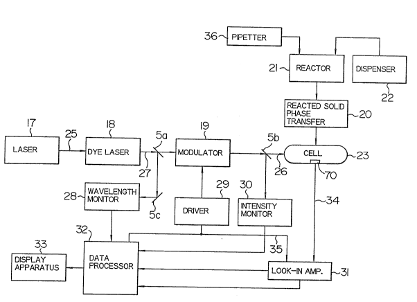
An outlined construction of an analytical ~-
apparatus for practicing one embodiment of the present
invention is shown in Fig. 1.
Light source for excitation 17 is a light
source for argon laser having an output of lOW. Laser
light 25 from argon laser light source has oscillation
rays in several wavelengths at 488 nm, 514.5 nm and over
the ultraviolet to visible regions. Depending upon the
size of the reaction product to be analyzed, an appro-
priate dye in dye laser 18 is subjected to pumping. For
example, when the size of the reaction product is 0.6 to
0O7 ym, the wavelength of the excited light 25 from the
argon laser light source 17 is set at 514.5 nm and
rhodamine is used as the dye for the dye laser 18. The
laser light 27, its wavelength being modified in
response to the size of the analyte, is in part divided
_ g _
: . .
: . .
, .. . .
' ' . ' ' ' :
' ~ "~ ' ~: ` . , '
: :: , ,,
:,;: - .
~00~8~
1 by half mirror 5a. The wavelength of incident light via
reflection mirror 5c is confirmed by wavelength monitor
28. The remaining laser light is modulated to a
periodic rectangular wave by a modulator 19 comprising
light chopper of rotary blade type and is converted into
the excited light 26. The excited light 26 is cast on a
measuring cell 23. In the cell, the sample absorbs the
light to give a photoacoustic signal. The photoacoustic
signal 34 generated in the measuring cell 23 is detected
by means of a microphone or piezoelectric element 70 and
amplified by a lock-in amplifier 31, by referring to
reference signal 35 synchronized with light intensity
modulation output from a driver 29 of the modulator 19.
A part of the excited light 26 is divided by the half
mirror 5b and its intensity is monitored by a light
intensity monitor 30. Information on the intensity of
the photoacoustic signal through line 72 and the phase
of the photoacoustic signal through line 73 is input to
a data processor 32; further information on the
wavelength, modulated frequency and intensity of the
excited light from a wavelength monitor 28, the
modulation driver 29 and the light intensity monitor 30
is also input to the data processor 32 through line 71.
The data processor 32 displays parameters on conditions
~or the measurement, for example, information on
wavelength of the excited light, modulation frequency,
etc. on a display apparatus 33 or performs data
processing of the measurement results and displays a
-- 10 --
. ,. , , .. :.
.. . . . . . .
~ . . ,, , :- , ~. ;
~ , . . .
.. . .
0~317
1 calibration curve of the reaction product or quantita-
tive results on a sample having unknown concentration,
and the like on the display apparatus 33.
Body fluids such as plasma are placed with a
sample pipetter 36 on a disk-like apparatus within a
reactor 21. Reagent solution for forming the antigen-
antibody complex is added through a reagent dispenser
22. The reacted solid phase on which the immune complex
is formed in the reactor 21 is inserted into the photo-
acoustic cell 23 by means of a reacted solid phasetransfer 20. The cell is sealed in such a state that
the air is present. The measuring cell 23 consists of a
glass container equipped with a light incident window,
inside of which a microphone is placed. An open-close
cover provided in the cell is closed and sealed when the
reacted solid phase is encased in the cell.
An example of the construction of the measur- -
ing cell 23 used in the apparatus shown in Fig. 1 is
shown in Fig. 6. The cell 23 has a cylindrical room,
the upper part of which can be opened by a reactor
forming material 1. The room 2 is sealable with a
quartz glass-made cover 3. The microphone 70 is
embedded in the side wall of the room 2. A stand 4 of
transparent glass is mounted to the bottom of the room.
The disk-like solid phase 5 can be placed at the center
of the stand 4~ In the example, the solid phase has a
size of 10 mm in diameter and 2 mm in high. However,
the solid phase can be changed to various sizes. When
;;: . , ~
. ~ :
~'~ 1 " ~, .. . .
... . . ~ . . - . . .
:::.. . . :: -
.. . .
.. : ,, . : . :
.
.: . ~
:,., : .
~000~3~7
1 the room ls covered, the volume of the space i5 O. 5 to
1.0 ml. The excited light is cast on the solid phase 5
through the cover 3. Therefore, the cover 3 functions
as a light incident window. For the construction of
such measuring cell 23, there are various modifications.
Fig. 5 shows a pretreatment equipment within
the analytical apparatus in Fig. 1. The pretreatment
equipment includes the reactor 21, the reagent dispenser
22 and the sample pipetter 36 in Fig. 1. A pipetter 40
lo in Fig. 5 corresponds to the pipetter 36 in Fig. l.
Distributor 37 in Fig. 5 corresponds to the dispenser 22
in Fig. 1.
In Fig. 5, a sample table 10 on which a
plurality of the standard substances having different
` 15 concentrations are placed for respective items for
measurement is provicled. A plurality of standard sub-
stances can be placed on the sample table 10 continuous-
ly for every item for measurement. A reaction table 121
has reactors 122, on the circumference of which the
reacted solid phase 5 having bound thereto a plurality
of antibodies for analyses of a plurality of items is
placed. The reaction table is constructed to be freely
rotatable. Transfer of the standard material and sample
is performecl by means of a sampling probe 41. Dispense
of the reagent is effected by a moving distributor 37.
The reactors 122 in which a plurality of the
solid phases in the kind and number are encased are
constructed to make continuous line on the reaction
- 12 -
~081~
1 table 121 or every item. A necessary solid phase is
supplied to the corresponding reactor through a reacted
solid phase supplier 42. On the reactor line, a dis-
charge apparatus 129 and a washing apparatus 124 are
5 placed. Details of the photoacoustic signal measuring
apparatus 49 are shown in Fic~. 1. A sealed type photo-
acoustic cell 23 is so constructed that a pressure
change generated as the result of the exposure of the
solid phase placed within the cell to the light source
17 can be detected by means of a microphone.
A controlling apparatus is e~uipped with a
multiplexer 53, an A/D converter 54, a read only memory
(hereafter referred to as ROM), a random access memory
(hereafter referred to as RAM), a printer 55, an
operation panel 52 and a driving circuit for mechanism
135. The A/D converter 54 is further connected to a
central processor 51 via an interface 50. The central
processor 51 functions to control the whole apparatus
including the mechanism, prepare a calibration curve and
perform data processing such as operation of the
concentration, etc. For the central processor, a
microcomputer is used.
The operation of the pretreatment equipment is
described below.
Firstly, when a plurality of the solid phases
are set on the reacted solid phase supplier 42, neces-
sary numbers of the solid phase are continuously
supplied to the reactors 122 on the reaction table 121
- 13 -
... ,,, .. ~ , . I , .. ~ . - ,. . ..
, . ~ . . .
. ... . .
.. , . :, .",.,,",
: ,: . . . ~
.
::. . . .
~08~
1 for every item. Next, sample containers 44 in which
sera (samples) to be analysed containing hormones,
cancer markers, infection-associated substances, etc.
are encased are supplied on t:he sampling position on the
sample table 10. Then, the tip of the probe 41 held in
a movable arm 12 in the pipetter 40 is dipped in the
sample containers and sucks a definite amount of the
serum and holds in the probe 41. Thereafter~ the probe
41 moves to the discharge position 45 on the reaction
table 121 and discharges the serum held in the reactor
122 including the solid phases of kinds corresponding to
the items to be measured, with the probe 41.
When this sampling operation is completed, the
reaction table 121 goes counterclockwise by 1 pitch of
the xeactor and stops at the position. When a time from
the rotation to the stop of the reaction table is, for
example, 20 seconds, the above operation is repeated as
one cycle for 20 seconds. As the cycle proceeds~ the
specific sample to be measured goes counterclockwise by
one pitch of the reactor at the position where the
reaction table 121 is in a standstill state. Discharge
of the reagent from the moving distributor 37 is made in
such a state that the reactor 122 stops at the discharge
position 47 on the reaction table 121. The distributor
37 selects a necessary reagent solution from a line of
reagent containers 39 to discharge into the reactor.
Taking as an example a specific sample, a first reaction
is initiated between the sample added at the discharge
;. .: ,~ ~ , . ,
,.
"'' ,.~ .,,, ;.".. , ;'' ,
",, : . . . .
)8~7
1 position 45 and the solid phase in the reactor and a
second reaction is initiated with the reagent at the
discharge position 47.
When the reactor 122 in which the reaction
s proceeds stops at a washing position 48, the discharge
and suction of reagent for washing are repeated by a
washing apparatus 124, whereby the solid phase is washed
as it stands in the reactor. When the reaction table
121 rotates and stops at the stop position 46, only the
reacted solid phase in the reactor is taken into the
photoacoustic cell in which the photoacoustic signal is
detected by means of a photoacoustic apparatus 49. Xn
output of the photoacoustic apparatus 49, a signal of
measurement wavelength currently required is chosen by
the multiplexer 53, incorporated into the central
processor 51 through the A/D converter 54, and memorized
on RAM. The central processor operates following the
program of ROM, extracts the measurement data in RAM and
performs data processing. The reaction table 121 is
placed in a thermostat 123, whereby the reaction in the
reactor can be proceeded at a controlled temperature.
For example, 5 or 6 standard substances per
one item which are required for preparing a calibration
curve are aligned in a continuous line on the sample
table 10. Accordingly, a plurality of the standard
substances having different concentrations for a
specific item are automatically transferred to the
reaction table 121 by a plurality of times (for example,
-: . . . .
;; . - ~
: ~ . . ' '
."
~:()001317
1 by twice each) through the sampling probe 41. In the
case of preparing a calibration curve on a sample which
has no linear relationship wlth its concentration, it
is indispensable to perform sampling the standard
substances having different concentrations by a
plurality of times and measure them. The present
apparatus enables to prepare such calibration curve.
These measurement data are collected for each item and
provided for the preparation of calibration curve. The
measurement results are displayed on the printer 55 and
CRT 56. Next, in order to verify the effects of the
present invention, an example of analyzing a serum
sample by appropriately modifying the apparatus of Fig.
~ l is shown below.
;15 Measurement Example l
A removable rotary holder was used instead of
the reaction table 121 shown in Fig. 5. A plurality of
solid phase disks composed of a porous material capable
of filtering liquid therethrough were set on the holder
After the cellulose membrane having bound thereto anti-
human HCG (human chorionic gonadotropin) antibody was
adsorbed to the porous reacted solid phase disks, the
holder on which the disks were placed was put on the
reaction apparatus 21. Through the sample pipetter 36,
lO0 ~1 of the serum sample was dispensed on the xeacted
solid phase disks. Five minutes after, lO0 ~l of anti-
human HCG antibody solution labeled with FITC
- 16 -
. . :,:: - - .. . .
,, - : ,. .
.~ , . ... .. .,. "
: , .. . .
.,, ~ .,.
: . . .: . .
.;. :
~ .
. .
~)0081~
l (fluorescein isothiocyanate) was added to the solid
phase disks and superfluous water was absorbed into the
porous material. After allowing to stand at room
~ temperature, Tris-hydrochloride buffer solution (pH 7.8)
was added to the solid phase disk. By doing so, the
unreacted excess FITC-labelecl anti-human HCG antibody
and water were absorbed into the porous material to
remove them from the cellulose membrane surface. The
antigen-antibody complex was formed on the cellulose
membrane of the solid phase disk.
Next, the reacted solid phase in the reaction
apparatus 21 was transferred to the photoacoustic cell
23 by means of the reacted solid phase transfer 20. The
photoacoustic cell was covered with a cover for sealing.
A laser light from the light source 17 was then cast
into the cell 23 to measure the photoacoustic signal
based on the antigen-antibody complex formed on the
cellulose membrane surface. Thus, HCG in the sample was
measured; an example of calibration curve in this case
is shown in Fig. 2.
.,~,.
Measurement Example 2
Filter paper (Toyo No. 51) was cut into a size
of l x 2 cm and made a raw material for the solid phase.
About l g of the paper solid phase was suspended in 20
ml of 5 M potassium phosphate buffer (pH 12.1) and lO ml
of water was added to the suspension. While stirring
with a stirrer, lO ml of BrCN solution (0.1 g/1) was
- 17 -
~. . ~ : .
.i- ~. , .. ... . :
: , ~ :. ~:. , ,,. :,.
, ,. ~ . .
. .
:: . .
:.: . . ,
... . .
0~1~
1 gradually added to the mixture over 2 minutes followed
by reacting for further 6 minutes. The reaction was
carried out at 4C. Immediately after the reaction~ the
reaction mixture was washed with a large quantity of
0.005 M ice-cooled NaHCO3 solution, and 5 mg of purified
anti-human AFP antibody and purified anti-human CEA
antibody were reacted at room temperature or 8 hours.
After further reacting with 1 M ethanolamine (pH 8.0) at
4C overnight, the reaction mixture was sequentially
washed with 0.5 M NaHCO3 solution, 0.1 M acetate buffer
solution (pH 4.0) and 0.015 M PBS ~pH 7.2), and then
freeze dried in 1.5 ml of PBS (pH 7.2).
After the solid phase having bound thereto
anti-human AFP antibody and anti-human CEA (carcino-
embryonic antigen) antibody was set on the reactionapparatus 21, 100 ~1 of a serum sample was added to the
solid phase. Immune reaction was carried out at room
temperature for 10 minutes. The solid phase was then
washed twice with 1 ml each of o.9% NaCl and the washing
liquid was removed from the system.
A first reagent dispersion of yellow latex
particles having bound thereto anti-human AFP antibody
and a second reagent dispersion of blue latex particles
haviny bound thereto anti-human CEA antibody were
prepared as the reagents. To the solid phase were added
250 ~1 each of the first and second reagent dispersions.
The mixture was reacted at room temperature for 10
minutes. The reaction mixture was then washed twice
- 18 -
.. v, . . . . - : . ~ . ,.: : . ...
': .' . ;~':: .. " ' , . ..
. : . -
.. . :
: : .,, : .
:. , . . : :
- ~ :
.
~0C)~)8~7
1 with 1 ml of 0.9~ NaCl. Ater the washing liquid was
removed, the solid phase was withdrawn from the reaction
apparatus 21 and placed in the photoacoustic cell 23.
After the cell 23 was sealed, the solid phase was
exposed to 2 kinds of laser light having different
wavelengths, whereby the photoacoustic signal was
measured. According to such a method, AFP and CEA can
be quantitatively determined concurrently on one reacted
solid phase disk. An example of the calibration curve
for simultaneous quantitative determination of AFP and
CEA used in this measurement method is shown in Fig. 3.
In this case, AFP and CEA are measured using the
wavelength corresponding thereto.
Measurement Example 3
As the solid phase, a disk-like glass piece
was used. Anti-T5H (thyroid stimulating hormone)
antibody was previously bound to the glass wall surface
on the solid phase. The reactor in which the solid
phase was encased was placed on the reaction apparatus
21. Through the sample pipetter 36, 20 ~1 of the serum
sample containing TSH was dispensed in the reactor
followed by reacting with the anti-TSH antibody on the
solid phase at 37C for 5 minutes.
Next, 100 ~1 of a dispersion of sheepanti-
rabbit IgG antibody-bound red latex particles was added
to the reaction mixture through the reagent dispenser 22
followed by reacting at 37C for 5 minutes. Then, the
-- 19 --
..
1 ,; ~ . , ~,
... . .
~.:. , . .: . . :
: . . .
. . .. . .
.: - ~ . . :,
~ .
1 unreacted liquid was resnoved. After washing with 0.05 M
phosphate buffer solution (pH 7.5), the washing liquid
was discharged. The washed solid pha~e was introduced
into the photoacoustic cell 23 and the cell 23 was
sealed. A laser light was then cast onto the solid
phase to measure the photoacoustic signal. An example
of calibration curve for quantitatively determining TSH
is shown in Fig. 4.
20 -
, " , . . .
:. . . . . . . . . .
.,;, . ~ . , :, ~ .. . .
-: . . , .. : . ..
:.: . ~ . . ' : '
. ::. ': , ., ~
:. .. . : ... .