Note: Descriptions are shown in the official language in which they were submitted.
2001503
Catheter for measuring motility and peristalsis in tubular
organs that transport their contents by means of
simultaneous, multiple impedance measurement
At present there are two techniques available for
diagnosing illness-related motility or peristalsis
disorders, namely
a) manometry by means of perfusion measuring techniques or
by means of semiconductor pressure sensors, and
b) image-producing methods which make use either of X-ray
representation or scintigraphy of contents provided
with contrast media.
With manometry, a pressure change is used to
characterize the contraction sequences in a narrow section
of the organ to be examined. Perfusion measuring
techniques are most widely used for this type of
measurement. These techniques use a catheter to measure
pressure changes resulting from the contraction of an
organ. A fixed inflow of fluid into the catheter insures
that any obstruction of outflow of the organ creates these
pressure changes. Since, however, it is necessary here to
use tubes with an internal diameter of more than 1 mm for
each channel, a multicatheter can have a maximum of only
eight channels in order to maintain an acceptable outer
diameter. A further important disadvantage is that fluid
is introduced by a perfusion catheter into the organ which
200~503
-- 2
can substantially impair the function of the organ,
particularly with a long-term measurement.
Image-producing methods are principally suitable for
showing motility, for example in the esophagus. However,
diagnostics in the intestinal area are problematic due to
the many overlappings. Moreover, the high cost outlay for
acquisition and operation of the equipment and for the
evaluation of the images obtained therefrom must be taken
into account.
The object of the invention is to provide a method and
a measuring device which is relatively simple to realize
for evaluating motility and/or peristalsis in tubular
organs that transport their contents, which can be used
universally and leads to reliable diagnostic results.
For obtaining signals for evaluating motility and/or
peristalsis in tubular organs that transport their
contents, or for determining the dynamic resilience of
tubular conveying organs, by means of simultaneous,
multiple impedance measurement, the invention provides for
the use of a catheter which is essentially made of a
flexible, non-electroconductive elongated member which
bears in the measuring area a plurality of electrodes
arranged at defined distances from one another and
enclosing the member at least partially in annular fashion,
the signal leads of which electrodes extend along or are
embedded in the interior of the member.
The catheter according to the invention permits signals
and data to be obtained which serve to evaluate the type of
motility and peristalsis in terms of space and time, namely
from stationary, propulsive or repulsive waves, from
contraction spreading etc. The measuring technique used as
a basis can be used for determining the characteristics,
such as the passage time, the passage speed, the
contraction frequency and the frequency of contractions of
individual organ sections.
In order to sense these characteristics, the catheter
2001503
-- 3
is introduced into an organ and fixed, with respect to
longitudinal displacement, in a defined position. The
signals to be used according to the measuring technique are
obtained by a simultaneous or quasi-simultaneous recording
of the impedence of at least three channels, 32 or more
channels being used typically.
Advantageous embodiments of the invention and further
details thereof are described in greater detail in an
exemplary embodiment below with reference to the drawings,
in which:
Fig. 1 shows the enlarged perspective view of a
measuring area section of a tubular catheter
with features according to the invention;
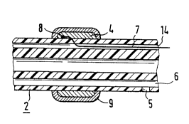
Fig. 2 shows an enlarged sectional representation in
longitudinal section through a measuring area
of a catheter of the type according to the
invention;
Fig. 3 illustrates a method for simultaneously
registering the esophagus activity with a
multiple measuring catheter of the type
according to the invention;
Fig. 4 shows, also in connection with Fig. 3, a
particular embodiment of a catheter of the
type according to the invention;
Fig. 5 shows a diagrammatic block circuit diagram for
illustrating a measuring construction using a
catheter according to the invention; and
Fig. 6 shows various possibilities for arranging the
circuits of individual measuring channels.
Evident in Fig. 1 is a part of the measuring area of a
tubular, preferably flexible catheter 1 according to the
invention, which is essentially a tube of non-electro-
conductive plastic. Possible materials here are, for
example, polyurethane, polyamide, polyethylene, poly-
tetrafluoroethylene, polyvinylchloride or certain silicon
rubber compounds.
-- 4
Placed around the outside of the tube 2 are annular
electrodes 4 which, as can be seen from Fig. 2, are
produced from a flat strip material and are firmly
adhesively bonded to the tube 2. The edges of the
electrodes 4 are rounded and designed in such a way that
when incorporated in the tube material no injuries can
result. Suitable materials for the electrodes are metals,
electrode materials of a second type and conductive
plastics. Further characteristics of the electrodes 4 are
their low-resistance, low polarization voltage in the top
electrode layer and their long-term stability. Since an
electrode 4 forms a junction to an electrolyte, it must, in
order to have the characteristics required, be provided
with a coating 9, for example of silver-silver chloride.
The distance between the electrodes 4 may be constant or
may be designed according to a spatial pattern. Electrode
width may vary, as well as the mutual distance between a
pair of electrodes which is assigned to one measuring
channel for the impedance measurement. The width of the
electrodes 4 may be designed to be be different it depends
on the diameter of the tube 2 and the measurements to be
taken.
The electrode leads extend inside the tube 2, for
example into a separate channel 14 which is separated by
partition walls from further individual channels 6.
A respective lead 7 is connected to the associated
- electrode 4 at an inner contact point 8.
The catheter 1 can be provided with one or more lumen
5, through which, at various places, substances can be
removed from the organ for analysis purposes or substances
or fluids can be introduced as contrast media or for
function stimulation. However, such a function stimulation
cannot only be carried out with a liquid, but also
electrically by means of the measuring electrodes 4
themselves. In addition, the electrodes may be used for
receiving electrical biosignals.
~001503
The basic representation of Fig. 3 illustrates the
simultaneous registration of esophagus activity with a
multiple measuring catheter 1 of the type according to the
invention.
In this case, a measuring channel is designed either
between two electrodes 4 (El/E2 or E2/E3 or E3/
E4 of Fig. 4) for measuring the longitudinal impedance or
between an electrode on the catheter 1 and a central large-
area electrode (not shown) on the body.
As Fig. 6 illustrates, the longitudinal impedance can
be measured either (a) sequentially, (b) sample-wise or (c)
with overlapping.
In Fig. 3, MG denotes a reference channel while the
values Rl, R2, R3 and R4 marked by respective
distance arrows designate impedance changes in the channels
1, 2, 3 and 4. In the example shown, the signals Rl to
R4 characterize a propulsive contraction wave. The
passage time or passage speed in the organ examined can be
calculated from the transit times between two channels.
Fig. 4 illustrates further details of a modified
embodiment of a catheter with features according to the
invention. The representation shows that the catheter
tube 2 has a well-rounded closure at the front end 3 and
its leads 7 extend from a connector 10 into the interior.
The diameter d may be as small as a few mm. The catheter
is flexible, but the measuring part IM of the catheter 1
can be designed to be of varying flexibility, or even
rigid. The length of the measuring part IM and the leads
Iz may vary up to several meters. By virtue of the tube
interior, the catheter can be stiffened temporarily, for
example during the measurement.
The catheter 1 is provided with few (at least three) to
many (e.g. 32 and more) electrodes 4 (El, E2, E3,
E4, ...). The electrodes are connected in an electro-
conductive manner via the leads 7 to the connector 10 of animpedance measuring arrangement, such as the one illustrated
Z001503
-- 6
in Fig. 5. This arrangement may be connected to a plotting
or recording device 18 and/or a signal processor 17 with
associated display.
Apart from the lumen 5 of Fig. 1 and Fig. 2, the
catheter 1 can be provided with one or more temperature
probes (not shown) along its longitudinal axis.
In addition to and at the same time as impedance
measurement, the catheter 1 can also be used for receiving
EMG signals which precede the mechanical contraction of an
organ.
By virtue of the electrodes, the catheter also permits
a simultaneous electrical function stimulation or excitation
of the organ examined.
Moreover, further biosignal transducers 15, shown in
Fig. 5, can be integrated in the measuring catheter for the
simultaneous reception of electrical biosignals (e.g EMG,
ECG). Amplifiers can be provided per channel for signal
separation or for stimulation devices in individual or all
channels. A current source 16 may be used for electrical
stimulation. In addition pressure balloon stimulation and
sensing devices can be used.
With a measuring catheter according to the invention
and the method based thereon, it is possible to register
the motility and peristaltic processes in both healthy and
unhealthy organs, e.g. in the esophagus, in the intestines,
in the urethra, etc. The movement of the contents of these
organs is as a rule orientated in one direction, although
numerous mixed processes do occur and are known, e.g. in the
intestines, in which the contents are transported to and
fro. For moving the contents, the contractions need to be
strictly synchronized. The weakening or a total failure of
the spontaneous activity of even a small secion can lead to
disorders of the entire organ. With the method presented
here and the catheter according to the invention, the
sections of the organ with a functioning disorder can be
readily diagnosed.
~001503
-- 7 --
Besides the use of the method in the medical field, a
technical application is also possible, for example, for
measuring the dynamic resilience of tube walls by applying a
pulse pressure wave for determining the dynamic stressing of
the walls. It is thus possible to recognize and eliminate
age-induced changes in a tube wall.
