Note: Descriptions are shown in the official language in which they were submitted.
;~0(~ '3
RADIOFRE(2UENCY ABLATION CATHETER
Th;s application is a continuation-in-part of U.S. Application Serial No.
07/276,294, Catheter with Radiofrequency Heating Applicator, filed November
25,1988.
Technical Field
Th;s invention pertains to a catheter designed to couple radiofrequency
(RF) energy to biological tissue surrounding the catheter tip. Typical
application is in thermal ablation of cardiac tissue. This invention further
pertains to an apparatus used to guide a cardiac ablation catheter to ablate
arrhythmia-causing myocardial tissue and to monitor the ablation procedure by
detecting, processing, and displaying endocardial EKG signals.
Ba~kground Art
Percutaneous thermal destruction (ablation) of problem myocardial tissue
(arrhythmogenic focus) is a therapeutic procedure used with increasing
frequency for treatment of cardiac arrhythmias (e.g., ventricular tachycardia).
Medically, ablation is covered in Ablation in Cardiac Arrhvthmias,
G. Fontaine & M. M. Scheinman (Eds.), Futura Publishing Company, New
York, 1987. A recent review of the ablation field is given in a chapter by D.
Newrnan, G. T. Evans, Jr., and M. M. Scheinman entitled "Catheter Ablation of
Cardiac Arrhythmias" in the 1989 issue of Current Problems i Cardiology, Year
2s Book Medical Publishers. Catheter ablation of ventricular tachycardia was first
described in 1983 as a nonsurgical method of destroying an arrhythmogenic
focus. Typically, a pacing catheter is introduced percutaneously and advanced
under fluoroscopic guidance into the left heart ventricle. It is manipulated until
the site of earliest activation during ventricular tachycardia is found, indicating
30 the location of problem tissue. One or more high voltage direct-current pulses
from a standard defibrillator are then applied between the distal electrode of the
catheter and an external large-diameter chest wall electrode. This procedure
works by destroying cardiac tissue responsible for the arrhythmia.
2(~036~9
Although this treatment is effective in some patients, there are serious
drawbacks to high voltage direct-current pulses as an ablative energy source.
The shock is painful, so general anesthesia is required. More importantly, the
discharge produces arcing and explosive gas formation at the catheter tip. The
resultant shock wave is responsible for serious side effects. The scar created by a
direct-current pulse tends to have a large border zone of injured but still viable
tissue that may serve as a new focus for ventricular tachycardia.
These problems have prompted a search for alternatives to direct-current
pulse as a source of ablative energy. Radiofrequency (RF) energy is a promising
o method being investigated. (RF without qualifiers refers here to the
electromagnetic spectrum from 10 kHz to 100 GHz, as per ANSI/IEEE
Standard 100-198~.) Laser energy is also being considered for catheter ablation
of arrhythmias (see Cohen, U.S. Patent No. 4,685,815) but is not pertinent to the
RF implementation considered here.
RF ablation using electrosurgical power units is in clinical investigation,
as a safer ablation alternative to high voltage direct current pulses. At present,
continuous, unmodulated current in the range of 0.5 MHz to 1.2 MHz, such as
that supplied by an electrosurgical RF power supply, is applied to the
endocardium via an electrode catheter in the same manner as with a direct-
current pulse. Ablative injury is produced by heating, generated by an electric
field emanating from the catheter electrode. There is no gas or shock wave
formation, and therefore no risk of serious barotraumatic side effects. However,as discussed in more detail later, the small size of the resulting lesion remains a
problem even with RF ablation.
In order to discuss and evaluate the technical state of the art of RF
ablation catheters and to compare it with embodiments of this invention, one
must first establish pertinent performance requirements. A general geometrical
requirement of catheter-based applicators is that they must be confined in a
slender cylindrical structure with a radius commensurate with the catheter
diameter. Subcutaneous insertion into the heart dictates that the catheter body
must be a flexible tube no more than 2 mm in diameter and about 1 meter long.
The diameter is constrained by the size of blood vessels used for catheter
insertion into the heart. The length is dictated by the length of the catheter
inside of the patient's body plus the length of the catheter between the patientand the external equipment.
~003G~9
In the discussion of catheter performance which follows, it is convenient
to adopt a cylindrical coordinate system with the z-axis coincident with the
catheter axis and pointed toward the distal end. The radial component is in the
direction normal to the catheter z-axis, and the circumferential component has as direction around the z-axis. Radius is measured from the catheter axis.
A simple cylindrical wire heat applicator antenna is shown in FIG. lA.
Applicator antenna 10 is a conductor immersed in a lossy dielectric medium
which has electrical properties typical of muscle tissue. The radius of applicator
antenna 10 is "a" and its height is "h". In spite of the simple geometry and lowlO frequency approximation used in the description, FIG. 1 retains the salient
features of a radial-field coupling of pacing catheters used as an RF antenna.
In FIG. lA, RF potential V14 is applied in a unipolar manner between
applicator antenna 10 and a remote boundary 15 which corresponds to a neutral
electrode applied to the skin. The exact location of boundary 15 is not important
lS to the shape of the radial electric field E near applicator antenna 10. Electric
field E16 coincides with current density vector Jr = aEr in the tissue, where a is
the conductivity of the tissue.
Continuity of current in the cylindrical geometry in FIG. lA results in
current density Jr which decreases with the inverse of the radius r: Jr = JOa/r
20 for r < h and power dissipation P = Jr2/a = (J20/a) (a/r)2. For r > h, the
spherical geometry is a more approlpriate approximation and results in
Jr = J0(a/r~2, and the corresponding electrical power dissipation is
P = Jr2/a = ~J20/a) ~a/r)4. The result is that the heating of tissue, decreases with
the radius within the bounds of the second to the fourth power of a/r. This
25 behavior of the electric field applies to a conducting medium below the
microwave region. In the microwave region (f > 900 MHz), the radial
attenuation of electric field is even faster due to the "skin depth" attenuation.
f~OO~ 9
Clinical experience indicates that in order to effectively ablate ventricular
tachycardia, it is desirable to thermally destroy (ablate) tissue over an area of 1-
2 cm of the myocardium (e.g., see Moran, J. M., Kehoe, R. F., Loeb, J. M.,
Lictenthal, P. R., Sanders, J. H. & Michaelis, L. L. "Extended Endocardial
Resection for the Treatment of Ventricular Tachycardia and Ventricular
Fibrillation", nn Thorac Sur~ 1982, 34: 538-43). As mentioned earlier, in order
to accomplish percutaneous insertion into the left ventricle, the heating
applicator radius is limited to 1 mm. In order to heat a 2 cm area, a 2 cm long
applicator can be used provided an effective heating diameter of 1 cm can be
reached. To overcorne present shortcomings of the RF ablation method, the size
of the lesion must be increased and this requires the minimization of the radialattenuation of the electric field and the associated heat dissipation.
The destructive ablation of tissue requires a temperature of
approximately 50C; this temperature defines the outer radius R of the ablation
region. It is therefore desirable to heat tissue to 50C up to 5 mm from the
catheter axis. Yet at 100C, undesirable charring and desiccation takes place.
So, ideally, the maximum temperature at the applicats)r electrode boundary
should be under 100C.
Ignoring for a moment heat conduction in the tissue, the rise in tissue
~o temperature is proportional to the electric power dissipation which in turn is
proportional to the square of the current density. In order to maintain a
100C/50C or a factor of 2 temperature ratio between the temperature at a
radius of 1 mm and the temperature at a radius of 5 mm, the ratio of the power
dissipation ratio should be 2 at these two distances. Yet the performance of thecurrent density in FIG. lA gives at best a power dissipation at the catheter
surface of ~R/a~2 or 25, and at worse (R/a)4 or 625 times more intense than
heat dissipation at a 5 mm radius.
In order to examine the effect of this wide range of heat dissipation, it is
useful to divide the lossy medium in FIG. lA into three cylindrical shells: first
shell R11 adjacent to the applicator antenna 10, followed by shell R12, and R13
beginning at the 10 mm radius. Since the shells are traversed by the same
current, and the potential drop across the shells is additive, power delivery can
be schematically represented hy three resistances R11, R12, and R13, as shown
in FIG. lB, connected in series with the source of RF potential V14.
~003~i~lg
The heat required to obtain adequate ablation at a Smm radius tends to
desiccate blood or tissue close to the applicator antenna 10, increasing the
resistivity of R11. This in turn further increases the relative power dissipation in
R11 in comparison with R12 and R13, until effective impedance of the
s desiccated region R11 becomes, in effect, an open circuit shutting off the flow of
RF power to the tissue beyond R11.
This indeed is the problem with state-of-the-art RF ablation catheters
which severely limits the effective heat delivery to more distant tissue. The
currently used RF ablation technique, based on a surgical RF power supply and
a pacing catheter, suffers from a steep temperature gradient, reportcd to decay
sharply (Haynes, D.E., Watson, D.D.: PACE. June, Vol. 12:962-976, 1989), and
has the associated problem of charring which disrupts and limits heating and
ablation.
Insulation of the applicator antenna 10 from the tissue does not reduce
lS the heat dissipation gradient: If the applicator antenna 10 is insulated from the
lossy medium by a thin dielectric tube, the effect of the dielectric can be
represented by capacitor (not shown) in series with the source of RF potential
V14. Now the applicator must be operated at a *equency high enough so that
the impedance of the sum of resistances R11 and R12 and R13 must be higher
than the capacitive impedance of the dielectric tube. Rl 1 still dominates the
heat distribution.
Effective ablation heating also requires that the heating along the heat
applicator axis should be as uniform as possible. Heating should then rapidly
attenuate to a negligible value along the portion of the catheter acting as a
transmission line.
A key improvement requirement is therefore the ability to ablate areas
significantly wider than the catheter diameter, confined only to the region of the
heat applicator. Heating should not be limited by charring and desiccation at
the catheter boundary.
Therefore, there is a need for a catheter-compatible RF energy delivery
system which dissipates heat more uniformly in the radial direction and is well
defined in the z direction, thereby leading to a more accurately controlled and
larger ablated region. It is also desirable to eliminate the effect of desiccation of
tissue, adjacent to the electrode, on heat dissipation to surrounding tissue.
~(~C)~ 3
An effective cardiac ablation catheter must satisfy three additional
performance re~uirements:
(1) The body of the catheter should act as an efficient and reproducible
RF power transmission line with the heat applicator transforrning the impedance
s of tissue (electrically a lossy medium) to match the characteristic impedance of
the transmission line.
(2) The detection of an endocardial potential, needed for mapping of
location of the arrhythmogenic tissue to be ablated, must coexist, without
interference, with the heating function.
o (3) All of the above must be accomplished in a flexible catheter, about
2 mm in diameter so as to allow percutaneous insertion into the left ventricle.
U.S. Patent No. 4,641,649 issued February 10, 1987 to P. Walinski, A.
Rosen, and A. Greenspon describes a cardiac ablation catheter consisting of a
miniature coaxial line terminating in a short protruding inner conductor
s applicator. This system operates at 925 MHz. To applicant's knowledge, no heat
dissipation profiles for the Walinski catheter are published. However, the smallarea of the stub-like applicator results in an E-field attenuation which is evenmore precipitous than in the case of the pacing catheter electrode discussed in
conjunction with FIG. lA.
Microwave ablation catheter experiments have been reported by K. J.
Beckman, & J. C. Lin et al, "Production of Reversible Atrio-Ventricular Block
by Microwave Energy" abstracted in Circulation 76 (IV): IV-405, 1987.
Technical details of a folded dipole applicator catheter used by Beckman have
been described by J.C. Lin and Yujin Wang in "An Implantable Microwave
Antenna for Interstitial Hyperthermia" in Proceedings of the IEEE, Vol. 75 (8),
p. 1132, August, 1987. The heating profile indicates an unacceptably high heat
dissipation along the transmission line. Neither of the two Lin references
address the all important issue of integration of monitoring of endocardial
potential with the folded dipole heat applicator.
X003G~9
There is a large body of technical knowledge concerned with the RF
catheter heating developed for oncological applications. The catheters are
inserted typically to the depth of a few centimeters into a cancerous tumor and
heat the tumor tissue by a few degrees centigrade. It was found that heated
tumor tissue is more susceptible to chemotherapy.
A variety of oncological applicators have been proposed including:
- ahelix:
(LeVeen, U~ 4,154,246 22,4,1986; Pchelnikof SU 1,266,548-A-1, 30.10.1986;
and Hines et al, US 4,583,556, 22.4.1986);
- a helix and a gap:
(Stauffer et al, US 4,825,880, 5.2.1989);
- linear dipoles:
(B.E. Lyons, R.H. Britt, and J.W. Strohbehn in "Locali~ed Hyperthermia in the
Treatment of Malignant Brain Tumors Using an Interstitial Microwave Anterma
s Array:, IEEE Trans on Biomedical Engineering Vol. BM~-31 (1), pp. 53-62,
January, 1984;
- folded dipoles:
(J.C. Lin and Yujin Wang "An Implantable Microwave Antenna for Interstitial
Hypertherrnia" in Proceedings of the IEEE, Vol. 7S (8): 1132, August, 1987); and - co-linear arrays:
(Kasevich et al, US 4,700,716, 20.10.1987).
RF cardiac ablation and oncological applications have the common
objective of uniform heating of tissue. There are, however, a number of
differences in the requirements for ablation vs. hyperthermia.
2s Ablation applications require uniform heating, combined with accurate
monitoring of the endocardial potential, without interference and preferably
Owithout introduction of any additional catheter wires. None of the oncological
references quoted above address the issue of monitoring of endocardial
potential. Other differences between hypertherrnia and ablation are:
(a) Ablation heating must create significantly higher temperature
differentials (30C for ablation vs. 5C for hyperthermia) and must operate in the
presence of rapid blood flow, and therefore requires significantly higher power
levels. The capabilities of the power supply and the power carrying capability of
transmission lines must therefore be higher.
~oo~ 9
(b) The problem of charring and desiccation, described earlier, is absen
in hyperthermia, but it can be a very important obstacle in ablation.
(c) Power leakage on the outside of the catheter transmission line is
unimportant in hyperthermia, yet it is unacceptable in cardiac ablation.
s (d) Typically in heating of a tumor, an array of antennas is used and sothe interaction of the antennas is important. In ablation, only a single element is
used so interactive properties are unimportant.
o Disclosure of the Invention
Accordingly, a principal object of the invention is an RF cardiac ablation
catheter, optimized for deep and uniform heat dissipation, and incorporating
means for accurate pickup of an endocardial lEKG potential in the proximity to
the catheter tip. This applicator exhibits deeper and more uniform heat
S dissipation and is less subject to power reduction from desiccation of tissue in
the proximity of the applicator, typical of state-of-the-art devices.
A further object of the invention is a cardiac ablation system which
provides monitoring and control of RF power supplied to the catheter and which
also provides endocardial signal processing and monitoring, and an electrogram
display of the endocardial signal, optimized for convenient mapping of
arrhythmogenic tissue.
Yet another object of the invention is an improvement in hyperthermia
catheters for application such as hyperthermia treatment of cancer where the
catheter with RF energy applicator offers adjustable depth of heating compatiblewith a tumor size.
Further advantages of the invention will become apparent from the
consideration of the drawings and the ensuing description thereof.
200;~ 9
Brief Description of the Drawin~s
FIG. lA shows a radial electric field of an antenna represented by a
conductor immersed in a lossy dielectric medium.
FIG. lB is an equivalent circuit describing the heat delivery of the radial
s electric field antenna in FIG. lA.
FIG. 2A shows a solenoidal antenna in the form of a helix imrnersed in a
lossy dielectric medium and generating an azimuthal electric field.
FIG. 2B is an equivalent circuit describing the heat delivery of an
azimuthal electric field in FIG. 2A.
o FIG. 3 shows details of a solenoidal antenna mounted on a catheter tip,
with endocardial signal monitoring capability.
FIGS. 3A and FIG. 3B show magnified details of FIG. 3.
FIG. 4 is a block diagram of an RF heating and intracardiac electrogram
monitoring catheter ablation system.
FIG. 5 shows a tri-axial catheter constructed from plated plastic.
FIG. 6 shows an embodiment of the invention utilizing a helix having a
variable strip width.
FIG. 7 shows an embodiment of the invention utilizing a variable gap
helix.
FIG. 8 shows an embodiment of the invention Itilizing a bifilar helical
antenna.
FIG. 9 shows an embodiment of the invention utilizing a cross-wound
helical antenna.
FIG. 10 shows an embodiment of the invention utilizing a variable surface
impedance catheter antenna.
20~)3~ 9
Modes for Carrs in~ Out the Invention
FIG. lA shows a radial electric field (E16) antenna represented by a
conductor immersed in a lossy dielectric medium. FIG. lB is an equivalent
circuit of heat delivery of the radial electric field antenna. Both figures haves been discussed in the Background Art section above.
FIG. 2 shows a conductor in the form of a helix 2~ traversed by RF
current I24. The radius of helix 20 in a catheter application is typically
a = 1 mm and the maximum desired radius of tissue heating for cardiac ablation
isR = Smm.
o Generally, a helix can support two modes of operation: transverse
electric (TE) and transverse magnetic (TM) mode. In the transverse electric
mode (E field transverse to the z-axis), shown in FIG. 2A, the dominant
component of the electric field is the azimuthal E~, component shown as E21,
E22 and E23. The corresponding magnetic field lines H21, H22, and H23 have
S axial Hz and radial Hr components. In the transverse magnetic mode (not
shown), the lines of E and H are interchanged: magnetic field H~ circles the
axis and the electric field forms arcs with Er and Ez components. FIG. 1 is a
special case of the TM mode showing only the radial component of the electric
field.
The azimuthal electric field Ee in the TE mode, and the associated
current density J~, = (JE~3, is unique in the sense that it does not be begin or end
at the catheter surface but in effect circulates around it. In FIG. 2B, the tissue
(electrically a lossy medium) is, as in FIG. lB, divided into three regions: Theshell of the lossy medium adjacent to the helix is energized by E21, the shell at
2s the intermediate distance energized by E22, and the shell corresponding to the
boundary of the ablation region is energized by E23. The resulting current pathsare parallel to each other and so appear in FIG. 2B as parallel resistances R21,R22, and R23 respectively, fed by the current source I24.
Now, if desiccation occurs adjacent to the helix, resistance R21 increases.
This reduces power dissipation in R21 and increases power dissipation in
resistances R22 and R23. In general then, as power is increased to a point of
desiccation at a catheter surface, the heat delivered to a desiccated volume
decreases in a TE mode antenna while it increases in a TM antenna. Thus, the
azimuthal electric field in a TE mode antenna is much less likely to cause
3~ excessive desiccation but even if desiccation occurs, it will not lead to a decrease
in power d;ssipation in more remote tissue.
~003~i~t~
ll
The TE mode dissipates significant amounts of power in the tissue at 915
MHz or above. The TM mode has the advantage that it is effective even at
much lower frequencies. The Ez field in the TM mode has somewhat better
radial heating penetration capabilities than the Ee field. Since there is no clear
5 advantage between the Ee in the TE mode and Ez in the TM mode of operation,
the choice depends on the application and both the TE, TM and hybrid mode
. designs are considered here.
A solenoidal antenna is defined here as a heating applicator antenna
comprising one or more helical windings. One embodiment of the solenoidal
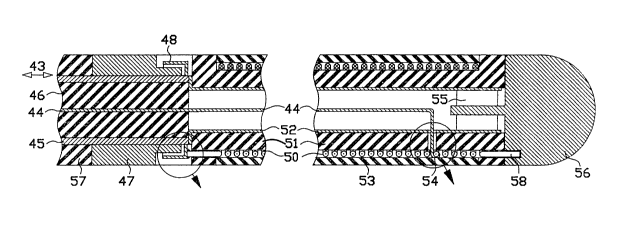
n antenna in an ablation catheter, with a wire wound helix, is shown in FIG. 3.
The antenna in FIG. 3 consists of a helix 50 with three terrninals: a proximal end
terminal 49 (FIG. 3A), a feed terminal 54 (FIG. 3B), and a distal end terminal
58 (FI~. 3). A heat-shrunk PTFE (also known under trademark TEFLON)
plastic sleeve 53 covers the helix 50.
In some applications, it may be desirable to distort the axisymetrical form
of the induced E-field. This can be accomplished by partially covering the
dielectric sleeve 53 with a metal screen (not shown). Currents induced in the
screen will modify the shape of a heating pattern and so serve as an aperture
antenna. An asymmetrical field pattern can also be accomplished by a loop
20 antenna, e.g., located in the r-z plane.
A transmission line which connects the distal end of ~he catheter to
external equipment has the form of a coaxial line 43 shown in FIG. 3. In a
preferred embodiment, coaxial line 43 includes a center conductor 44
(approximately 0.16 mm diameter), a dielectric 46 (approximately 1.35 mm
25 outside diameter), a metal braid shield 45 and an insulating sleeve 57
(approximately 1.8 mm outside diameter). A small diameter and flexible
construction make the coaxial line 43 suitable for biomedical catheter
applications.
~0~3Gf~9
A distal monitoring electrode 56 is connected to a distal end terminal 58
of helix 50 and to bypass capacitor 55. Bypass capacitor 55 is connected to shield
45 through metallized coating 52 inside of core 51. The function of the bypass
capacitor 55 is to ground RF power. Thus when the RF power is applied to the
5 helix 50, distal monitoring electrode 56 has little RF voltage thereby preventing
distal monitoring electrode 56 from acting as a heat applicator. Distal
monitoring electrode 56, in conjunction with a proximal monitoring electrode 47,picks up an endocardial potential. In this embodiment, the distance from a
beginning of proximal monitoring electrode 47 to an end of the distal electrode
l0 56 is approximately 20 mm.
When operated in a TE mode the number of turns of helix 50 is chosen so
that at an operating frequency of 915 MHz, the helix is somewhat short of being
at a quarter wavelength resonance. Helix 50 is wolmd on a dielectric core 51.
The proximal end terminal 49 of helix 50 is connected to a variable tuning
capacitor 48 (FIG. 3A). Variable tuning capacitor 48 is moved with respect to
proximal monitoring electrode 47 during manufacture for tuning to a resonance
at operating conditions. Tuning capacitor 48 is controlled by adjusting a space
40 between capacitor electrodes 47 and 48. At lower frequencies, the
capacitance of inter-electrode space is insufficient and the capacitor is
~o implemented by a discrete component.
RF power is coupled to a helical resonator by connecting the center
conductor 44 to helix 50 at the feed terminal 54 (see FIG. 3B). The position of
feed terminal 54 on the helix is selected for a good match between the
characteristic impedance of the coaxial line 43 and the impedance of the
25 resonator under typical operating conditions. Under some circumstances the
best match can be obtained when the feed terminal 54 and the distal end
terminal 58 coincide, and the helix 50 is fed at its distal end terminal. The
choice of an axial quarter wavelength resonator is by no means unique. One
could just as well select any multiplicity of quarter wavelengths, such as a half-
30 wavelength resonator.
~OO~ 9
13
When in operation in the TM mode, the frequency of operation can bemuch lower, e.g., 27 MHz. Helix 50 can then be viewed as a discrete inductance,
tuned into ser;es resonance by a discrete component capacitor 48. In the TM
mode, core 51 on which heiix 50 is wound, can be made from a ferrite. At
s 27 MHz, a ferrite core can significantly increase inductance of the helix and
decrease losses in the tuned circuit. In order to use the Ez electric field
component in the TM mode, sleeve 53 is removed to allow direct contact
between the winding of the helix and the surrounding tissue.
In cardiac ablation, it is essential to be able to rnonitor endocardial
o potential just before and after the application of heat. Before application ofheat, it is necessary to locate the arrhythmogenic tissue to be ablated.
Afterward, endocardial potential is used to assess effectiveness of destruction of
arrhythmia-causing myocardial tissue. FIG. 4 shows a block diagram of a system
which combines controlled heat delivery by a solenoidal antenna, with
monitoring of endocardial potential.
Distal monitoring electrode 56, in conjunction with the proximal
monitoring electrode 47, picks up a local endocardial potential and feeds this
signal through coaxial line 43 to capacitor 62. Capacitor 62 represents a short
circuit for the RF power and an open circuit for a much lower frequency band
20 (typically 0.1 Hz to lO0 Hz) associated with endocardial signals. An endocardial
signal travels unobstructed on lines 63 and 64 to an input to a low-pass filter 61.
Low-pass filter 61 has a high input impedance to the RF power and
therefore blocks the transmission of RF power to switch 60 while allowing
passage of the endocardial signal. Switch 60 is closed simultaneously with
2s application of RF power, thus providing additional protection for monitor 59.Intracardiac signal processing, display, and recording is accomplished by monitor
59 which displays the intracardiac electrogram. Existing equipmen~ is suitable
for application as monitor 59.
RF power is generated in an RF power source 41. The RF power is
30 controlled and monitored in controller 42 which couples the RF power to the
coaxial line 43 through capacitor 62, which for RF represents substantially a
short circuit.
~0~3~,~9
14
Fig. 5 shows an alternative implementation of a catheter using metal
plating on plastic, such as silver on PTFE. Such plating offers a number of
advantages over the design shown in FIG. 3. One advantage is a unitary design:
the plating process can in one step create coaxial shield 69, helix 71, and disk 82
serving as a capacitive coupling electrode. In microwave application, shield 69
may be used alone or in conjunction with a secondary shield made from a metal
braid (not shown).
Another advantage is that helix 71 made from a metal strip provides a
more effective use of the metal cross-section than the circular cross-section wire
such as used in the helix 50 in FIG. 3. For silver or copper, the RF current
penetrates only .01 mm at 27 MHz and 0.002 mm at microwave frequencies.
This so called "skin depth" is so small in good conductors that plating thickness
easily exceeds it. In round wires, the current flows only on the surface, yet the
wire adds two diameters to the diameter of the catheter, without any
contribution to conduction.
Fig. S shows a tri-axial design of the catheter. A coaxial RF transmission
line is formed between coaxial shield 69, plated on the outside of the plastic tube
72, and an inner conductor 73 plated on the outside of a smaller plastic tube 74.
A stranded small-gauge center wire 75, along the axis of plastic tube 74, is
shielded from the RF by plated inner conductor 73. Center wire 75 is used to
transmit endocardial signals from distal monitoring electrode 80. Optionally a
ferrite bead 83 acts as a RF choke to further decouple RF from distal monitoringelectrode 80. A proximal monitoring electrode 76, in the form of a ring, is
seated on and makes electrical contact with the shield 69.
A proximal end terminal 81 of the plated helix 71 seamlessly joins with
the shield 69. A distal end terminal 77 of the helix 71 seamlessly joins with
plated disk 82, plated on an end surface of plastic tube 72. Metal disk 79
connects along its inside diameter to inner conductor 73. Dielectric disk 78
separates the metal disk 79 from the plated disk 82. The three discs 82, 78 and
79 form a capacitor between inner conductor 73 and the helix 71. The role of
this capacitor is to tune the inductance of the helix 71 to resonance so that under
operating conditions, the transmission line sees a resistive load equal to a
characteristic impedance of the coaxial line.
~003~.~9
A capacitance between the turns of the helix 71 in the plated strip design
is much smaller than a comparably spaced circular cross-section wire. It is
therefore possible to make the gap 84 between turns significantly smaller in a
plated strip design. This narrow-gap geometry generates an intense electric field
s between turns, primarily z-axis oriented across the gap, with a rather steep
attenuation in the radial direction. As a result, most of the Ez field passes
through the dielectric cover tube 70 without penetrating into the outside tissue.
The dominant component of the electric field in the tissue is the azimuthal field
E~3 induced by current in helix 71. The advantages of the Ee field have been
o discussed earlier.
Yet another advantage of metal-on-plastic plating is that a variety of
antenna patterns can be readily and accurately implemented. For example, a
helical strip 85 in FIG. 6 has a variable width constant-gap winding. A helical
strip 86 in FIG. 7 has a constant width variable-gap winding. This type of helical
S strip (85 or 86) design allows control of the electric field distribution in the z-
direction.
An antenna in FIG. 8 consists of two interspaced helices 87 and 88,
wound in the same sense and defining a bifilar antenna geometry. The bifilar
helices have two proximal end terminals and two distal terminals. The proximal
20 end terminals can be connected to the transmission line and the distal end
terminals can be shorted or preloaded with an RF impedance to optimize the
power flow.
An antenna in FIG. 9 consists of a helix 89, plated on a plastic sleeve 90
(shown partially cut), and helix 91 plated on a plastic tube 92. The two helices25 89 and 91 are wound in an opposite sense and therefore cross over each other,defining a cross-wound antenna geometry. Like the bifilar antenna, a cross-
wound antenna has two proximal end terminals 93 and 94 and two distal end
terminals 95 and 96. The proximal end terminals can be connected to a
transmission line and the distal end terminal can be shorted or preloaded with
30 an RF impedance to optimize the power flow. It should be noted that in this
configuration, unlike the bifilar configuration of FIG. 8, current entering at
proximal end terminal 93 and flowing up through helix 89 circulates around the
axis in the same direction as the current flowing down through helix 91 and
~03
16
exiting at proximal terminal 94. An effect on induced azimuthal fields Ee is
therefore additive. The polarity of the Ez field caused by the up current in helix
89 and the down current in helix 91 is opposite, and thus tends to cancel each
other. The cross-wound antenna is therefore an efficient source of the azimuthals Ee field.
All of the antennas described thus far are of the solenoidal variety, i.e.,
include one or more helices. The antenna shown in ~;IG. 10 is different. FIG. 10shows a proxirnal ring electrode 25 and a distal tip electrode 26, mounted or
plated on a catheter tube 24 and shaped very similarly to the currently used
l0 pacing catheters. An electrical connection is maintained by a twisted pair
transmission line 27. Unlike currently used catheters where the electrodes are
made from plain metal, proximal ring electrode 25 and distal tip electrode 26
have their metallic surface coated with control coatings 28 and 29 respectively.Optionally, the gap between proximal ring electrode 25 and distal tip electrode
s 26 can be filled with gap coating 30. (Thickness of coatings is exaggerated in
FIG. 10 for the sake of clarity.)
The control coatings vary in thickness as a function of the axial distance
from the inter-electrode gap, being thickest along the edges of the inter-
electrQde gap and thinning away from the gap. Without the coating, the
20 strongest Ez field is adjacent to the inter-electrode gap. The coatings, by
changing the surface impedance, equalizes the external electric field and
improve radial penetration of the field.
The coatings 28, 29, and 30 can be made from a resistive material or from
a dielectric. A resistive coating, introduces the highest resistance close to the
25 inter-electrode gap. As a result, the external field adjacent to inter-electrode
gap is reduced, the external field intensity is equalized and the radial penetration
is improved. A capacitive coating, made from a dielectric, exhibits a smallest
capacitive impedance near the inter-electrode gap and accomplishes field
equalization similar to the resistive coating. There is, however, significantly
30 less heat dissipation in the capacitive coating than in the resistive coating.
While certain specific embodiments of improved electrical catheters and
systems have been disclosed in the foregoing description, it will be understood
that various modifications within the scope of the invention may occur to those
skilled in the art. Therefore it is intended that adaptations and modifications
35 should and are intended to be comprehended within the meaning and range of
equivalents of the disclosed embodiments.