Note: Descriptions are shown in the official language in which they were submitted.
vom- ~a
2t~~'~"~'~6
NIM-4 PATENT
PIiA.SE MODULATED SPECTROPIiOTOMETRY
BACKGROUND OF T~iE INVENTION
The application of the basic dual wavelength principle to
detect hemoglobin and myoglobin changes in tissue began with the
work of G.A. Millikan in his studies of the cat soleus muscle, and
~cm~r r~rb
NIM-4 PATENT
the work of Millikan and Pappenheimer who detected hemoglobin
deoxygenation in the human ear lobe. Multiwavelength instruments
have been developed; these instruments use either a
multiwavelength laser diode light source or a time shared filter
technique, in which high precision is sought through various
algorithms which deconvolute background signals, oxidized and
reduced cytochrome signals, and oxy- and deoxyhemoglobin signals.
Such instruments are oxy-complex and often have difficulty
obtaining light sources with wavelengths appropriate to the
algorithms that have been developed, or they have such low light
levels that photon counting is necessary. They are generally in
the price range of $80,000 and have produced much experimental
data in the literature on neonates and adults. The basic problem
of such methods is that the optical pathlength -is not known ab
initio but is calculated by reference to animal models where the
hemoglobin can be removed and cytochrome directly studied.
Transferability of such data from the animal model to the human is
one difficulty that had to be overcome prior to the invention of
time-resolved spectroscopy, where the pathlength is measured
directly.
Continuous wave spectroscopy (CWS) of tissue hemoglobin has
the demonstrated advantages of great simplicity and sensitivity,
as well as affording an "early warning" of tissue hypoxia. The
-2-
NIM-4 PATENT
application of picosecond-pulse time-resolved spectroscopy (TRS)
to tissue in order to determine optical pathlengths, quantify the
changes in hemoglobin concentration, and determine the actual
concentration values of hemoglobin and cytochrome has great
applicability to clinical studies of tissue hypoxia. Moreover,
time-resolved spectroscopy used in conjunction with continuous
light spectrophotometry offers a means of calibrating the optical
pathlength which photons travel as they migrate through tissue.
While trend indication can be of great value in many situations,
the capability to quantify hemoglobin concentration for both
continuous and pulsed light techniques greatly extends their
applicability to clinical studies. Ihese problems have
been discussed at length elsewhere.
SUMMARY OF THE INVENTION
It has now been found that the principles of dual wavelength
spectrophotometry may be applied to time-resolved
spectrophotometry choosing a carrier frequency at a value in which
the time characteristic is compatible with the time delay of
photon migration from input to output through a scattering medium.
-3-
~aA~7~6
't'lre pxe~prtl: i.ttvartl:j.ott yt:ov_lde~r nteLltocl~r and ~t~t~tat~~~tts
wltet:ehy
rt modal.7l:ed waivefot:m i.n t:nattemf.l:l:ecl l:o d !~c~el:t:pr:Log
tnACll.ttm attd
cle~ecl:ed aC~er ml.gr_al:i.ott I:Iterel:ltt-otaglt. '.t'Itp clc~l:ected
wctvefor.m 471.1.:1. '
Itave ~Jeetl d.l.het-pd artcl tn~ty t:lot~s bn c~oml.~~tt:ed t:o t-_Itp
~.nl.l;~.a:l.
w7ve1'or_m. r'or. nxrtm[tJ.e, a w7vr, rotvm Ln plts~trxa !~It I.J'l:ed l.~y
~Iw cla l.vy
3_rt m~.gra~J_vo ~Itlvttgit Llte ec~tL.l:et:l.ttg mAd I_um. '.t'Itua, ~.rt a
pr.efPr.t:ed
emhocl.l_mPn~s lrlte plttyse of I:lte wrlvefot:m :Le mvdttl.af:ed attd t:lie
plt7ee
eit 1. f fi iq det;ec~ecl . '.I'Itr, d l f l~pr. Attoe l.tt pltnee r~It 1. f
l: hel:weert l:wct
w7vel:ot:mg o1' ernl.l:~PCI pt_pet:t:omt,gttel:l.c~ t_ecll.el:i.ott lieu l.ttg
dl.ft:erettl:,
I;ttowt) w~tve:l.ertgl:lte rcttt l:ltr,tt lie prc.m~ppr~ed l:o dt~l:eom~.ne
rite
Co11Ce11l:Y.rllr:lotl yr t'ttt ilbt~orpl::l.vA rottel:.1_latwttt: surlt art
ltemogl.nlo.l~rt.
'I'ltus,a_t: J:r~ nit crh )ect, of ~:Ite L~r.erettl: .f.mveml.i.ort Lo
K,r.ov.l.dc
mel:ltvda arid ~t~par~tt:u~: for. el~uclyi.ttg pltol:.oit td.grril:l.ott
ttr~i.rtg a l.gttst 1.
moclu:lal:i.ort ~eCltn~.s~uc~s ~:uclt as t:f.mra, frPqttettc~y attd ptta~tn
modul.at:.loo. T.t~ 1_s 7 aypcl.f ~.c oltyc'L o1' rlte y.r.A~tettl:
:Lttvettt:l.ott l:n
pr:ov.l.de mel.ltvdt~ arid a~pat:al:ue wlteYehy pltace moclu 1_at:ed
epecl.r.o~ltorome~r_y ( >~'1~I5) tray Ire ul:l..l J_~erl l.tt
c:olt~uttel~.l.ort w 1.1:lt
cort~.i.ttuottr3 wave eperl:t:vmr~t:r: y ( C'I~IS ) ~o clel:erm.l.rte the Cr
i.l: i.r~a l_ v71 rte
oL art ahcrot:p~~.ve pigment: c~vclt es Itemog l.olo i.rt, et: trite pod.ttl:
I:ItA '
)?Cr/E'x xaE;~.v hpgl.ttq l:o clect:R7~P. Ll. Lct s~ttorltAr ob)aCt, of I:Ite
Lt t:eseoL j. overtW_ott Lo Ltt:ovi.de ar a spPCl.f l.v pmlrod.i.mertLs a
tltta.l ,
wave:t.ettg~lt pltaee mocltt l.al: S.ott by~tl:t~m witi.c:lt w 1.1..1_ a 1.
l.vw elm c l.Lo i.c~.v 1.
r,pp~.lcat::l.om of l:tie ctclveol~st~aeb of 1-.Lmr~ oeeol.vecl apecC~eor~ooyy
f.tt sttt
ecottotnl_r71_ attcl c~onwrer:c~L~.I. l y !'n.va 1. lal.e ~mlno~i S mnmt:.
_. ,t ...
In one embodiment, the invention provides a system for
examination of biological tissue of a subject, comprising:
an optical input port constructed to introduce photons of
electromagnetic radiation into the biological tissue; an '
optical detection port, spaced several centimeters apart
from the input port, constructed to acquire the photons that
have migrated over migration paths in the examined tissue
from the input port, the scattering and absorptive
properties of the examined tissue being determined by
photons migrating between the optical input port and the
optical detection port; a first oscillator connected to
waveform generating means for generating a first carrier
waveform at a first frequency on the order of 10g Hz, the
first frequency having a time characteristic compatible with
the time delay of photon migration from the input port to
the detection port; a light source, operatively connected to
the first oscillator including waveform generating means,
constructed to generate electromagnetic radiation of a
selected wavelength modulated by the first carrier waveform,
the wavelength being sensitive to an absorptive or
scattering constituent of the tissue; a detector constructed
to detect, at the detection port, radiation of the
wavelength that has migrated over migration paths in the
examined tissue from the input port; and a phase detector
constructed to compare the detected radiation with the
introduced radiation and determine therefrom the phase shift
of the detected radiation at the wavelength, the phase shift
corresponding to
an optical pathlength between the input and detection ports
and being indicative
-4a-
of the scattering and absorptive properties of the examined
tissue.
In a further embodiment, the invention provides a
spectroscopic method for examination of biological tissue
comprising the steps of: positioning an optical input port
at a selected location relative to the biological tissue;
positioning an optical detection port, spaced several
centimeters apart from the input port, at another location
to acquire the photons that have migrated over migration
paths in the examined tissue from aid input port, the
scattering and absorptive properties of the examined tissue
being determined by photons migrating between the input port
and the detection port; generating a first carrier waveform
at a f irs t f requency on the order of 10R Hz , the f i rs t
frequency having a time characteristic compatible with the
time delay of photon migration from the input port to the ,.
detection port; introducing at the input port
electromagnetic radiation of a selected wavelength, the
radiation having been modulated by the carrier waveform, the
wavelength being sensitive to an absorptive or scattering ,
constituent of the tissue; detecting radiation of the
wavelength that has migrated over migration paths in the
examined tissue from the input port to the detection port;
and comparing the detected radiation with the introduced
radiation and determining therefrom the phase shift of the
detected radiation at the wavelength, the phase shift
corresponding to an optical pathlength between the input and
detection ports and being indicative of the scattering and
absorptive properties of the examined tissue.
BRIEF DESCRIPTION OF THE DRAWINGS
-4b-
E
2t~~~7'~'~~
NIM-4 PATENT
Figure 1 illustrates a simplified single wavelength phase
modulated spectrophotometer made in accordance with the present
invention.
Figure 1A is a block diagram of an embodiment of a dual
wavelength phase modulated spectrophotometer made in accordance
with the present invention.
Figure 2 is a block diagram of another embodiment a phase
modulated spectrophotometer made in accordance with the present
invention.
Figure 3 is a block diagram of the preferred embodiment of
the spectrophotometer of the present invention.
Figure 4 is a block diagram of an alternate embodiment of the
spectrophotometer of the present invention.
DETAILED DESCRIPTION
The time, frequency, or phase of a signal may be modulated.
Phase modulation appears to be a convenient implementation of the
time-released spectroscopy (TRS) technique discussed above. In
Figure 1A, a single wavelength spectrophotometer using the
principle of phase modulation is shown. In this embodiment, a
frequency generator 17, operating at 200 MHz, excites a 4mW laser
diode 11, which emits light at a wavelength of 760 nm. The light
is conducted to the subject 20 via optic fiber 15. After the
light has migrated through the tissue, it is detected. Preferably
this detector is comprised of a photomultiplier tube and its
-5-
NIM-4 PATENT
associated voltage supply 16; one such device is the Hamamatsu'~
8928.
The frequency generator 17 also receives an input from a 50
kfiz oscillator 19, transmitting a 200.05 MHz reference waveform,
which is input into the detector 16. Accordingly, the output
waveform 22 from the detector 16 is at a carrier frequency equal
to the difference, i.e., 50 kHz. The waveform 22 from the
detector 16 and a reference waveform from the oscillator 19 are
fed into a phase and amplitude detector 24. In this embodiment,
the phase and amplitude detector 24 is a lock-in amplifier. The
output of the lock in amplifier are signals representative of the
phase shift and amplitude of the detected signal. These signals
are then processed and related to the relative concentration of an
absorbing constituent, such as hemoglobin.
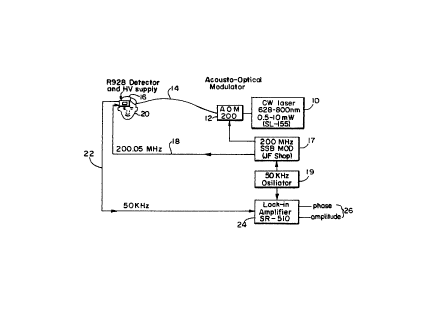
In the embodiment of Figure 1, a helium-neon laser light
source 10 is connected to a wide band acousto-optical modulator 12
operating at 200 MHz. The acousto-optical modulator 12 frequency
modulates the light emitted by the laser 10. The light is
conducted via a fiber optic light guide 14 to the forehead of the
subject 20 as shown, or other region to be studied. Signals about
3-6 cm from the location of the input waveform are received by a
detector 16, for example a Hamamatsu 8928. The dynodes are
modulated by a 220.050 MHz signal 18 so that a 50 Hz hetrodyne
signal 22 will be obtained and can be fed into a lock-in amplifier
24, such as a PAR SR510. As above the reference frequency for the
*Trade Mark -6-
NIM-4 PATENT
lock-in amplifier is obtained from the 50 Hz difference between
the two frequencies. The phase shift between the transmitted and
detected waveforms is measured with high precision and the output
waveforms, shown at 26, are plotted as an analog signal on a strip
chart recorder tv allow the user to follow the variations in the
propagation of light through the brain or other tissue. A
logarithmic conversion of the signal is then obtained. The result
is linearly related to the change in concentration of an
absorptive pigment, such as hemoglobin.
Referring to Figure 2, there is shown a block diagram of a
simplified embodiment of a dual wavelength phase modulation
spectrophotometer made in accordance with the present invention.
Unlike the single wavelength system of Figure 1A, this embodiment
allows the determination of the.cvncentration of an absorptive
constituent on an absolute basis. The embodiment of Figure 2 is
similar to that depicted in Figure lA, except that light is
transmitted to the subject at two discrete wavelengths.
Figure 2 illustrates a second embodiment of the apparatus of
the present invention. In this embodiment, the laser diode light
is amplitude modulated and the phase shift caused by photon
migration is measured by an optical detector, a mixer, and a phase
detector. The dual frequency time sharing system is comprised of
stable oscillators 30,32, such as Kenwood Model #321 for 220 Hz;
the oscillator system preferably used can generate waveforms from
144 to 440 MHz (Kenwood''TM721A). Continuous variation of the
'Trade M,ark. -7-
2~~~~~6
NIM-4 PATENT
frequency is possible, although, as will be understood by one of
ordinary skill, the three frequencies mentioned, 144, 220 and 440
MHz, are adequate for the purposes of initial studies and other
applications. The oscillators 30,32 are set 50 Hz apart and the
difference frequency is detected by a mixer 34 to obtain a
reference phase signal 36, as shown. A 200 Hz electronic switch
38 alternately excites laser diodes 40,42, nominally operating at
between about 750-60 nm and 800-10 nm, to emit 220 MHz modulated
light which is conducted by fiber optic guides 44,46, preferably
about 3 mm in diameter, to the surface of the head of the subject
20, or other region to be examined.
In order to achieve satisfactory operation at 220 MHz, it has
been found that the most cost effective detector 48 for this
purpose is Hamamatsu 8928. A more advantageous device, however,
is the Hamamatsu R1645u, which is a microchannel plate tube having
120 picosecond transit time spread, and a high gain; that is, a
two-stage microchannel plate photomultiplier 48. This tube, which
is capable of current amplification of 5 X 10⁵ (57 dB) is similar
to those used for pulsed time measurements in time resolved
spectroscopy (TRS) studies, and is considered to be ideal for
these purposes. The photomultiplier
_g_
~~l ~~~~~
NIM-4 PATENT
tube 48 is connected to a high voltage supply 50 which has an
output of about 3400 volts, in order to ensure high gain. The
photomultiplier tube 48 can be connected to the brain or other
tissue area by the fiber optic guides 44,46 or may be directly
connected and placed in a housing isolated from ground potential,
as illustrated.
As above, the detector 48 is attached to the subject 20 and
is connected to a mixer 52, which down converts the 220 MHz output
of the detector 48 to a 50 kHz signal by mixing with a 220.050 MHz
signal from the oscillator 32. A lock-in amplifier 54 determines
the phase of the exiting waveform. The lock-in amplifier 54 also
obtains the logarithm of the signal. This signal is then fed to a
second phase detector/lock-in amplifier 56 which determines the
difference between the signals at each of the two wavelengths,
this signal 58 is directly proportional to the concentration of an
absorptive pigment, such as hemoglobin. This embodiment may be
used on neonate, as well as adult brains.
A preferred embodiment of a time-shared, dual wavelength
laser diode phase modulation spectrophotometer is illustrated in
Figure 3. In this embodiment, a pair of laser diodes 100,102 are
excited in parallel by a stable frequency generator 104 (Kenwood
321) at 220 MHz. Each of the diodes 102,104 generates
electromagnetic radiation of a different wavelength, preferably
760 nm and 800 nm. The electromagnetic radiation is time shared
by a vibrating mirror 105, which illuminates a single fiber optics
_g_
2~~~~~6
NIM-4 PATENT
probe at a modulating frequency, preferably about 60 Hz. The
synchronization of the motion of the mirror 105 and the 60 Hz
phase detector 120 (explained below) is accomplished using an
electrical coupling of the reference voltage in the 60 Hz lock-in
amplifier 120. Thus, electromagnetic radiation at each wavelength
is synchronized between emission and detection.
One of ordinary skill will note that the spectrophotometer of
Figure 3 differs from the embodiment depicted in Figure 2 in that
the latter embodiment uses a carrier modulation system to code the
excitation power of one laser from another, while the embodiment
of Figure 3 continuously switches between the output light from
two lasers excited at the same frequency.
The time shared 760/800 nm light is applied to the subject 20
via an optic fiber 106. Several centimeters away, an output probe
108, preferably comprising a second fiber of relatively large area
will pick up the light which has migrated through the subject and
illuminates a photo detector 110, which is a suitable
photomultiplier tube (Hamamatsu 928) or a microchannel plate
detector (Hamamatsu R1645u). The light collected is phase shifted
from input oscillations by the time delay in photon migration
between input and output.
A second oscillator 114 which generates a 220.030 MHz
waveform is connected to a mixer 112. The 220.000 MHz output of
the detector 110 is also connected to the mixer. As a result, the
phase modulation frequency is downshifted to 30 kHz, which is a
-10-
NIM-4 PATENT
convenient frequency for lock-in detection. This signal is input
to a phase detector 116, which is preferably a lock-in amplifier.
A second input to the phase detector 116 is obtained by connecting
an input from the 220.000 MHz oscillator 104 and the 220.030 MHz
oscillator 114 to a mixer 118 to obtain an unshifted 30 kHz signal
which is used as a phase reference. Thus, the lock-in amplifier
116 operates with a reference phase obtained directly from the
frequency generators 104,114 and a phase modulated input obtained
by photon migration through the subject 20.
The phase of the signal output will vary between the phase
due to light propagation at 800 nm and the phase due to light
propagation at 760 nm. The output of the lock-in amplifier 116 is
thus a 60 Hz waveform, the amplitude of which bears the phase
information at the two wavelengths. The output of the phase
difference detector 116 then is connected to the same waveform as
that which drives the 60 Hz vibrating mirror 105. The output of
the phase detector may be obtained by using switch contacts on the
vibrating reed modulation which alternatively connects opposite
phases of the 60 Hz waveform to the integrating network, each one
at the peak of the waveform of the output phase detector. The
output is put into a differential amplifier to record the
difference of the amplitude of the two parts of the 60 Hz
waveform, corresponding to the 760 nm and 800 nm phase shift.
This phase difference output is suitably filtered from 0.05-1 Hz
-il-
~~~~~~6
NIM-4 PATENT
and provides a running time record of the changes in hemoglobin
concentration by dual wavelength time-resolved spectroscopy.
The advantage of the system illustrated by Figure 3 is that
it affords a single light guide input to the subject operated from
two laser diodes which are continuously operated at the same
oscillator frequency. Thus, spurious phase differences in
frequencies associated with excitation are minimized. That is, no
differential phase shift is expected between the 760 nm and 800 nm
signals. Thus, the 30 kHz difference signal would represent the
true phase delay between these two wavelengths. Moreover, phase
noise in this region would be minimized by the differential
detector 116. The photomultiplier tube detector 110 can be of any
adequately fast type, since the mixing function is separated from
the detector. The lock-in amplifier technique obtained to derive
the difference of the phase and amplitude of the two signals has
the highest signal to noise ratio possible for this type of
equipment.
The principles of time-shared dual wavelength
spectrophotometry, together with lock-in technology, follows the
principles employed in dual wavelength spectrophotometry
generally. However, the present invention provides a vastly
improved device, since the carrier frequency of 220.000 MHz is
sufficiently fast to measure photon migration times between input
and output with a characteristic time of about 5 nanoseconds to be
observed. Therefore, the sensitivity of the system disclosed is
-12-
~s~~~~s
NIM-4 PATENT
high, approximately 70° per nanosecond or 3° per centimeter
change
of pathlength, as observed in experimental models.
The application of the principles of dual wavelength
spectrophotometry to time-resolved spectrophotometry involves the
choice of a carrier frequency at a value in which the time
characteristic is compatible with the time delay of photon
migration from input to output. The device disclosed achieves the
result of precisely measuring the absorbance changes in photon
migration, over a specified distance, e.g., over approximately one
meter, as contrasted, to the continuous light method in which
photon migration is measured over all possible path lengths. A
path length of approximately one meter is preferably selected in
order to ensure exploration of all parts of the brain for brain
bleeding studies. Obviously, higher frequencies would select
smaller portions of the brain which are more localized to the
input-output configuration.
For a multiple-scattering medium such as human tissue, the
only known method for determining the path length of transmitted
photons is the measurement of the time of flight and of the
refractive index, from which the distance travelled may be
calculated. Since this path length in the brain is on the order
of centimeters, the transit time is on the order of nanoseconds or
less. A direct measurement of such periods in this time domain
has several fundamental drawbacks. As the required time
resolution becomes finer, the detection bandwidth must increase;
-13-
~~~~~~6
NIM-4 PATENT
signal power at best remains constant, while noise power increases
proportionally with the increasing bandwidth. For sources such as
laser diodes, where average output power for both pulsed and
continuous operation are nearly the same, signal power typically
declines when the pulse width is reduced. Since the time between
probe pulses must be long enough for the returning light to decay
to approximately zero, the duty cycle of the pulse train is
typically low; this implies low average signal power or the use of
high peak power, which may endanger the skin covering the tissue
being studied. Finally, both the expense and difficulty of
constructing suitable electronic circuits is considerably greater
for pulsed than for continuous-wave systems. As an alternative to
time-domain measurement, a CW system may be employed with phase
measurement taking the place of time intensity, a simple
calculation based on measurement of the phase shift between probe
and return light at a single frequency yields the characteristic
decay time. Such a system has the advantages of narrowband
modulation and detection and high average power in the probe
signal, yielding a considerable advantage in signal-to-noise ratio
and therefore in data acquisition time. There is a considerable
body of literature on this technique of time measurement,
particularly as applied to radar, time standards, and
spectroscopy. Perhaps the most relevant to this application is
the literature on the phase-resolved measurement of fluorescent
decay kinetics.
-14-
~~~~~~6
NIM-4 PATENT
Another alternate embodiment of the apparatus of the present
invention is depicted in Figure 4. This system relies more upon
communications technology rather than NMR technology, and is
essentially a single sideband system where the sidebands are
displaced in proportion to the modulation frequency shift
required. This design places more reliance upon the existing
radio frequency transmitter/receivers which, at prices of about
$300 per frequency for transmit/receive, is a significant
advantage.
A block diagram of a system as described directly above is
shown in Figure 4. In this embodiment, a first standard
communications transmitter-receiver (transceiver) 200, operating
at 220 MHz, is used in the transmit mode to generate a waveform
which excites a laser diode 202. The transceiver 200 is used in
the single side band (SSB) mode to provide SSB modulation at 3
kHz. This carrier signal is fed back to the transceiver 200 and
into a phase detector/filter 208, which also receives an input
from a second transceiver 204. As in the previous embodiments,
the laser diode 202 emits light which is conducted to the subject
20 via optic fibers 216.
The SSB modulated signal is phase shifted by the delay in
migration through the brain. The light is scattered and absorbed
as it migrates through the subject 20 and is received by an
optical coupler/fiber assembly 218. The received light is then
transmitted to a detector 220, either of the photomultiplier tube
-15-
2~~~~~6
NIM-4 PATENT
or the microchannel plate type, both of which are discussed above
in reference to other embodiments.
The output of the detector 220 is coupled to the RF input to
the second transceiver 204, i.e., the transceiver is used in the
receive SSB mode and a phase shifted 3 kHz tone is obtained and
connected to the phase detector filter 208. The output is a 3 kHz
phase shifted signal which is input to the second SSB transceiver
204. In order to ensure phase coherence, the first transceiver
200 and the second transceiver 204 form a phase locked loop. The
3 kHz carrier waveform is also locked to 220 MHz by frequency
dividers 206, thereby locking the 220 MHz and the 3 kHz phases and
allowing the phase shift to be determined with high precision. As
seen in Figure 4, an output of the transmitter oscillator 200 is
frequency divided by about 7X10⁵, to yield a 3 kHz signal. The
output of the phase detector/filter 208 is thus related to the
phase shift and, accordingly, is representative of the absorption
within the subject.
The carrier frequency is initially chosen to be 220 MHz; this
is sufficiently high to give a detectable phase shift for decay
times of a few nanoseconds, but low enough to be within the
bandwidth of a number of commercially-available active mixers.
Although diode-ring mixers are readily available up to 36 GHz,
they have significantly less dynamic range than active (transistor
bridge or linear multiplier) designs; a large dynamic range is
crucial for this type of spectrophotometer system. A heterodyne
-16-
~~0'~'"'l~: 6
NIM-4 PATENT
system is chosen to allow multiple optical wavelengths to be
transmitted and detected in parallel on individual subcarrier
frequencies, and to allow phase detection to be carried out within
the frequency range of commercial phase sensitive detectors, i.e.,
"lock-in amplifiers." These devices have a superb noise figure,
linearity, dynamic range, and phase and amplitude accuracy: their
performance is very much superior to any phase detector operating
directly at the RF carrier frequency. Generation of the reference
signals for the lock-in amplifiers by frequency division from the
master RF oscillator provides adequate phase coherence of all
subcarriers and all demodulated signals with respect to the
carrier; no phase calibration between wavelengths is required.
Frequency generation by division also provides minimum possible
phase noise for a given master oscillator. Should additional
carrier frequencies be required, such as for the measurement of
multi-exponential decays, the only changes required in this design
would be the addition of a one-by-N RF switch and additional RF
oscillators.
Laser diodes are chosen over thermal sources, for their much
higher radiance, ease of coupling to optical fiber, narrow output
spectrum and wavelength stability, long life, and ease of
modulation at RF frequencies. In order to maximize the signal-to-
noise ratio of the system, and to avoid problems of
intermodulation distortion due to laser nonlinearities, single-
sideband suppressed-carrier modulation is used. The intermediate
-17-
NIM-4 PATENT
frequencies are chosen within the range of 10 to 100 KHz; they
must be high enough to allow a realizable Q of the single side
band filters, but low enough to be within the range of low-cost
commercial lock-in amplifiers.
The heat sinks of the lasers are preferably temperature-
controlled using Peltier coolers and feedback control.
Temperature control is necessary in order to stabilize the
wavelength of the lasers, and to allow sufficient tuning of the
output wavelength to cover the tolerance (approx. ~ 10 nm) of
commercial diodes. However, it should be noted that post-
demodulation detection of phase shift substantially eliminates
this consideration, since neither constant wavelength or amplitude
are required.
The optical system consists of one optical isolator per
laser, lens assemblies for coupling the laser light into the
optical fibers, a fiber bundles) for transmitting the light to
and from the subject, a fiber-subject coupler on the distal end of
the bundle(s), and a light detector assembly.
The isolators are necessary in order to prevent optical
feedback into the laser cavity due to reflections from the optics
or subject: such feedback, even at levels as low as -60 dB, are
well known to cause both amplitude and phase noise in laser
sources.
The fibers chosen for fabrication of the bundles must have
sufficiently small dispersion that the phase uncertainty
-18-
~s~~~~s
NIM-4 PATENT
introduced at the nominal 100 MHz modulation frequency is much
smaller than the phase shifts of interest. At the same time, the
maximum possible core diameter and numerical aperture are desired.
This simplifies and makes more robust the laser-fiber coupling,
and greatly increases the return light signal collected from the
subject, which is approximately a Lambertian radiator.
For this reason, single-mode fibers are ruled out, despite their
extraordinary bandwidth-length products. For multimode fiber,
only modal dispersion is significant for the sources, lengths, and
bandwidths considered here; we therefore disregard waveguide and
material dispersion. Considering first step-index fiber, simple
ray optics will show that it is numerical aperture, and not core
size, that determines modal dispersion. For an allowable time
uncertainty of 100 picoseconds, and a'total step-index fiber
length of two meters, a numerical aperture of approximately 0.17
or less would be required: all commercial step-index multimode
fibers have a much larger numerical aperture. For an ideal
graded-index multimode fiber, however, modal dispersion for ,
meridional rays is zero, from Fermat's principle, and actual
bandwidth-length products in commercial graded-index fibers exceed
100 MHz-km at the wavelength of interest for this study. We
therefore choose a graded-index fiber with 100 micron core size,
0.3 numerical aperture, and 100 MHz-km bandwidth-length product.
-19-
~(e~'7'~'~6
NIM-4 PATENT
This fiber is also cheap enough ($0.50/meter) to be considered for
fabricating bundles.
The detection optics consist of a fiber bundle of cross-
sectional area matched to the active area of the detector, an
optical bandpass or comb filter to pass only the laser wavelengths
and prevent detector saturation by room light, and the detector
itself. Initially, a photomultiplier tube with a GaAs(Cs)
photocathode will be used this detector has a gain advantage of
about thirty compared to a PMT with silicon photocathode, and
about 300 compared to a silicon avalanche photodiode, not
including the much smaller active area of the avalanche
photodiode. However, the photomultiplier tube also has a marginal
bandwidth for this application, and extraction of the signal from
the middle of the dynode chain may be required, thus reducing the
gain. Should the signal-to-noise ratio prove sufficient, the
system may be easily modified later by substitution of an
avalanche photodiode for the PMT, reducing the cost and increasing
bandwidth, reliability and ruggedness. The avalanche photodiode
detectors are also to be used for imaging experiments due to their
smaller size and lower cost. Conversely, if greater detection
bandwidth and high gain are needed,simultaneously, a microchannel
plate photomultiplier may be used; the disadvantages here are the
much greater cost and lower photocathode sensitivity,
approximately a factor of 30 compared to GaAs(Cs) available.
-20-
~~~'~'~'~6
NIM-4 PATENT
After a variable gain stage, the signals out of the detector
are heterodyned back to the IF frequencies and fed into commercial.
two-phase lock-in amplifiers; one lock-in per optical wavelength
(If frequency) is used. While this increases the cost, it reduces
data acquisition time by a factor of about 1.41 (SQRT 2)
(comparing two amplifiers to one), and avoids undesirable
assumptions about the relative absorption kinetics at each
wavelength.
The entire system may be controlled by an IBM-'compatible
portable computer, with IEEE 488, analog-digital,.digital-analog,
and bidirectional digital interfaces. All of the latter can be
provided by two low-cost plug-in cards for the IBM. The use of.
the computer control has a number of significant advantages, among
them the possibility of automatic operation for testing and
phantom experiments, simplified reconfiguration as the system is
refined, speed, accuracy, and ease of post-processing of data, in
particular for statistical analysis. The choice of a portable
machine greatly simplifies clinical trials of the present
invention.
The technology of multiwavelength phase modulation each
encoded by its own subcarrier can readily be carried out as
illustrated in the preferred embodiment of Figure 3. The output
of such a system would then replace the continuous wave technology
of existing systems, and at the same time, take advantage of the
algorithms for decoding various states of hemoglobin and even
<Trade Mark -21-
~~~~s
NIM-4 PATENT
cytochrome. The great advantage is that the optical pathlength is
known and not assumed. Thus, phase modulation is a convenient
implementation of the TRS technique, since it can be built to
emphasize delay times on the order of 5 nanoseconds where the
decay is exponential and long path migrations are involved.
-22-