Note: Descriptions are shown in the official language in which they were submitted.
2~7895
GC268a
JANTHINOMYCIN
Cultivation of a strain of the microorganism
Janthinobacterium lividum which has been deposited
in the American Type Culture Collection as A.T.C.C.
No. 53,857 yields a novel class of anitbiotics
hereinafter referred to by the trivial chemical
names "janthinomycin A, B, and C." Janthinomycin
A, B, and C have activity against Gram-positive and
Gram-negative bacteria and anaerobic bacteria.
Janthinomycin has been analyzed and found to have
the following general chemical structure:
I.
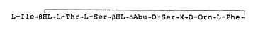
L-Ile~BHL-L-Thr-r.-Ser-~HL;~Abu-D-Ser-X-D-Orn-L-Phe
wherein ~-Abu is dehydro-a-aminobutyric
acid;
~HL i~ D-erythro-~-Hydroxyleucine;
and further wherein X is ~-Hydroxy-
tryptophan for janthinomycin A;
X is ~-ketotryptophan for janthinomycin B;
X is dehydrotryptophan for janthinomycin C.
7~395
GC268a
--2--
In solution, the B-keto-tryptophan residue
of janthinomycin B exists as a mixture of geometric
isomers, that is as the E and Z enols of B-keto-
tryptophan, hereinafter janthinomycin Bl and B2.
The two isomers are separable chromatographically,
but 510wly interconvert to give a pH and solvent
dependent equilibrium mixture. The two isomers are
referred to in the text as janthinomycin ~1 and B2.
FIGURE 1 shows the ultraviolet spectrum or
janthinomycin A in water and in O.OlM HCl and O.OlM
NaOH.
FIGURE 2 shows the infrared spectrum of
janthinomycin A in potassium bromide.
F'GURE 3 shows the 67.5 MHz carbon NMR
spectrum of janthinomycin A in deuterated
acetonitrile-deuterated water (1:4).
FIGURE 4 shows the 400 MHz proton NMR
spectrum of janthinomycin A in deuterated
acetonitrile-deuterated water (g:l).
FIGURE 5 shows the ultraviolet spectrum of
janthinomycin B in water.
FIGURE 6 shows the infrared spectrum of
janthinomycin B in potassium bromide.
FIGURE 7 shows the 67.5 MHz carbon NMR
spectrum of janthinomycin 3 (31 92 about 3:1) in
deuterated acetonitrile-deuterated water (1:4).
FIGURE 8 shows the 400 MHz proton NMR
spectrum of janthinomycin B (B2:BI about 4:1) in
deuterated acetonitrile-deuterated water (1:1, pH
7.1 with Na2DPO4~.
21~ 395
GC268a
--3--
FI~URE 9 shows the 400 MHz proton NMR
spectrum of janthinomycin B ~82:BI about 1:4) in
deuterated acetonitrile-deuterated water (4:1).
FIGURE 10 shows the ultraviolet spectrum of
janthinomycin C in water.
FIGURE 11 shows the infrared spectrum of
janthinomycin C in potassium bromide.
FIGURE 12 shows the 67.5 M~z carbon NMR
spectrum of janthinomycin C in deuterated
acetonitrile-deuterated water (4:1).
FIGURE 13 shows the 400 MHz proton NMR
spectrum of janthinomycln C in deuterated
acetonitrile-deuterated water (4:1).
The Microorganism
The microorganism used for the production of
janthinomycins A, B and C is a strain of
Janthinobacterium lividum isolated from stagnant
water collected in Tyler State Park, Newtown, PA.
A subculture of the organism can be obtained from
the American Type Culture Collection, ~ockville,
MD. Its accession number in this repository is
A.T.C.C. No. 53,857.
In addition to the specific microorganism
described and characterized herein, it should be
understood that mutants of the microorganism
produced through the use of chemical or physical
mutagens can also be cultivated to produce the
product.
~ 7~95
GC268a
--4--
The culture of Janthinobacterium lividum can
be isolated from the stagnant water sample by
preparing a suitable dilution in a medium
consisting of the following:
Gram
NaCl 8.5
KH2 PO4 . 3
Na2HPO4 0 . 6
Gelatin 0.1
and distilled water to 1 liter.
0.1 ml of this material was plated onto agar
plates containing:
Measure
Peptone 1 g
K2HPO4 0.2 g
Glucose 1 g
1% Crystal Violet0.1 ml
Soil extract 1 liter
Agar 15 g
The pH is adjusted to 6.8 and the mixture
autoclaved at 121C for 15 minutes. Ten ml of a 1
(W/V) cycloheximide solution is then added to a
liter of medium. The soil extract is prepared as
ollows: 1000 ml soil ls boiled in 2 liters of
water for 1 hour. The solids are filtered out
through cheesecloth and the solution then
centrifuged for 20 minutes at 28,000 rpm. The
supernatant i8 then iltered through Whatman paper
and autoclaved for 30 minutes at 121C.
The organism is a motile Gram negative
bacterium that is rod-shaped with sub-polar to
lateral flagella. Colonies on nutrient agar are
2Q~7B95
GC268a
-5~
gelatinous and dark purplish-black in color. The
gelatinous material is extracellular
polysaccharide; the pigment produced is violacein.
Glucose is utilized oxidatively. Acid is produced
from trehalose but not from 1-arabinose or
d-xylose. The organism is negative for arginine
dihydrolase, production of HCN and esculin
hydrolysis. The bacterium is identified as an
aberrant strain of Janthinobac~erium l ividum.
The Antibiotic JanthinomYcin
The antibiotic janthinomycin can be produced
by cultivating Jan~hinobacterium lividum, A.T.C.C.
No. 53,857 at, or near, 25C under submerged
lS aerobic conditions in an aqueous nutrient medium
containing assimilable carbohydrate and nitrogen
sources. The fermentation is carried out until
substantial activity is imparted to the medium,
usually about 24 to 28 hours.
After fermentation solid ammonium sulfate is
added (25% wt/v) to the whole broth, ths broth is
centrifuged and the resulting pellet is extracted
with methanol. The methanol-pellet mixture is
centrifuged and the resulting methanol extract is
made approximately 10% aqueous by the addition of
water, and then extracted with carbon
tetrachloride. The layers are separated and the
methanol extract delivered for isolation.
The methanol extract is added to MCI gel
CHP20P (CXP20P) in water and the mixture is stirred
for one hour. The charged resin is collected by
vacuum filtration and washed with methanol, water,
2(~7895
GC268a
-6-
and acetonitrile. The char~ed resin is then pack~d
in a column and the antibiotics are eluted with
acetonitrile-water-formic acid. Further
purification is achieved by chromatography on
Sephadex LH-20 in acetonitrile-water. Partial
resolution of the three antibiotics, janthinomycins
A, B, and C, is effected by chromatography on
CH~20P eluting with a gradient of acetonitrile-
water-formic acid. Final separation and
purification of janthinomycins B and C is achieved
by chxomaography on CHP20P eluting with an
acetonitrile-a~ueous ammonium dihydrogen phosphate
buffer gradient, followed by desalting on CHP20P,
eluting with acetonitrile-water-formic acid, to
give the pure antibiotics as off white powders.
The mount of janthinomycin C in the fermentations
is variable, and its presence may be an artifact of
the isolation conditions (janthinomycin A is
converted to janthinomycin C under acidic
conditions). When janthinomycin C is present, it
co-elutes with janthinomycin B in each of the
initial chromatographies.
The ultraviolet spectrum of janthinomycin A
is given in FIGURE 1 and shows: Amax (El% 1 cm)
337(~), 287(30), 276(36), 204 nm (270). The
infrared spectrum of janthinomycin A in potassium
bromide is shown in FIGURE 2. The following peaks
are evident: 3342, 2962, 1743, 1659, 1602, 1525,
1383, and 1126 cm 1. The FAB mass spectrum of
janthinomycin A in dithiothreitol-dithioerythritol-
dimethylsulfoxide-glycerol with added sodium iodide
2~7~395
GC268a
--7--
shows the following ions: (M+Na)+ 1215, (M+H)+
1193 (weak), (M+H-H2O)+ 1175, (M+I) 1319. Without
added sodium iodide, only the (M+H-H2O)+ 1175,
and (M-H-H20) ions are seen. The 67.5 MHz ~3C NMR
spectrum of janthinomycin A in deuterated
acetonitrile-deuterated water (1:4) is shown in
FIGURE 3. The 400 MHz lH NMR spectrum of
janthinomycin A in deuterated acetonitrile-
deuterated water (4:1) is shown in FIGURE 4. Thin
layer chromatography of janthinomycin A on Merck
silica gel-60 using chloroform-methanol-70S aqueous
ethanol, 7:3:5, gives an ~f value of 0.38. High
performance liquid chromatography of janthinomycin
A on a Hamilton PRP-l column (150 x 4.1 mm),
eluting with 3uffer A at 1 ml/min., and monitoring
the absorbance at 220 nm, gives a retention time of
3.90 min. Bu~fer A is CH3CN-H2O (33:67), 1~ in
N~4H2PO4 adjusted to pH 3.6 with 85% H3PO4.
The ultraviolet spectrum of janthinomycin B
is given in FIGURE S and shows: ~max (El% 1 cm)
312(82), 260(sh), 242(120), 204 nm (350). The
infrared spectrum of janthinomycin B in potassium
bromide is shown in FIGURE 6. The following peaks
are evident: 3322, 3066, 2967, 1742, 1655, 1522,
1384, 1116 cm 1 The FAB mass spectrum of
janthinomycin B in dithiothreitol-dithioerythritol-
dimethylsulfoxide-glycerol with added sodium iodide
shows the following ions: (M+Na)+ 1213, (M+H)+
1191, (M+I)- 1317. Without added sodium iodide,
0 only the (M+H)+ 1191 and (M-H) 1189 ions are seen.
The high resolution FAB mass spectrum shows an
(M+H)+ of 1191.6142 consistent with the molecular
formula C5~H83NI20l6 (1191.6050). The 67.5 MHz 13C
2(~7~9S
GC268a
--8--
NMR spectrum of janthinomycin B in deuterated
acetonitrile-deuterated water (1:4) is shown in
FIGURE 7. The ratio of janthinomycin Bl:B2 in this
sample is approximately 3:1. The 400 MHz lH NMR
S spectrum of janthinomycin 8 (Bz:Bl approximately
4:1) in deuterated acetonitrile-deuterated water
(1:1, pH 7.1 with Na2DPO4) is shown in FIGURE 8.
The 400 MHz IH NMR spectrum of janthinomycin B
(32:Bl approximately 1:4) in deuterated acetonitrile-
deuterated water (4:1) is shown in FIGURE 9. Thin
layer chromatography of janthinomycin B on Merck
silica gel-60 using chloroform-methanol-70~ aqueous
ethanol, 7:3:5, gives an Rf value of 0.35 for
janthinomycin 82 and 0.39 for janthinomycin ~1.
High performance liquid chromatography of
janthinomycin B on a Hamilton PRP-l column (15 x
4.1 mm), eluting with 8uffer A at 1 ml/min, and
monitoring the absorbance at 220 nm, gives a
retention time of 1.74 min. for Bl and 2.76 min.
for B2.
The ul~raviolet spectrum of janthinomycin C
i5 given in ~IGURE 10 and shows: ,~max (El% 1 cm)
339(100), 220(250), 195 nm (400). The infrared
spectrum of janthinomycin C in potassium bromide is
shown in FIGURE 11. The following peaks are
evident: 3342, 3066, 2968, 1744, 1654, 1602, 1526,
1384, cm 1, The FAB mass spectrum janthinomycin C
in dithiothreitol-dithioerythritol-dimethylsulfoxide-
glycerol shows the following ions: (M+H)+ 1175,
(M-H)- 1173 and the high resolution EAB mass
spectrum shows an (M+H)+ Of 1175.6205 consisten~
2~ 5
GC268a
_g_
with the molecular formula Cs,H83NI2Ols
(1171.6101). The 67.5 MHz 13C NMR spectrum of
janthinomycin C in deuterated
acetonitrile-deuterated water (4:1) is shown in
FIGURE 12. The 400 MHz IH NMR spectrum of
janthinomycin C in deuterated
acetonitrile-deuterated water (4:1) is shown in
FIGURE 13. Thin layer chromatography of
janthinomycin C on Merck silica gel-60 using
chloroform-methanol-70~ aqueous ethanol, 7:3:5,
gives an Rf value of 0.37. (Janthinomycin C is not
resolved from Bl when both are present.) High
performance liquid chromatography of janthinomycin
C on a Hamilton PRP-l column (150 x 4.1 mm), eluting
with ~uffer ~ at 1 ml/min and monitoring the absorbance
at 220 nm, gives a retention time of 2.06 min.
Compounds of Formula I, and pharmaceutically
acceptable salts thereof, can be used as agents to
combat bacterial infections (particularly
Gram-positive infections) in mammalian species,
such as domesticated animals (e.g., dogs, cats,
cows, horses and the like) and humans. They can be
administered u~ing modes of administration which
have been used in the past to deliver penicillins
and cephalosporins to the site of the infection.
Such methods of administration include intravenous,
intramuscular and as a suppository. The dosage of
the antibiotic of formula I used will, of course,
vary with the particular antibiotic, the size of
the host and the severity of the infection. For a
2Q~37~95
GC262a
--10--
human adult, daily doses of about 250 milligrams to
about 2 grams are exemplary. Further information
about the potency of the compounds of this
invention is set forth below under the heading
"~iological Activity".
The following examples further illustrate
the preparation and utility o~ janthinomycin.
2(~7~3g5
GC268a
ExamDle 1
Janthinobacterium lividum was maintained on
the following sterilized medium (A):
Grams
Yeast extract 5.0
Glucose 5.0
MgSO4-7H2O 0.1
FeSO4 7H2 O . 1
Soil extract filtrate* 200 ml
Agar 17.5
Tap H2O 300 ml
Media was sterilized at 121C for 15
minutes.
*Soil extract filtrate-l vol soil + 2 vols. H2O
extracted at 100C for 1 hour and filtered.
A loopful of surface growth from an agar
slant (medium A) of JantAinob~cterlum lividum was
used to inoculate each of three 500 ml Erlenmeyer
flasks each containing 100 ml of the following
sterilized medium (B):
Grams
Yeast extract 5.0
Glucose 5.0
MgS04 7H2 O . 1
FeSO4-7H20 0.1
Tap H2O to 1 liter
Media was sterilized at 121C for 15
minutes.
After inoculation, the flasks were then
incubated at 25C on a rotary shaker (300 rpm; 2
inch stroke) for approximately 96 hours with a
2Q~7139~
GC268a
12-
resulting broth pH of 8.0 - 8.5. After the
appropriate incubation, as described above, 2%
(vol/vol) transfers were made from the grown
culture flasks to two hundred 500 ml Erlenmeyer
flasks each containing 100 ml of sterilized medium
(B) as described above. After inoculation, the
flasks were once again incubated at 25C on a
rotary shaker (as previously described) for
approximately 24-28 hours with a resulting broth pH
of 7.1 - 7.5. (NH4)2S04 (5 kg, 25% wt/vol) was
added to the pooled broth (approx. 19-20 l) and the
mixture was stirred for one hour. The broth-
(NH4)2S04 mixture was then centrifuged and the
supernate discarded. The pellet (800-900 g) was
extracted with methanol (2.5 L, 1.5 hours) and the
mixture again centrifuged. The methanol supernate
was made approximately 10~ aqueous by addition of
0.2L of water, and then extracted with 0.8L of
carbon tetrachloride. The layers were separated,
and the upper layer added to 0.6L of water and 0.6L
of CHP20P. This was stirred for 1 hour and the
resin collected by vacuum filtration. The charged
resin was washed (in the funnel) with 2 L of
methanol, 1 L of water and 2 L of CH3CN. The
charged resin was then packed in a column (5 x 50
cm) and the active components eluted as a purple
band (60 ml) at the acid front with CH3CN-H20-
HC02H, 70:30:1. These fractions were taken to
dryness ln v~cuo (109.2 mg). This material, a
~0 mixture of janthinomycin A and B, as well as other
impurities, was chromatographed on a 2.5 x 23 cm
Sephadex LH-20 column in CH3CN-H2O, 8:2. The
7~95
GC268a
-13-
active components co-eluted between 105 and 180 ml.
The active effluent was concentrated in vacuo to
give 53.5 of material. Partial resolution of
janthinomycin A and B was achieved by
chromatography on CHP20P (1.5 x 34 cm, 2 ml/min)
eluting with a linear gradient prepared rom
CH3CN-H2O-HCO2H, 20:80:0 and 60:40:1 (220 g each).
Fractions containing predominately janthinomycin B
(eluting between 138 and 152 ml) or janthinomycin A
(eluting between 166 and 192 ml) were pooled
separately and concentrated to dryness in vacuo to
give 5.5 mg of crude janthinomycin B and 22.8 mg of
crude janthinomycin A.
Exam~le 2
Final purification of crude janthinomycin A
(84.8 mg), obtained from 60 L of broth as decribed
in Example 1, was achieved by a repetition of the
chromatography on C~P20P (1.5 x 36 cm, 2 ml/min)
with a linear gradient of CH3CN-H2O-HCO2H, 20:80:0
to 60:40:1 (225 g each). Janthinomycin A eluted
between 124 and 148 ml, and was nicely separated
from a small amount of janthinomycin B and also a
yellow impurity. The active fractions were taken
to dryness in vacuo, the residue dissolved in 0.5
ml of water, and CH3CN was added until a
precipitate forms. Once again the solvent was
removed in vacuo to give 58.5 mg of janthinomycin A
as an off-white powder.
~0
2(~ 395
GC268a
-14-
Exam~e 3
The partially purified janthinomycin a (5.5
mg) obtained from chromatography on CHP20P
(described in Example 1), was combined with
comparable material (101.1 mg) from earlier
fermentations. ~inal purification was achieved by
a repetition of the chromatography on CHP20P (2.5 x
35 cm) with a linear gradient prepared from
CH3CN-H2O-HCO2H, 20:80:0 and 60:40:1 (640 9 each).
Janthinomycin B eluted between 250 and 376 ml. The
active fractions were combined and concentrated to
dryness in vacuo. Janthinomycin B was obtained as
an off white powder by dissolving the dried residue
in a minimum amount of water, adding CH3CN until a
precipitate formed and concentrating the sample to
dryness in vacuo (86.1 mg).
Example 4
Crude antibiotic obtained from several 20 L
fermentations as described in Example 1, that
contained both janthinomycin B (a mixture of B1
and B2) and C (125.1 mg) was suspended in a buffer
made by adding 3.3 ml of CH3CN to a solution of 6.7
ml of water and 0.1 g of (NH4)2HPO4 adjusted to pH
7.1 with 85% H3PO4. The pH of this sample was
adjusted to 3.6 with 85~ H3PO4 immediately before
chromatography on CHP20P eluting with Buffer A (50
ml), followed by a linear gradient of Buffer A to
Buffer B (220 g each). Buffer A was made by adding
330 ml of CH3CN to a solution of 670 ml of H2O and
10.0 g of NH4H2PO4, adjusted to pH 3.6 with 85~
H3PO~. Buffer B was made by adding 600 ml of CH3CN
2(~ 95
GC268a
-15-
to a solution of 400 ml of H2O and 10.0 g of
NH4H2PO4, adjusted to pH 3.6 with 85% H3PO4.
Janthinomycin C eluted between 63 and 75 ml while
janthinomycin B2 eluted between 150 and 225 ml.
The activities were pooled separately and taken to
dryness in vacuo. Each was partially desalted by
partitioning between BuOH-H2O (3 times, 3 ml each
of BuOH and H2O), combining the BuOH layers, and
taking them to dryness in vacuo, giving 44.8 mg of
janthinomycin 3 (as a mixture of 31 and ~2 ) and
39.5 mg of janthinomycin C. Final purification was
achieved by desalting on CHP20P (1.5 x 20 cm)
eluting with a linear gradient of CH3CN-H2O-HCO2H,
20:80:0 to 60:40:1 (120 g each), to give 37.0 mg of
janthinomycin 8. Desalting of a combined pool of
like samples o~ janthinomycin C (94.1 mg) gave 53.5
mg of pure material.
8iological ActivitY
The following methodology was used to
determine ~he minimum inhibitory concentration
(hereinafter referred to as MIC) of the compound o~
this invention.
The aerobic test organisms were grown in
approximately 15-20 ml of Antibiotic Assay 3roth
(Difco) by inoculating (in tubes) the broth with a
loopful of the organism from a ~HI (Difco) agar
slant. The inoculated tubes were incubated at 37C
for 18 to 24 hours. These cultures are assumed to
contain 109 colonly forming units ~CFU) per ml.
The cultures were diluted 1:100 ~o give a final
inoculum level of 107 CFU; dilutions were made with
Yeast ~eef Broth (Difco).
2~37895
GC268a
-16-
Janthinomycin was dissolved in an
appropriate diluent at a concentration of 1,000
~g/ml. Two-fold dilutions were made in Yeast 8eef
Broth (Difco), resulting in a range from 1000 ~g/ml
to 0.05 ~g/ml. 1.5 ml of each dilution was placed
into individual petri dishes to which 13.5 ml of
K-10 agar was added. The composition of K-10 agar
was:
Grams
Beef extract 1.5
Yeast extract 3.0
Peptone 6.0
Dextrose 1.0
Agar 15.0
Distilled water q.s. to 1 liter.
The final drug concentration in the agar
ranged from 100 ~g/ml to 0.05 ~g/ml. organism
growth control plates containing agar only were
prepared and inoculated before and after the test
plates. The organisms were applied to the agar
surface of each plate with a Denly Multipoint
Inoculator (which delivers approximately 0.001 ml
of each organism) resulting in a final inoculum of
104 CFU on the agar surface.
The plates were incubated at 37C for 18
hours and the MICs determined. The MIC was the
lowest concentration of compound inhibiting growth
of the organism.
The results of the agar dilution assays are
as follows:
2~
GC268a
-- 17--
~_
~
~ '
,~ ,~ 1 ~ o ~1 o o o u~ o o o o o o o o
5 ~ c~ _1 o u~ ~ ~ o o o o o o o o
'~
m
~ --
.,, ~
10 ~ ~
~ U ~ ~ ~ O O ~ u) ~ ~ ~ u~ ~
.,1 o ~ ,t o _, o o U~ <~, .0 .0 0 U~ O O ~ ~ ~ O
~ ~ V N ~ U~ ~1 ,0~
I~
~1
15 ~,3 ~:
^
~; ~ 1
E~ U ~i
`
~: a
0~ ~ ~D ~ ~O ~ ~ ~ ~ u~ ~ ~ u~ u~ Ul
~ C.~ O ~ i O O m N ~D 0 ul 11~ O O ~1 ~ ~ U
O ~ o u~ O ~ O ~ u~
Z t` ~ o ~ o ~ ~ u t~
~ ~ ~ _1 0 ~ ~ ~`I ~ C~ ~ ~ u~ a~ ~ ~ ~I ~ '`
u ,~I o a~ D O O O O a~
25u~
u~
u~
~ ~ ~ ~ ~ ~ ~ ,~ .~ .~ .~ ~ ~ ~ ~ vl o o
3 û ~ o o ~o ~o s.,
~ ~ U
O O O O ~ Uu ~
u ~ u u o o t~
e o o o O
In ~ ~ ~ ~ o o o ~ 0 o~
.t ~ ~ ~ ~ o ~ 0 ~ O
~ ~ u ~ U t~ o o o
3 5 O c~
7~39S
GC268a -
- 18--
~_
.
.,~ o o o o o u ~
S ~C~ ooooo~ ~
~ s~ ~ .~
1: -
10 '~
~J ~
~ ~ o
,~ o u~ o o o u~ ~
~3 U In ~ U~ U9 0 ~ O
'~ ~
_~ _l
15 ,~ ~ O
~ ul u~ u~ O O c~ E3
20 ~i ~ ~ o ,1 u~
* 0~
o co ~ ~ u) a~ ~ u 3
Z t` ~ ~ ~ N ~ ~1 ~
O IO r~ n ~ ~ F Z
~ o a~ a
tq S
u ~ ~
.. ~ ~ a~
o
,~
o o
~ O~ ~ ~
3 0 ~ ~ ::
0 ~ U
U oq ~q
~ ~ o ~ ~ o
tn ~ ~ O u~
,~ o ~ ~ o o ~ o
1:: ~ Z
c~
. ~ ~ 1~ u~
O ~ ~ CQ
2(~7895
- 19- GC268a -
m
,~
5 C ' ,~
~ u o o o o o o o o o o
l~
. ~
~ a --
15 ~ ~ u O O O O O O O O O O
~ ~ 5E
E~ ~,
o ~ ~ ~ r u~ o o
:Z t~ ~ 0 co o ~ ~ o
~ o o o ~ U~ ~ ~ o
v~ a~ ' ~ o <~ ~1 o N
U~
~n
~ ~ .
U~
0 Q~ 0 0 0 0
~1 0 ~ D lt 0 0 0
.~ -- 0
3 0 u~
u
u~o_~o_~o_1o,1o_1oo_1o~o_~
C~ U O
~ o~ o ~ o t) o ~ o ~ o o o o t~ o ~ o ~
U~ ~ r~ ~ ~rl ~ ~ r~ ~ h ~ rl
~ tJ ~ _ r~ _ ~ ~ ~ _ ~ _ ~ ~ _ ~ _ ~ _
S~ ~ ~ ~ ~ ~ ~ ~ ~ ~ ~
3 5 o
39S
GC268a-
-- 20--
~ _
. ~
O ' ~ ~ d~ ~ ~ N
--
O O O O O O O O ~ ~ O
I~
~ ~
o, ~ a~
~; --
~ '~ v o o ,~ o o o o oo o _~
~ ~ :~
E~
o ~ q~ ~p ~o a~ o ,~ D 0
Z ~ ~ a~ ~1 ~ ~ ~ a~ ~I t`
u~ a~ ~1 oo ~ 0 ~D O ~ a~
c~ a~ ~ ~ o o ~ o ~ a~ ~ o
u
s~
3 0 _
U rl t~
~ ) U
o ~1 o _~ o ,t o ~1 o ~1 o ,~ o o o o ~
u c~ o o o
ouoUo~1o~1o.4o~o~o~
~a ~ ,t ~ ~ ~ ~ ~ ~ ~ ~ o o o
,1; 0
P~
~ ~ U ~ ~ ~ ~ ~ ~ 1~ ~ ~1
o ~
2~7~95
- 21- GC268a -
o o .~
~ ~ ~
N C -- ~ ~ ~0 CD D U
15 ~ ~ oooo ~ C
0
~ C~-cC
o u~ O ~ O o
Z 0 ~ N ~I ~
O ~ 0~ N ~S) U 3 S S
25 v O ~ ~~ o u x
30 ~ ~ ~ ~ ` 3
~o~ ~c~ .
~ '. U ~ ~
~n o o~ Z
35 o ~ ~ O ~ u~ ~ #
37~3~5
GC268a
-22-
The susceptibility of a number of anaerobic
bacteria to a mixture of janthinomycins A and B was
also determined by an agar dilution technique.
Test organisms were prepared from 24-48 hour
cultures grown in Chopped Meat Broth (Scott
Laboratories, Fiskeville, R.I.), or from washings
from chocolate agar slants. These slants were
prepared by adding hemoglobin to Protease #3 agar
(Difco) to a concentration of 1 percent. The
growth was washed off the slants with Brain Heart
Infusion Broth (BBL Microbiology Systems~ and
diluted to a density of 1 x 10⁸ CFU/ml. The
trifluoroacetate salt of a mixture of
janthinomycins A and B was dissolved in the
appropriate diluent at a concentration o~ 1,000
~g/ml. Two fold dilutions were made in Yeast Beef
Broth (Difco) resulting in a range from 1,000 ~g/ml
to 0.5 ~g/ml. A 1.5 ml sample of each dilution
was placed into individual petri dishes to which
13.5 ml of DST agar (Oxoid USA, Inc. Red Branch
Road, Columbia, Md.) containing 5% lysed sheep
blood and 0.5 ~g/ml vitamin K was added. The final
drug concentration in the agar ranged from 100
~g/ml to 0.05 ~g/ml. organism growth control
plates containing agar only were prepared and
inoculated before and after the test plates. The
organisms were applied to the surface of each plate
with the Denly ~ultipoint Inoculatox (which
delivers approximately 0.001 ml of each organism)
resulting in a final inoculum level of 105 CFU on
the agar surface. Plates were incubated at 37C
for 1~ hours in an anaerobic chamber (Forma
Scientific, Marietta, Ohio) and the MIC values then
determined. The MIC is the lowest concentration of
antibiotic inhibiting growth of the organism.
7~395
GC268a
-23-
The results of the agar dilution assays are:
Mixture of
Janthinomycin A
and B
5 Orqanism SC No.* MIC ~ ~/ml)
Bacteroides 9005 6.3
thetaiotaomicron
Bacteroides fnagilis 9844 25.0
Bacteroides fragilis 10277 50.0
Bacteroides 10278 25.0
tAetaiotaomicron
Bacteroides fragilis 10279 25.0
Bacteroides fragilis 10280 50.0
Bacteroides fragilis 11085 50.0
Bacteroides 12885 ----
melaninogenicus
Clostridium 8572 0.4
histoly~icum
Clostridium 11256 0.4
perfringens
Clostridium 1780 0.2
septicum
Clostridium 2372 0.05
sporogenes
Clostridium 11251 50.0
difficile
Hemophilus 8568 0.4
vaginalis
Hemophilus 9640 O. 1
vaginalis
Bifido~aeterium11260 0.2
dentium
*SC No. is the number of the microorganism in the
collection of E. R. Squibb & Sons, Inc.,
Princeton, New Jersey.
;~Q~ 395
GC268a
-2~-
Mixture of
Janthinomycin A
and B
Organism _ SC No.~ MIC (~q/ml~__
S "
Fubacterium lentum 11261
Fusobacterium 10338 ----
necrop~orum
Peptococcus 11264 0.8
variabilis
Pep~os~reptococcus 11263 0.8
anaerobius
Propionibacterium 4020 0.4
acnes
*SC No. is the number of the microorganism in the
collection of E. R. Squibb ~ Sons, Inc.,
Princeton, New Jersey.