Note: Descriptions are shown in the official language in which they were submitted.
2011320
This invention concerns a shaft-part implantable into a
tubular bone for use in a joint-end prosthesis or as a bone-
shaft replacement.
A large number of shaft parts for joint-end prostheses
used in uncemented implants are known which require
automatically locking the shaft in the marrow cavity. As an
illustration, the patent document W0 86/06954 discloses a
hip-prosthesis shaft with medial and lateral legs joined to
one another by link structures. The shaft enlarges its
cross-section upon bending stress and thereby fastens the
shaft against the marrow cavity. Even though this and
similar systems of the state of the art do clamp the shaft,
the contact with the bone always remains restricted to three
points. But these point contacts entail high local stresses.
The object of the invention is palliation. Its purpose
is to create a self-locking shaft-part for a joint-end
prosthesis of which the surface lends itself to making areal
contacts with the bone so that the local stresses shall be
relatively slight.
In one aspect, the invention provides a shaft for an
adaptable bone prosthesis comprising first and second sets of
relatively movable fibers crossing each other and braided
together to form a hollow, frustoconical body having an
adaptable length and an adaptable diameter, said body having
a central axis, an outer surface, a proximal end and a distal
end, said proximal end being larger than said distal end,
-
la 2011320
said first and second sets of fibers each subtending an angle
with said central axis of said body and wherein said angle is
substantially the same for both sets of fibers; said body
being dimensioned for insertion into a bone to form a joint-
end prosthesis or bone shaft replacement and being adaptable
to conform to the shape of the environment in which it is
used.
In a further aspect, the invention provides a shaft for
an adaptable bone prosthesis comprising first and second sets
of relatively movable fibers crossing each other and braided
together to form a hollow, frustoconical body having an
adaptable length and an adaptable diameter, said body having
a central axis, an outer surface, a proximal end and a distal
end, said proximal end being larger than said distal end, and
means defining an axially extending slit through said body,
said body being dimensioned for inserting into a bone to form
a joint-end prosthesis or bone shaft replacement and being
adaptable to conform to the shape of the environment in which
it issued.
In a still further aspect, the invention provides a
shaft for an adaptable bone prosthesis comprising first and
second sets of relatively movable fibers crossing each other
and braided together to form a hollow, frustoconical body
having an adaptable length and an adaptable diameter, said
body having a central axis, an outer surface, a proximal end
and a distal end, said proximal end being larger than said
- - ` 2011320
lb
distal end, and a first annular end member, said distal ends
of said fibers being attached to said end member; said body
being dimensioned for insertion into a bone to form a joint-
end prosthesis or bone shaft replacement and being adaptableto conform to the shape of the environment in which it is
used.
Preferably, said body further comprises a second annular
end member, said proximal ends of said fibers being attached
to said second end member.
More preferably, longit~ nAl sections of said outer surface
of said body have concave shapes.
Essentially the advantages offered by the
invention are that, because of the shaft-part
construction, maximum adaptation of the shaft surface
to the anatomy of the marrow cavity is achieved and
e~
20~ zo
that both implantation and removal of the shaft-part
can be carried out rapidly and without complications.
Because of the open and preferably frusto-conical
design of the shaft, the marrow cavity remains open --
- 5 contrary to the case of conventional systems -- so that
the bone maintains its blood circulation and bone
metaplasia continues.
Another advantage lastly is that because the shaft
cross-section is essentially circularly or elliptically
symmetric, any rotational forces -- of the kind that
predominantly arise in a rapid, intermittent manner in
hip prostheses -- are significantly better absorbed and
shunted in comparison to the mostly leaf-like shafts of
the state of the art.
The drawing shows an illustrative embodiment of
the invention and also elucidates the principle of
operation. Details are provided below.
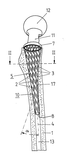
Fig. 1 is a lateral perspective of the shaft
designed as the femur part of a hip prosthesis,
Fig. 2 is a cross-section along line II-II of the
shaft of Fig. 1,
Fig. 3 is a cross-section in the manner of Fig. 2
of a longitudinally slotted shaft part of the
invention,
Fig. 4 is a perspective of the shaft part of the
invention as yet not clamped, and
Fig. 5 is a perspective of the clamped shaft part
of the invention.
As shown by Fig. 1, the femur part designed as a
hip prosthesis includes a frusto-conical braid 5
tapering from the proximal to distal ends in harmony
with the anatomy of the marrow cavity and is
constituted by two mutually crossing series of fibers
2, 3. Preferably the frusto-conical braid 5 assumes a
-
201~320
. 3
- nearly circular cross-section in the distal zone and a
nearly elliptical cross-section in the proximal zone.
As indicated in Fig. 1, the external contours of the
braid 5 are longitudinally concave, i.e., the cross-
section is narrower at the center than a regular
frustrum of cone which would have straight outer
contours. The fibers 2, 3 consist of a body-compatible
metal, or of a suitable metal alloy or a plastic.
Carbon fibers, illustratively of Pyrocarbon, are
preferred. The fibers 2, 3 subtend an angle to the
longitudinal axis 4 of the hollow frustrum of cone
formed by the braid 5. As a rule the angle is about
the same for both sets of fibers 2, 3. However, in
order to achieve a better adaptation or fit of the
braid in the proximal femur part, the angle ~ also may
be variable along each fiber 2 or 3 and preferably
becomes smaller from the distal toward the proximal
zones.
In a preferred embodiment mode, the gaps 17 in the
frusto-conical braid 5 made of fibers 2 and 3 are
filled as shown in Fig. 2 with an elastomer 9 such as
silicone rubber or Silastic in order to achieve a
smooth shaft surface. This circumstance is significant
because it has been found that newly formed bone
material 1 easily grows into the coarse-structured
surface of the bare braid 5, and removal of the shaft
in the event of a new operation would thereby be
seriously hampered.
As shown by Fig. 3, the frusto-conical braid 5 may
comprise an axial slit 6 in a further and preferred
embodiment mode.
The frusto-conical braid 5 comprises an annular
member 7 in the proximal zone for fastening the
proximal ends of the fiber sets 2 and 3, and an annular
201132~
member 8 for fastening the distal ends of the fiber
sets 2 and 3. These annular members 7, 8 -- which may
be circular or elliptical -- make it possible upon
exertion of traction on one of the two rings with
s simultaneous fastening of the other ring to change the
geometry of the braid 5 or of the hollow frustrum of
cone formed thereby, in particular the length and the
diameter of the shaft. This ability of the shaft part
of the invention to be changed in shape leads to its
application to implantation or removal of the
prosthesis so outfitted and is discussed in detail
below in relation to Figs. 4 and 5.
The shaft part of the invention is implanted
conventionally by inserting the shaft 10 into the
marrow cavity 13 of the femur 1. As soon as the shaft
10 has attained a first and preliminary wedged position
in the marrow cavity 13, then, as illustratively shown
by Fig. 4, the distal annular structure 8 is engaged by
a suitable long-stem instrument 14 and is further
displaced distally by the application of axial
pressure. In this process, tension is applied to the
fibers 2, 3 that are fastened by their proximal ends to
the annular member 7 fixed in the marrow cavity 13, and
this tension results in an extension of the hollow,
frusto-conical braid 5 with concurrent tapering. The
angle between the fibers 2 or 3 and the longitudinal
axis 4 of the hollow frustrum of cone formed by the
braid 5 then becomes less.
This stretching of the frusto-conical braid 5
allows attaining optimal contact with the marrow cavity
13 as shown in Fig. 1. This areal anchoring is
retained even following implantation because any
possibly "sinking" of the shaft would perforce lead to
widening the frusto-conical braid 5.
XO~ 2~
The implantation instrument 14 is very easily
handled because the frusto-conical braid 5 is open
throughout, and therefore the tip of the implantation
instrument 14 which is shaped to engage member 8 may be
inserted directly through the inside space 16 of the
frusto-conical braid 5 to engage the annular member 8.
Contrary to the case of the implanted shafts of
the state of the art that are cemented or structured
and cementless, the removal of the shaft part of the
invention takes place in exceedingly simple manner. A
suitable instrument 15 is designed to engage and seize
the proximal annular member 7 and to pull it further
proximally. In this procedure traction is exerted on
the fibers 2, 3 which are fastened by their distal ends
to the annular member 8 fixed in the marrow cavity 13,
such that the hollow, frusto-conical braid 5 is
extended while tapering, so that the shaft part easily
detaches from the marrow cavity 13 and is removed from
it.
