Note: Descriptions are shown in the official language in which they were submitted.
2014833
TITLE
RADAR TOMOGRAPHY
BACXGROUND OE THE INVENTION
The present invention relates to medical imaging
apparatus and method, and particularly to tomoqraphy
utilizing radar pulses.
A variety of medical imaging modalities are kno~n and
include nuclear magnetic resonance, ultra-sound,
sonography, positron emission, digital subtraction
angiography, and x-rays. Computed tomography is a
well-known method for ~anipulating data to produce
medical images. For example, ultra-sound, positron
emission, and X-rays may utilize computed tomography
techniques to produce images for diagnosis. A recent
article, "III Imaging With Photons", by Edward
Rubenstein, appearing in the December, 1988, edition of
CURRENT TRENDS IN MEDICINE, explains ~everal of these
imaging methods and is incorporated herein by
reference.
2C~ 333
However, all known medical imaging modalities are
considered to be either too expensive or may be at
least somewhat harmful to the patient. For example, a
nuclear magnetic resonance machine may cost $2.5
million and require almost one-thousand dollars to
produce an image. On the other hand, the use of X-rays
is disadvantageous in that repeated use may result in
harm to the patient.
Furthermore, known imaging techniques can create an
image by passing energy through the patient to produce
a projected image or a cross-sectional image of the
patient. The p~wer required to pass certain types of
energy and energized particles through a patient is
expensive to produce and may cause harm to patient
tissue.
Thus, the medical practitioner often is presented with
the dile~ma of choosing between the desire to perform a
thorough diagnosis and excessive cost or patient harm
resulting from such thorough diagnosis. In fact,
medical insurance companies are demanding greater use
of medical imaging equipment, while patients are being
informed by the media and various consumer advocates
that increased use of, for example, X-rays is
unnecessary and harmful. Therefore, medical personnel
are placed in the difficult position of trying to
satisfy both their patient's needs and their insurer's
requirements.
Accordingly, what is needed is a simple, fast, low-cost
medical imaging technique which causes no harm to the
patient.
20~4833
It is known that radio waves will penetrate human
tissue, and that radio wavelengths of electromagnetic
radiation are considered non-ionizing, thus causing no
radiation damage. For example, current technologies
employ short-wave and microwave radiation to treat deep
~uscle injury with controlled heat. No tissue damage
occurs even when the radio waves are applied steadily
for periods of up to 30 minutes. U.S. Food and Drug
Administration (FDA) guidelines for use of such
modalities are currently available. Furthermore, radar
technology is relatively well developed in military and
civilian aviation. In addition, the proliferation of
radar guns and related equipment in traffic enforcement
is well-known.
Radar uses a wavelength of several meters to several
millimeters. Radar can also be focused into more
concentrated beams than X-rays. In addition, sensitive
radar receivers are available which can image an object
at great distances registering a small fracti~n of the
radiated energy. Radar also produces an image by
reflecting energy from an object, thus requiring less
power and producing less ti6sue damage in the patient
than known techniques. ~hus, it appears that radar
signals may be useful in medical imaging.
SUMMARY OF THE INVENTION
An object of the present invention i5 to provide a
medical imaging method and apparatus utilizing radar
signals.
In order to achieve the above object, the present
invention is directed to a method and apparatus for
emitting a plurality of radar or radio pulses toward a
~ubject with an antenna, providing the radar pulses to
- 4 - 2~4833
the antenna with a transmitter, and receiving the
plurality of radar pulses reflected from the subject
with a receiver. A timer/gate circuit is used to
select predetermined radar pulses from among the
S received, reflected radar pulses. The radio pulses
selected are those which correspond to a predetermined
area, at a predetermined depth, of interest within the
subject.
Preferably, the timer/gate circuit can be controlled in
order to scan the predetermined area throughout the
subject.
If desired, a three-dimensional image of a
predetermined volume within the subject can be produced
by generating relative movement between the antenna and
the subject. This produces a sequence of scans at
differing depths within the target volume within the
patient. A processor then stores and manipulates the
view data in order to produce a three-dimensional vieh
of the predetermined volume within the subject.
In order to more accurately focus the emitted and
reflected radar pulses, the present invention may
include a matrix filter, coupled to the antenna, which
reduces noise by eliminating unwanted reflection and
diffraction components. The matrix filter may include
a plurality of radar absorbing tubes disposed to form a
grid in cross-section.
Of course, the present invention may also include
display means for displaying the predetermined two and
three-dimensional areas within the ~ubject.
The advantageous structure and functions according to
the present invention will become readily apparent to
- 5 - 2(~1~833
those of ordinary skill in thi~ art from the following
detailed description of t~e preferred embodiment, taken
together with the accompanying drawing.
BRIEF DESCRIPTI~N OF ~HE DRAWING
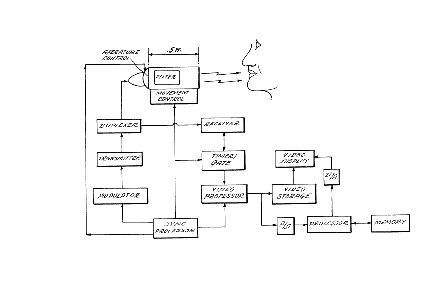
FIGURE 1 is a schematic block diagram of the apparatus
according to the preferred embsdiment; ~nd
FIGURE 2 is a perspective view showing the matrix
filter of Figure 1.
DETAILED DESCRIPTION OF THE PREFERRED EMBODIMENT
The principle of radar is relatively simple. Radio
wave energy is emitted tow~rd an object and its
position and relative ~ovement may be determined
through the return radio echo. The frequency of the
radio pulses and the intensity of each pulse may be
varied in accordance with the type of echo desired, the
relative distance to and movement of the subject, and
the type of antenna used. Fro~ the return echo, the
distance to the object may be readily calculated by
well-known Doppler techniques. The signal-to-noise
(SNR~ ratio of the return echo pulses may be diminished
by resonance, diffraction, or off-phase interference.
Techniques for reducing re60nance (artificial wave
amplification), and off-phase interference are well-
known and could be implemented in the preser.t
invention.
Diffraction may reduce the SNR by causing scattering of
the return pulses into the receiver. As will be
discussed below, the present invention proposes a
matrix filter in order to reduce diffraction noise.
- 6 - ~ 8~
Producing a medical image from the return echo pulses
can be a matter of applying existing technology. h'ell-
known computed tomography techniques ~ay be used to
process the return radar signals in order to produce
usable images for medical diagnosis. For example, a
timer/gate device may be used to gate the receiver so
that it receives only pulses from a selected distance.
Another technique is to utilize a so-called range
filter in which a plurality of range bins are disposed.
A return radar signal entering a particular range bin
indicates t~at the 6ub~ect is at a predetermined
distance from the antenna. Such techniques are known
in the radar field and need not be described in greater
detail herein.
Referring n~w more particularly to the drawing, Figure
1 is a block diagram of a preferred embodiment of the
present invention. This embodiment is a radar
tomography device adapted for use in dentistry to
examine a patent's teeth, although the principles of
the present invention may be adapted to a wide variety
of medical imaging applications and devices.
In Figure 1, the patient or subject 2 is exposed to
pulsed radio signals 4 emitted from an antenna head 6.
As schematically shown there, antenna head 6 includes
an antenna 8, an aperture control device 10, a matrix
filter 12, and a cone or cylinder spacer 14. A
~tandard dental X-ray cone is usually 8 or 18 inches
long, ~nd therefore, an 18 inch cone or cylinder spacer
14 would be quite normal for use with the patient and
by medical personnel. In addition, nn 18 inch spacer
14 would provide approximately a 1 meter path for rays
emitted from the antenna and reflected from the
subject.
- 7 - 2~1~833
Antenna 8 ma~ comprise any well-known or conventional
radar anten~a. For example, parabolic, Cassegrain,
dipole, or flat semi-conductor antennas may be used.
The antenna should ~e simple, light-weight, and
inexpensive. The antenna should also be ~mall enough
to fit into the antenna head 6 and allow for ease of
operation by medical personnel.
The aperture control device 10 i~ used to control the
aperture of the antenna 8. This device 10 may include
synthetic aperture control circuitry, or mechanical
means such as two plates of radar-absorbing materials
with slits ~oving in opposite directions allowing
synchronous radiation emission and reception through
one aperture at a time. Additionally, while the
aperture control 10 is shown located between the
antenna 8 and the filter 12, it may be located between
the filter 12 and the patient 2. Again, such aperture
control devices are relatively well-developed and need
not be described in further detail here.
A matrix filter 12, as mentioned earlier, is used to
reduce diffraction noise from the reflected return
signal, and to properly focus the emitted signal on the
area of the patient of interest. The matrix filter 12
may be designed in a predetermined pattern to
correspond to the number of 6cans desired, and the
location of the area of interest within the subject. A
detailed description of one preferred embodiment of a
matrix filter 12 will be provided below with reference
to Figure 2.
A duplexer 16 is provided to switch the antenna between
a transmitting mode and a receiving mode. In the
~5 absence of the duplexer, the transmitted energy may
harm a receiver 22 connected therethrough to receive
Z(3~33
the reflected radiation. Again, duplexers are very
well known and are readily available. Of course, two
antennae (one for transmitting, one for receiving) may
be used in the present invention, thus eliminating the
need for a duplexer.
A transmitter 18, also connected to the dupluxer 16, is
a high~power oscillator which generates the radar
pulses at a predetermined frequency, amplitude, and
phase. A modulator 20 provides pulses of input power
to activate the transmitter 18. For the duration of
the input pulse from the modulator 20, the transmitter
18 generates a high-power radio frequency wave,
converting a DC pulse to a pulse of radio frequency
energy. The exact frequency of the emitted energy may
be tuned to any appropriate range, as desired. The
generated radio wave pulses are then transmitted to the
antenna 8 through the duplexer 16.
The receiver 22 receives the reflected radar pulses
from the antenna 8 through the duplexer 16. Typically,
the receiver 22 is a superheterodyne receiver which
translates the received signals from their frequency to
a lower, intermediate frequency at which they can be
filtered and amplified more conveniently. Translation
is usually accomplished by adding the received signals
to the output of a low-power local oscillator in a
mixer. The output of the mixer is usually amplified
and then filtered to reduce interfering signals,
electrical background noise, resonance, and off-phase
interference noise. Finally, the amplified received
signals are output to a video processor 26 through a
timer/gate 24 discussed below in detail. Radar
receivers as described above are well known and need
not be explained in further detail.
21~14~33
The timer/gate 24 is a device which selects
predetermined pulses from among the received pulses in
order to effect spatial control. For example, as the
radar pulse~ are reflected back from the lower jaw of
the patient 2, the timer/gate 24 ~elects only those
return pulses timed to return from a desired depth (for
example, 2 centimeters from the forward edge of radar
head 6). Accordingly, only the gated pulses would be
accepted for imaging. Preferably, timer/gate 24
controls the receiver 22 so that it only receives radar
pulses from the desired location. By varying the
return-plane distance within the patient by moving the
antenna head toward or away from the volume of the
patient under study, or by varying the time of
acceptable pulse return, readings can be obtained for
any desired tissue depth within the patent 2. The
timer/gate 24 must be very sensitive since the patient
2 will be positioned close to the radar head 6. Timers
capable of measurinq picoseconds are now known. For
example, such a timer identified by Model No. DG-535 is
available from Stanford Research.
~y moving radar head 6 relative to the patient 2, and
then scanning in the depth direction through operation
of the timer/gate 24, information may be derived in
three-dimensions. Such techniques are well-known in
the computed tomography field. This method will allow
volumetric information to be obtained from the subject.
The video processor 26 receives the selected output
from receiver 22 and processes the 6ignal to produce a
video si~nal capable of being 6tored in a video storage
device 28, and/or displayed on video display 30.
Apparatus 6uch as the video processor 26, video storage
3S 28, and video display 30, are known and available.
- 1 o - 2(~14:8~3
A synch processor 32 synchronizes the operation of the
apparatus. Specifically, the transmitter 18 and video
processor 26 are synchronized by generating a
continuous stream of very short, evenly spaced pulses.
They designate the tiues at which ~uccessive radar
pulses are to be transmitted, and are ~upplied
simultaneously to the modulator 20 and video processor
26. In addition, synch processor 32 controls
timer/gate 24 to effect proper 6canninq control. Such
synch processors are widely used in radar devices, and
in computed tomography ~pparatus, and therefore, can be
readily adapted to the prese~t invention.
A high-resolution image of the area or volume of
interest may also be obtained by providing relative
movement between the antenna head 6 and ~ubject 2.
Thus, the movement control device 34 may be coupled to
the antenna head 6 to move it with respect to patent 2.
In a manner si~ilar to a CATSCAN, the antenna head 6
may be moved in an arc around subject 2 in order to
take several "shots" or "views" of the subject 2. In
each view, the radar pulses are scanned in the X and Y
directions by use of the aperture control 10, and in
the depth direction by using the timer/gate 24. When
information regarding the plurality of ~views" is
combined, a higher resolution image of the volume of
interest may be obtained. Those having skill in this
field will understand that the principles of $mage
processing used in a CATSCAN device can be adapted to
t~e present radar tomography device.
The 6ignal output fro~ the video processor 26 is an
analog video 6ignal capable of being stored on the
video ~torage device 26 ~for example, a VCR), or
displayed on the video display device 30. However,
digital techniques offer significant opportunities for
X014~333
image enhancement. Therefore, the analog signal from
the video processor 26 may be provided to an analog-to-
digital converter 36 to digitize the signal. The
digitized signal is then provided to a digital
processor 38 which can manipulate the data in a variety
of well-known ways. For exa~ple, information from a
plurality of "views", as discussed above, may be
combined within the processor 38 to produce a high-
resolution, three-color, three-dimensional view of a
volume of interest within subject 2. Such images may
then be converted to an analog signal by a digital-to-
analog device 42 for display on the video display 30.
~he digital output from the processor 38 may also be
provided to a memory 40 which stores the information
for later retrieval and use. Imaging processors such
as those used in nuclear magnetic resonance imaging may
be adapted for use in the present invention.
Figure 2 is a perspective view of a preferred
embodiment of the matrix filter 12. The matrix filter
12 has the dual function of focusing the emitted radar
energy on the area of interest and eliminating
diffraction noise from the reflected return pulses.
Diffraction caused by 6cattering of the return waves is
avoided by the size of the matrix filter 12. Matrix
filter 12 is preferably a radar-absorbing 10 centimeter
square parallel filtering box, broken into a cross-
sectional grid of square tubes. The grid comprises a
plurality of perpendicularly disposed radar-absorbing
panels 121. The number and spacing of the panels may
be modified somewhat, depending upon the desired radar
frequency, phase, and power. Alternatively, the filter
may be made of a matrix of parallel cylindrical tubes
of radar-absorbing materials. Of course, the tubes may
be of other cross-sectional shapes. Again, the design
- 12 - 2~4833
of such filters is fairly well developed in the radar
field.
Thus, what has been described is a medical imaging
modality using radar-frequency signals tG produce
inexpensive, high-resolution images of a subject. The
apparatus utili~es existing technology, and therefore,
should be relatively inexpensive to manufacture,
market, and operate. Further~ore, medical insurers and
patients alike will welcome such a safe, low-cost
alternative to X-rays and nuclear magnetic resonance.
The specific structural details of the devices
represented by blocks in the sch~matic diagram of
lS Figure 1 are per se well-known or could be readily
constructed by the person of ordinary skill in this
field without undue experimentation. Therefore, the
exact structure of the blocks in the schematic is not
described in detail in order to more clearly describe
the present invention, and since such details are not
critical to the best mode of carrying out the present
invention.
While the present invention has been described with
respect to what is presently considered to be the
preferred embodiment, it is to be understood that the
invention is not limited to the disclosed embodiment.
To the contrary, the present invention is intended to
cover various modifications and equivalent arrangements
included within the spirit and scope of the appended
claims. The scope of the following claims is to be
accorded the broadest interpretation CO as to encompass
all such modifications and equivalent ~tructure and
functions.