Note: Descriptions are shown in the official language in which they were submitted.
20i~495
MODIFYING A MEMBRANE FOR USE AS A GRAFT
The present invention relates to a method for modifying a
membrane intended for use as a surgical graft. In
particular, the present invention relates to modifying a
membrane for use in tympanic membrane repair.
Membranes formed from both natural and synthetic
substances are known for use as implants and grafts for
repairing damaged natural tissue. For example, in the
surgical repair of perforated eardrums (i.e.,
tympanoplasty), a replacement membrane is placed on the
damaged eardrum to cover the perforation and the ear packed
with surgical sponge material to hold the graft in place.
In time, the natural tissue response hopefully incorporates
the graft into the surrounding tissue.
Because of the low adhesion properties of many materials
useful in tympanoplasty, problems can arise both during and
after the surgical procedure. During the surgery itself,
accurate placement of the graft is often difficult, the
slippery nature of the replacement membrane making it
difficult to handle. Furthermore, even though the ear is
packed with sponge material to keep the replacement
membrane in place, gross movements of the head may cause
slippage of the material with respect to the perforation.
Even the packing procedure itself may dislodge the
replacement membrane from its proper position. Also,
incorporation of the replacement membrane into the
surrounding natural tissue is slow and often incomplete.
Accordingly, the present invention is a method for
texturizing a replacement membrane for use in tissue repair
comprising perforating the membrane. One or more
perforations in the membrane permit natural tissue growth
inside the perforation, thereby anchoring the membrane to
the surrounding natural tissue. Membrane material
projecting from its surface as a result of the perforation
process also serves to mechanically anchor the membrane in
place on the natural tissue. The present invention is also
a method of modifying a replacement membrane for use in
tissue repair comprising providing the membrane with a
handle that projects from the surface of the membrane.
When the membrane is used to repair a perforated eardrum,
the handle provides a convenient means for inserting the
membrane through the perforation and accurately positioning
it.
Fig. 1 is an electron micrograph showing a membrane of
the present invention magnified 500x, and Fig. 2 is an
electron micrograph showing a perforated membrane of the
present invention magnified 3,000x. Figs. 3, 4, 5, and 6
are perspective views of different embodiments of the
membrane having a handle according to the present
invention.
Any cohesive membrane useful in natural tissue repair is
useful in accordance with th~ present invention. Such
membranes include transplanted tissue from the recipient
(i.e., an autograft), transplanted tissue from an
individual of the same species (i.e., a homograft), or
transplanted tissue from an individual of another species
(i.e., a xenograft). In addition to natural tissue, which
ca~ be glutaraldehyde treated, such as fascia, fat,
perichondrium, and cartilage, synthetic materials are also
useful, such as polylactic acid, silicone, polyurethanes,
dacron, and polytetrafluoroethylene. Preferably, the
membrane is an artificial membrane comprising a product
ma~e by cross-linking molecules of interpenetrating
denatured collagen coupled at their lysine epsilon amino
groups with a coupler through carbonyl groups, sulfonyl
groups, or combination thereof on the coupler wherein non-
coupled lysine epsilon amino groups are bonded to a
modifier wherein the modifier is a carbonyl, sulfonyl,
carbamoyl, or ~malic acid group. Tne preferred membrane
201~
used in accordance with the present invention is made by
denaturing coupled, and preferably modified, collagen
molecules. The coupled and modified collagen molecules
useful in accordance with the present invention and their
method of manufacture are disclosed in United States
Patents 4,713,466 and 4,883,864. The carbonyl groups,
sulfonyl groups, or combination thereof on the coupler are
bonded together through an R group wherein R is a C2-20
saturated or unsaturated aliphatic, aromatic, or aliphatic-
aromatic group that is unsubstituted or substituted withhalogen, or Cl_4 carboxy, alkyl, or alkoxy and having 0-5
heteroatoms wherein the heteroatom is oxygen, sulfur, or
nitrogen. Preferably, the coupler has the formula -CO-CH2-
CH2-C- or -CO-CH2-CH2-CH2-C~-. The modifier has the
formula RCO-, RNHCO-, RS02-, or COOR'CHOHCH~COOR')- wherein
R is a C2_20 saturated or unsaturated aliphatic or aromatic
group that is unsubstituted or substituted with halogen,
C1_4 alkyl or alkoxy, and having 0-5 heteroatoms wherein
the heteroatom is oxygen, sulfur, or nitrogen, and R' is H,
Na, K, or Li. Preferably, the modifier has the formula ~-
NH-CO-, more preferably CH3(CH2)3-NH-CO-. Coupling is
performed by reacting native collagen with a polyfunctional
amine-reactive agent selected from the group consisting of
a carboxylic acid halide, sulfonyl halide, anhydride, and
reactive active ester in aqueous media at a pH greater than
about 8 and at a temperature between 0 and 35C.
Preferably, the poly-functional amine-reactive agent is
succinic acid dichloride or glutaric acid dichloride.
Preferably, the ratio of poly-functional amine-reactive
agent used per weight of native collagen varies between
about 1/100 and 6/1, more preferably between about 1/50 and
2/1. Modification involves reacting the native collagen
molecules with a mono-functional amine reactive agent
selected frum the group consisting of an anhydride, acid
2~
halide, sulfonyl halide, active ester, isocyanate, and
epoxy succinic acid in aqueous media at a pH greater than
about 8 and at a temperature between about 0 and 35 C.
Preferably, the mono-functional amine-reactive agent is n-
butyl isocyanate or epoxy succinic acid. The weight ratioof mono-functional amine-reactive agent used per amount of
native collagen is between about 1/100 and 10/1, more
preferably between about 1/10 and 1/1. Coupling and
modifying are performed in any order, or simultaneously.
Denaturing is preferably performed by heating the
coupled and preferably modified collagen molecules in
aqueous media or non-aqueous media at a temperature ~etween
about 40 and 120C. Heating causes the normal collagen
helix to unwind, producing single stranded collagen
molecules coupled at their lysine epsilon amino groups.
Upon cooling, the solution forms a gel, which is believed
to contain an interpenetrating network of hydrogen bonded
~-helixes with segments of single stranded collagen
exposed.
Accordingly, the heated collagen molecules are cast into
a desired shape, such as a film, through the use of an
appropriate mold and then allowed to cool and gel.
Preferable thicknesses for the gel are between about 0.127
cm and 1.27 cm, more preferably between about 0.254 cm and
0.613 cm. After cooling, the interpenetrating, denatured
collagen molecules are cross-linked to form an artificial
membrane useful in tympanic membrane repair.
Preferably, the gel is dehydrated, which is helieved to
cause some cross-linking of the collagen molecules. After
dehydration, the membrane has a preferable thickness
between about 0.1 and 0.5 mm, more preferably between about
0.15 and 0.25 mm. Preferably, after dehydration the
molecules are further cross~linked to increase the burst
strength of the membrane. Further cross-linking is
2~5'~9~
preferably performed by treating the membrane with chemical
cross-linking agents or exposing the membrane to sufficient
actinic radiation. Useful cross-linking agents include
polyfunctional amine reactive agents such as a carboxylic
acid halide, sulfonyl halide, anhydride, and reactive
ester. Examples of such agents are disclosed in the
aforementioned United StateQ Patent 4,713,466. Methods for
using chemical cross-linking agents will be apparent to the
skilled artisan. Preferably, chemical cross-linking is
performed using non-aqueous systems in order to prevent
hydrolysis of the cross-linking agent. For example, the
membrane is immersed in succinyl chloride, either neat or
in pyridine or other suitable organic base that would
neutralize HCl evolved during the cross-linking reaction,
at an amount of about 0.001-0.1 moles of agent per gram of
membrane, preferably about 0.005-0.05 moles/g.
Alternatively, chemical cross-linking can be carried out in
aqueous media while maintaining a pH of 8-10 and usin~ an
amount of cross-linking agent between about 0.05 and 0.5
moles per gram of membrane, depending on the rate of
hydrolysis of the particular agent used. Useful forms of
actinic radiation include ultraviolet light, gamma
radiation, and electron beam radiation. Sources and
methods of applying radiation to the membrane will be
apparent to the skilled artisan. After cross-linking, the
membrane is preferably washed to remove unreacted agents,
and further sterilized, e.g., by autoclaving or exposure to
gamma radiation or ethylene oxide, before use in tympanic
me~brane repair.
The artificial membrane preferred for use in accordance
with the present invention is optionally cleaned and
purified before use. For e~ample, either before or after
heating, but prior to cooling, a solution of the coupled
a~d modified collagen molecules are filtered to remove
2C~ 9~i
particles. At any stage in the process after denaturation,
extraction purification, e.g., using sterile water for
injection, high-purity grade acetone, or other suitable
solvent or solvent mixture, can be employed.
The size of the membrane itself will vary depending upon
its intended use. When used in human tympanic membrane
(eardrum) repair, the membrane has a preferable thickness
between about 20 and 500 ~m, more preferably between about
45 and 155 ~m, and length and width dimensions slightly
larger than the perforation in the eardrum, which
preferably effects a surface area on one side of the
membrane between about 3 and 500 mm2. When a hydratable
material is used as the membrane, the foregoing ranges are
determined when the membrane is fully hydrated, i.e., after
immersion in physiological buffer at 37C until equilibrium
is reached. Perforations are made in the mèmbrane in
accordance with the present invention by a variety of means
using devices that will be apparent to the skilled artisan.
For example, the membrane can be initially cast in a dish
having holes through which spikes project upwardly. After
the membrane forms in the dish, the spikes are withdrawn
from the holes leaving the perforated membrane. For
thermoplastic membranes a heat-staking process (using hot
metal wires) can be used to create the perforations.
Preferably, the membrane is perforated after it is formed
by forcing it o~er a matrix of pyramidal spikes attached to
a plate. By perforating the membrane in this manner, the
rough edges created around the perforations in the
membrane, as shown in Figs. 1 and 2, act like hooks to
adhere the membrane to the tissue surface. ~he size,
number, and arrangement of the perforations vary based on
the considerations of tissue strength, desirability of
water and air impermeability, and creation of angular edges
for adhering the membrane to the tissue surface.
2~:~L5'~9~
Preferably, the perforations have a minimum cross dimension
greater than about 50 ~m, and a maximum cross dimension
less than about 400 ~m. More preferably, the spikes are
designed and pressure is applied to the membrane such that
holes that are large enough to permit fibroblast migration
and reproduction, yet small enough to permit the membrane
to act as a water barrier, are made in the membrane.
Accordingly, the holes are more preferably made in the
membrane to have a minimum cross dimension greater than
about 140 ~m, which is designed to accommodate optimum
rates of fibroblast migration and reproduction, and a
maximum cross diameter less than about 250 ~m, which
permits the membrane to act as a water barrier in the
absence of pressure. Any number of perforations are
present in the membrane, and they are arranged on the
surface of the membrane in any desired pattern.
Preferably, the perforations are arranged in a square grid
about 200-600 ~m apart as measured from the center of the
perforation, more preferably between about 300 and 500 ~m
apart. As shown in Figs. 1 and 2, the perforations have a
siæe of about 150 ~m, and are located about 500 ~m apart in
a square grid pattern. Preferably, the edges of the
perforations have a height, i.e., the distance projecting
from the surface of the membrane, between about 25 and 500
2S ~m, more preferably between about 50 and 150 ~m, most
preferably about 100 ~m.
The handle provided on the membrane in accordance with
the present invention has various forms, is made of various
tissue-compatible materials, and is attached to the
membrane in various ways depending upon the surgery
involved, membrane material used, and suryical tools
employed. Preferably, the handle is made of a
biodegradable material. Useful biodegradable materials
include polymers having at least one hydrolyzable ester,
2~15495
amide, or ester/amide linkage, such as polylactic acid,
polygalactic acid, lactic acid/galactic acid copolymers,
polyhydroxybutyric acid, polydioxanone, collagen, collagen
derivatives, catgut derivatives, polyurethanes,
polytetrafluoroethylene, glutaraldehyde-treated tissue, and
silicones. For example, the handle can be medical grade
suture thread that is passed through the membrane and back
again; by pulling on both ends of the thread the membrane
can be held in place, and by pulling on one end of the
thread, the handle can be removed from the membrane after
surgical implantation. When using the preferred membrane
in accordance with the present invention, a portion of a
suture thread is advantageously set in the denatured
collagen solution such that a useful length of the thread
projects axially from the center of the cooling solution
while a sufficient portion of the thread is imbedded in the
solution to provide an anchor once the membrane is formed.
After membrane formation, the suture is then an integral
part of the membrane. Suture handles are exemplified in
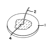
Figs. 3, 4, and 5. As shown in Fig. 3, one end 4 of suture
thread 2 is imbedded in membrane 1. Referring to Fig. 4,
both ends 4 of suture thread 2 are imbedded in membrane 1
to form the handle as a closed loop. A closed-loop handle
is also shown in Fig. 5, wherein part of continuous suture
thread 6 is imbedded in the membrane 1. Instead of a
suture thread, as shown in Fig. 6, the handle can also be
made of a strip 8 of the same material as the membrane, the
end 10 of which is imbedded in the membrane 1.
Alternatively, the membrane and handle are cast in one
piece from the same material to form a unitary structure,
i.e., a mold the shape of the membrane and handle is used.
As shown in the accompanying figures, the size and shape
of the handle vary. Generally, it is an elongated strip of
material having a length, i.e., the distance projecting
2~1i5'~95
from the surface of the membrane, between about 100 and
2000 ~m, more preferably between about 150 and 1000 ~m.
The handle can be located on any part of the lateral
surface of the membrane, but is preferably located as close
to the center of the membrane as possible.
To more clearly describe the present invention, the
following, non-limiting examples are provided. All parts
and percentages in the examples are by weight unless
indicated otherwise.
EXAMPLE 1
A membrane is prepared using modified collagen. Coupled
and modified collagen is prepared by addition of a chemical
coupling agent and subsequen~ly an amine modifying agent,
using aseptic technique under a laminar flow hood. Abou~
500 ml of chilled (4C) VitrogenTM collagen Type I solution
(Collagen Corp., Palo Alto, CA) is poured into a glass
reactor vessel. The pH of the solution is brought to 9 by
the addition of 5N and lN sodium hydroxide. At a
temperature between about 4 and 8~C, the solution is
vigorously agitated and 0.28 g of succinyl chloride is
added all at once to the solution. The reaction is allowed
to proceed for 20 minutes, and during this time the pH is
held within the range between 9.0 and 9.35 by the gradual
addition of lN sodium hydroxide solution as needed.
The product obtained above is modified with a reagent
that reacts with the exposed amine groups on the coupled
collagen molecules. The vigorous agitation of the solution
is continued while 0.35 g of neat butyl isocyanate is added
to the vessel as rapidly as possible. The reaction is
allowed to proceed for 1 hour, and during this time the pH
is held within the ra~ge between 9.0 and 9.25 by the
addition of lN sodium hydroxide solution as needed. As the
reaction proceeds, the solution is gradually allowed to
warm to room temperature. The pH of the solution is then
2C3~5~9~
decreased slowly by the addition of 6N hydrochloric acid to
precipitate out the modified collagen. The acid is added
until the cloudiness of the solution stops increasing
(generally at a pH of about 4.0-4.7). The solution is
allowed to continue mixing for 5 minutes. The resulting
collagen slurry is centrifuged at a temperature of about
4-C, at a speed sufficient to create a force of about
10,000 G (as measured at the bottom of the centrifuge
tube), and the supernatant removed.
The resulting collagen precipitate is washed by adding
pyrogen free water to a total volume of about 240 ml of
collagen suspension, and the p~ is adjusted to within the
range of 4.5 and 4.7 by the addition of lN hydrochloric
acid or lN sodium hydroxide as needed. The neutralized
collagen suspension is centrifuged at 4 D C, at a speed of
about 10,000 rpm (16,000 G at the bottom of the centrifuge
tube), for 10 minutes. After removing the supernatant,
this procedure is repeated three times for a total of four
washings. The final collagen concentration is adjusted to
approximately 2% by weight.
The washed and modified collagen product is then
denatured by heating at 60-80 n C for one hour in a water
bath. The material is then neutralized with lN sodium
hydroxide as needed to bring the pH to within the range of
7.0 to 7.2.
About 6.0 ml of the warm denatured collagen solution is
t r a n s fe r re d int o a sterile flat -b ottom
polytetrafluoroethylene dish ~ cm in diameter. The side of
which has been machined to about a 5-15 angle outward from
the bottom to facilitate subsequent membrane removal, and
the bottom of the dish has been machined roughly to create
a relief of about a 1.27 ~m in the surface of the membrane.
The dish is then covered with a sterile petri dish and
allowed to sit at amb~ent conditions (approximately 23C)
2~L5't~
for 25 minutes to allow the denatured collagen to slowly
cool and gel.
The covered dish is then transferred to a pre-purged,
nitrogen box having about 70 1 total volume. At a nitrogen
flow rate of about 12-15 l/minute, the material is dried
for about 24-36 hours to form a dehydrated membrane about
O.058 mm thick.
The membrane is laid on an aluminum foil at a distance of
15-16 cm from a 15 Watt, 254 nm, ultraviolet light source
for a period of about 4 hours.
The membrane is then purified by immersion in 150 ml of
high-purity grade acetone and allowed to sit for a minimum
of 2 hours under gentle agitation. The charge of acetone
is then decanted and the purification/extraction step
repeated twice.
The membrane is used for repairing a 3 mm perforation in
the tympanic membrane of a chinchilla. After the membrane
is wetted with sterile, pyrogen-free water and allowed to
hydrate for 5 minutes to facilitate cutting, a 1 cm
diameter disk is then cut from the membrane using a
stainless steel punch die and a small arbor press cutting
against a polytetrafluoroethylene plate. The disk is then
texturized to create perforations in the membrane by
forcin~ it o~er pyramidal titanium spikes fixed 500 ~m
apart in a square pattern on a plate, each spike being
about 150 ~m2 at its base and about 150 ~m high. The disk
is then allowed to dry for at least one hour. After drying
the membrzne is sealed in appropriate packaging and
sterilized by exposure to 0.5-0.6 Mrad gamma radiation.
Surgical repair is commenced by ma~ing a posterior
approach to the bulla of the anestheti~ed chinchilla. The
margin of the perforation in the tympanic membrane is
neatened and a re~ion 1.5 mm wi~e is d~nuded of epithelium
usin~ a half-Hough tool. The artificial membrane disk is
2~5 ~
removed from its sterilized packaging and trimmed to an
appropriate diameter, i.e., such that at least 3.4 mm of
the membrane contacts the intact borders of the ruptured
eardrum, or about 1.5 times the diameter of the eardrum
perforation. The trimmed membrane is positioned over the
perforation so that the side of the disk bearing the raised
areas formed during texturizing faces the eardrum tissue.
Ten minutes are allowed for fibrin formation to completely
proceed within the perforations on the disk. Although no
sponge packing of the bulla is used, gross movements of the
animal performed between about lO minutes and one hour
after surgery d~ not displace the disk. Tympanometry is
performed after several days using a model 6A typanometer
(Maico, Minneapolis, MN~, which shows near normal tympanic
membrane function. A histological evaluation of the
implants removed after 30 days shows fibroblast
infiltration of the areas, as well as the deposition of new
type I collagen within the graft matrix.
EXAMPLE 2
A texturized disk prepared as in EXAMPLE 1 is used to
close a 2.5 mm perforation in the tympanic membrane of a
monkey. An approach across the ear canal is made to the
damaged tympanic membrane of the anesthetized monkey, the
margin of the perforation is neatened, and the epithelium
peeled back around its circumference. The disk is trimmed
and positioned over the perforation from the middle ear
side as in EXAMPLE 1. The edges of the epithelium are
then folded over the top of the disk and ten minutes are
aliowed for fibrin formation to occur ~ithin the
perforations. Gross movements of the animal do not
dislodge the disk, even in the absence of sponge packing.
2~i5~
13
EXAMPLE 3
A membrane prepared as in EXAMPLE 1, without perforation,
to have a thickness of about 100 ~m is cut to make a strip
about 1 mm lonq and O.25 mm wide, which is then bent and
creased in the middle at a 90- angle to form a handle. One
leg of the handle is imbedded into a collagen solution that
is prepared and cast as in EXAMPLE 1 such that the other
leg projects at a right angle from the solution. While
maintaining the handle in this position, the solution is
allowed to gel and then dried, irradiated, and purified as
in EXAMPLE 1. The completed membrane can then be cut to a
desired size to form a membrane with a handle, such as
shown in Fig. 5.