Note: Descriptions are shown in the official language in which they were submitted.
20~L6,~7
-
DEVICE AND METHOD FOR DEPLETION OF THE
LEUCOCYTE CONTENT OF BLOOD AND BLOOD COMPONENTS
This invention relates to a method for depleting
the leucocyte content of whole blood and products
derived therefrom, particularly from human packed red
blood cells, and more particularly from anti-coagulated
human packed red blood (PRC) cells which have been
derived from whole blood freshly drawn from a blood
donor.
It has been the practice for 50 years or more to
transfuse whole blood, and more recently blood
components, from one or more donors to other persons.
With the passage of time and accumulation of research
and clinical data, transfusion practices have improved
greatly. One aspect of current practice is that whole
blood is rarely administered; rather, patients needing
red blood cells are given packed red cells (hereinafter
"PRC"), and patients needing platelets are given
platelet concentrate. These components are separated
from whole blood by centrifuging, the process
providing, as a third product, plasma, from which
various other useful components are obtained.
In addition to the three above-listed components, whole
blood contains white blood cells (known collectively as
"leucocytes") of various types, of which the most
important are granulocytes and lymphocytes. White
blood cells provide protection against bacterial and
viral infection.
In the mid to late seventies, a number of
investigators proposed that granulocytes be separated
from.donated blood and transfused into patients who
~:*
-- 1 -- ~
2016297
lacked them, for example, those whose own cells had
been overwhelmed by an infection. In the resulting
investigations, it became apparent that this practice
is generally harmful, since patients receiving such
transfusion developed high fevers, had other adverse
reactions, and often rejected the transfused cells.
Further, the transfusion of packed cells or whole blood
containing donor leucocytes can be harmful to the
recipient in other ways. Some of the viral diseases
induced by transfusion therapy, e.g., Cytomegaloviral
Inclusion Disease which is a life threatening infection
to newborns and debilitated adults, are transmitted by
the infusion of homologous leucocytes. Another life-
threatening phenomenon affecting immunocompromised
patients is Graft versus host disease (GVH); a disease
in which the transfused leucocytes actually cause
irreversible damage to the blood recipient's organs
including the skin, gastrointestinal tract and
neurological system. More recently, HTLVl virus has
become a threat. These viruses, and to a substantial
degree as well the HIV (AIDS~ virus, are resident in
the leucocytes, and for this reason removal of
leucocytes is regarded as beneficial.
Conventional red cell transfusions have also been
indicted as adversely influencing the survival of
patients undergoing surgery for malignancy of the large
intestine. It is believed that this adverse effect is
mediated by the transfusion of agents other than donor
red blood cells, including the donor's leucocytes.
Removal of leucocytes to sufficiently low levels
to prevent the undesired reactions, particularly in
packed red cells which have been derived from freshly
drawn blood, is an objective of this invention.
In the currently used centrifugal methods for
2016~97
separating blood into the three basic fractions (packed
red cells, platelet concentrate, and plasma), the
leucocytes are present in substantial quantities in
both the packed red cells and platelet concentrate
fractions. It is now generally accepted that it would
be highly desirable to reduce the leucocyte
concentration of these blood components to as low a
level as possible. While there is no firm criterion,
it is generally accepted that many of the undesirable
effects of transfusion would be reduced if the
leucocyte content were reduced by a factor of about 100
or more prior to administration to the patient. This
approximates reducing the total content of leucocytes
in a single unit of PRC (the quantity of PRC obtained
from a single blood donation) to less than about 1 x
107. Recently, it has become more widely perceived that
in order to prevent viral infection by transfused
blood, factors of reduction should be more than 100,
preferably more than 1000, and more preferably 30,000
or 100,000 fold or more, such as 1,000,000 fold.
One of the most effective means of reducing
leucocyte content that has been discovered hitherto is
disclosed in U.S. Patent No. 4,925,572 (U.S. Patent
Application No. 07/259,773, filed October 19, 1988),
which is directed towards the bedside filtration of
PRC. By contrast, this invention relates to the
filtration of freshly drawn whole blood and of fresh
PRC, that is, PRC that is filtered within 24 hours, and
more preferably within 6 hours, of the time the blood
was drawn. The behavior of fresh PRC is very different
from that of the 2 to 35 day old blood that is the
focus of the disclosures in U.S. Patent No. 4,925,572
(U.S. Patent Application No. 07/259,773, filed October
19, 1988). The standards for leucocyte depletion are
-- 3
- 20~6297
also very different; the above patent has, as its
objective, leucocyte reduction by a factor of up to
about 3000 to 10,000. While this is excellent for many
purposes, the objective of the present application is
leucocyte reduction by a factor in excess of about
30,000, and preferably of about 1,000,000 or more.
Defininq a Unit of Blood
and a Unit of Packed Red Cells:
Blood banks in the United States commonly draw
about 450 milliliters (ml) of blood from the donor into
a bag which usually contains an anticoagulant to
prevent the blood from clotting. Herein, the quantity
drawn during such a donation is defined as a unit of
whole blood.
While whole blood is to a degree used as such,
most units are processed individually by centrifugation
to prodlce one unit of PRC. The volume of a unit of
PRC var~es considerably dependent on the hematocrit
(percent by volume of red cells) of the drawn blood,
which is usually in the range of 37% to 54%; and the
hematocrit of the PRC, which varies over the range from
50 to over 80%, depending on whether yield of one or
another blood compound is to be minimized. Most PRC
units are in the range of 170 to 350 ml, but variation
below and above these figures is not uncommon.
Drawn whole blood may alternatively be processed
by separating the red cells from the plasma, and
resuspending them in a physiological solution. A
number of physiological solutions are in use. The red
cells so processed may be stored for a longer period
before use, and with some patients there may be some
advantages in the removal of plasma. "Adsol" is the
- ~016~97
trade name of one such procedure, and SAG-M is a
variant used in parts of Europe.
As used herein, the term "fresh blood product"
includes anti-coagulated whole blood, packed red cells
obtained therefrom, and red cells separated from plasma
and resuspended in physiological fluid, in all cases
processed including filtration within about 24 hours
and preferably within 6 hours of when the blood was
drawn.
In parts of the world other than the United
States, blood banks and hospitals may draw less or more
than about 450 ml of blood; herein, however, a "unit"
is always defined by the United States' practice, and a
unit of PRC or of red cells in physiological fluid is
the quantity derived from one unit of whole blood.
As used herein, PRC refers to the blood products
described above and to similar blood products obtained
by other means and with similar properties.
PreviouslY Available Means
to Remove Leucocytes from PRC
The Spin-Filter system for obtaining leucocyte
depleted packed red cells is described by Parravicini,
Rebulla, Apuzzo, Wenz and Sirchia in Transfusion 1984;
24:508-510, and is compared with other methods by Wenz
in CRC Critical Reviews in Clinical LaboratorY Sciences
1986; 24:1-20. This method is convenient and
relatively inexpensive to perform; it has been and
continues to be used extensively. However, the
efficiency of leucocyte removal, while generally about
90~ or better, is not sufficiently high to prevent
adverse reactions in some patients.
Centrifugation methods are available which produce
2016297
lower levels of leucocytes in red cells, but these are
laboratory procedures which are very costly to operate,
and sterility of the product is compromised to a degree
such that it must be used within 24 hours.
Other methods for leucocyte depletion, such as
saline washing or deglycerolizing frozen red cells,
have been or are used, but these have disadvantages for
economical, high reliability service.
A number of devices have been proposed in which
fibers are packed into housings, and whole blood passed
through them in order to remove microaggregates and a
portion of the leucocyte content. These devices have,
when reduced to practice, all required saline to be
applied either before or after use, or both before and
after use, and are very poorly suited for blood bank
use.
Characteristics Desirable
in a LeucocYte Depletion Device
An ideal device for leucocyte depletion intended
for use by blood banks would be inexpensive, relatively
small, and be capable of processing one unit of PRC
rapidly, for example in less than about one hour, and
reduce the leucocyte content to the lowest possible
level. Because of the high cost and limited
availability of red blood cells, this ideal device
would deliver the highest possible proportion of the
red cells present in the donated blood. Such a device
is provided by this invention.
Devices which have previously been developed in
attempts to meet this objective have been based on the
use of packed fibers, and have generally been referred
to a-s filters. However, it would appear on preliminary
20~62~7
review that processes utilizing filtration based on
separation by size cannot succeed for two reasons.
First, the various types of leucocytes range from
granulocytes and macrocytes, which can be larger than
about 15 ~m, to lymphocytes, which are in the 5 to 7 ~m
range. Together, granulocytes and lymphocytes
represent the major proportion of all of the leucocytes
in normal blood. Red blood cells are about 7 ~m in
diameter, i.e., in size they are in the range of one of
the two major components which must be removed.
Secondly, all of these cells deform so as to pass
through much smaller openings than their normal size.
Accordingly, and because it is readily observed by
microscopic examination that leucocytes are adsorbed on
a variety of surfaces, it has been widely accepted that
removal of leucocytes is accomplished mainly by
adsorption rather than by filtration. An unexpected
and surprising result of this invention, however, is
that filtration through certain filters having a
controlled pore size is critical to reach the target
levels of leucocyte depletion.
Blood Component Recovery
In the preceding section, reference was made to
the desirability of recovering a high proportion of the
red cells delivered to the separation device. There
are several causes for reduced recovery of red cells:
(a) Losses due to hold up within the connecting
tubing;
(b) Losses due to liquid which remains within the
device itself at the conclusion of the
filtration;
(c) Losses due to adsorption on the surfaces of
~U1~2~7
the device, or due to mechanical entrapment
within the device;
(d) Loss due to clogging of the filter prior to
completion of the passage of the full unit of
blood;
and (e) Losses due to contact with incompatible
surfaces, which can cause clotting.
CaPacity
As separated from whole blood in current blood
banking practice, packed red cells contain not only a
proportion of the leucocytes present in the blood as
drawn from the donor, but also contain some platelets
(which tend to be very adhesive), fibrinogen, fibrin
strands, tiny fat globules, and numerous other
components normally present in small proportions. Also
contained are factors added at the time the blood is
drawn to prevent clotting, and nutrients which help to
preserve the red cells during storage.
During the centrifuging process which concentrates
the red cells and partially separates them from the
remaining components, there is a tendency for
microaggregates to form in PRC. These may comprise
some red cells together with leucocytes, platelets,
fibrinogen, fibrin, fat, and other components. Gels,
which may be formed by fibrinogen and/or fibrin, may
also be present in PRC produced by blood banks.
If the leucocyte depletion device comprises a
porous structure, microaggregates, gels, and
occasionally fat globules tend to collect on or within
the pores, causing blockage which inhibits flow.
2()16297
Ease and Rapidity of Priminq
Ease of use is an important characteristic of any
leucocyte depletion system. As noted above, for
leucocyte depletion devices, ease of priming is a
particularly important factor. The term "priming time"
refers to start-up of flow of PRC from the bag through
the filter to the patient, and is the time required to
fill the filter housing from its inlet to its outlet.
An object of this invention is to maintain a short
priming time, preferably less than 30 to 120 seconds,
to conserve technician time.
Preconditioninq of LeucocYte
Depletion Devices Prior to Priminq
A number of devices in current use require
pretreatment prior to passing blood, usually consisting
of passing physiological saline. The necessity for
such an operation is very undesirable in blood bank
processing because it complicates the procedure,
requires technician time, and puts maintenance of
sterility at risk.
The reasons for using such pretreatment vary.
They include removal of acid hydrolysate developed
during steam sterilization of devices containing
cellulose acetate fibers, assurance of freedom from
foreign solids which may be present in natural fibers,
and if the fibers are hygroscopic to prevent hemolysis
(loss of the integrity of red blood cells with
subsequent loss of their contents to the external
milieu).
This invention provides a leucocyte depletion
device which requires no preconditioning prior to
processing PRC derived from freshly drawn blood.
_ g
- 2~1~297
Definition of Voids Volumes
The concept of "voids volume" is related to, but
distinguishable from, the term "bulk density." In
fact, the term "bulk density" is misleading when
referring to a broad spectrum of fibers with large
variations in specific gravity. For example, polyester
fibers may have a specific gravity of about 1.38 while
inorganic fibers prepared from zirconia may have a
specific gravity of greater than 5. Thus, in carrying
out the instant invention, references to voids volume
should not be confused with the term bulk density.
The concept of voids volume may be explained as
follows:
Calculation of Voids Volume,
Given Bulk Density and Fiber Density
Bulk density, D, is the weight of a given volume
of fibrous aggregate divided by its apparent volume.
Normally, this is expressed in g/cc.
By fibrous aggregate is meant one or more fibers
occupying a given or apparent volume, e.g., a mass of
non-woven intertangled fibers with a certain proportion
of voids or spaces within the mass.
In order to calculate the voids volume, V, the
density, d, of the fibers must be known. The density,
d, is also expressed in g/cc.
1. The volume of 1 gram of fibrous aggregate = 1
D
2. The volume of 1 gram of fibers = 1
3. The voids volume, V, is the total aggregate
-- 10 --
201629~
volume less the fiber volume or
_ 1
D d
Example:
Given: Volume of fibrous aggregate = 10 cc
Weight of aggregate = 1 g
Density of the aggregate D = 1 = 0.1 g/cc
Density of fiber d = 1.38
Volume of 1 g of the fibers is 1 = .725 cc
1.38
Hence voids volume
= 1 _ 1 = 1 _ 1 = 10 - .725
D d 0.1 1.38
= 9.275 cc
or, expressed as percent V = 9.275 x 100
= 92.75%
The following table illustrates the difference
between specifying voids volume and density. As
illustrated there, at constant density, a column of
glass fibers (glass being much more dense than, e.g.,
polypropylene) has a voids volume of 94% versus only
83.3% for a column of polypropylene.
D, Column Material Density of Voids
density of the the fiber, Volume
(q/cc) fiber (g/cc) (%)
O.lS glass* 2.5 94.0
0.15 polyester 1.38 89.1
0.15 polypropylene 0.9 83.3
* Glass varies in density from 2.3 to 2.7 g/cc. The
2~16~97
2.5 g/cc figure used here is in the mid range.
Definition of Pore Diameter
In the definition of various filter media, it will
be necessary to use the term "pore diameter". This
term as used herein is as determined by the modified
OSU F2 test described below.
Wetting of Fibrous Media
When a liquid is brought into contact with the
upstream surface of a porous medium and a small
pressure differential is applied, flow into and through
the porous medium may or may not occur. A condition in
which no flow occurs is that in which the liquid does
not wet the material of which the porous structure is
made.
A series of liquids can be prepared, each with a
surface tension of about 3 dynes/cm higher compared
with the one preceding. A drop of each may then be
placed on a porous surface and observed to determine
whether it is absorbed quickly, or remains on the
surface. For example, applying this technique to a 0.2
~m porous tetrafluoroethylene (PTFE) filter sheet,
instant wetting is observed for a liquid with a surface
tension of about 26 dynes/cm. However, the structure
remains unwetted when a liquid with a surface tension
of about 29 dynes/cm is applied.
Similar behavior is observed for porous media made
using other synthetic resins, with the wet-unwet values
dependent principally on the surface characteristics of
the material from which the porous medium is made, and
secondarily, on the pore size characteristics of the
2(~1629~
porous medium. For example, fibrous polyester,
specifically polybutylene terephthalate (hereinafter
"PBT") sheets which have pore diameters less than about
20 ~m will be wetted by a liquid with a surface tension
of about 50 dynes/cm, but will not be wetted by a
liquid with a surface tension of about 54 dynes/cm.
In order to characterize this behavior of a porous
medium, the term "critical wetting surface tension"
(CWST) is defined as follows. The CWST of a porous
medium may be determined by individually applying to
its surface a series of liquids with surface tensions
varying by 2 to 4 dynes/cm, and observing the
absorption or non-absorption of each liquid. The CWST
of a porous medium, in units of dynes/cm, is defined as
the mean value of the surface tension of the liquid
which is absorbed and that of a liquid of neighboring
surface tension which is not absorbed. Thus, in the
examples of the two preceding paragraphs, the CWST's
are, respectively, about 27.5 and about 52 dynes/cm.
In measuring CWST, a series of standard liquids
for testing is prepared with surface tensions varying
in a sequential manner by 2 to 4 dynes/cm. Ten drops
of each of at least two of the sequential surface
tension standard liquids are independently placed on
representative portions of the porous medium and
allowed to stand for 10 minutes. Observation is made
after 10 minutes. Wetting is defined as absorption
into or obvious wetting of the porous medium by at
least nine of the ten drops within 10 minutes. Non-
wetting is defined by non-absorption or non-wetting of
at least nine of the ten drops in 10 minutes. Testing
is continued using liquids of successively higher or
lower surface tension, until a pair has been
identified, one wetting and one non-wetting, which are
~U16297
the most closely spaced in surface tension. The CWST
is then within that range and, for convenience, the
average of the two surface tensions is used as a single
number to specify the CWST.
A number of alternative methods for contacting
porous media with liquids of sequentially varying
surface tension can be expected to suggest themselves
to a person knowledgeable of physical chemistry after
reading the description above. One such method
involves floating a specimen on the surfaces of liquids
of sequentially varying surface tension values, and
observing for wet-through of the liquid, or if the
fiber used is more dense than water, observing for
sinking or floating. Another means would be to clamp
the test specimen in a suitable jig, followed by
wetting with the test liquids while applying varying
degrees of vacuum to the underside of the specimen.
Appropriate solutions with varying surface tension
can be prepared in a variety of ways, however, those
used in the development of the product described herein
were:
Surface Tension
Solution or fluid range. dynes/cm
Sodium hydroxide in water 94 - 110
Calcium chloride in water 90 - 94
Sodium nitrate in water 75 - 87
Pure water 72.4
Acetic acid in water 38 - 69
Ethanol in water 22 - 35
n-Hexane 18.4
FC77 (3M Corp.) 15
FC84 (3M Corp.) 13
2016297
Wettinq of Fibrous Media bY Blood
In packed red cells, as well as in whole blood,
the red cells are suspended in blood plasma, which has
a surface tension of about 73 dynes/cm. Hence, if
packed red cells or whole blood is placed in contact
with a porous medium, spontaneous wetting will occur if
the porous medium has a CWST of about 73 dynes/cm or
higher.
Hematocrit is the percent by volume occupied by
red cells. The hematocrit of packed red cells ranges
from 50 to 80%. Thus, 50 to over 80% of the volume of
PRC consists of the red cells themselves and, for this
reason, the surface characteristics of the red cells
influence the wetting behavior of PRC. The surface
tension has been measured and is given in the
literature as 64.5 dynes/cm. ("Measurement of Surface
Tensions of Blood Cells & Proteins", by A.W. Neumann et
al., from Annals N.Y.A.S., 1983, pp. 276-297). The
lower surface tension of red cells affects the behavior
of PRC, for example during priming of filters and
during filtration, in ways which are not fully
understood.
The benefits conferred by preconditioning fibers
to CWST values higher than the natural CWST of PBT and
other synthetic fibers include:
(a) An important aspect of this invention is the
discovery that fibrous media treated to convert the
fiber surfaces to a particular range of CWST perform
better with respect to priming time, leucocyte
depletion efficiency, and resistance to clogging than
do fibrous media with CWST values outside of those
ranges.
(b) Synthetic fiber media whose CWST values have
201~297
been elevated by grafting have, when hot compressed,
superior fiber-to-fiber bonding and are for this reason
preferred for use in making the preformed elements used
in this invention.
(c) Detrimental effects such as occasional
clotting of blood associated with non-wetting as
described in previous sections are avoided.
(d) Devices made using unmodified synthetic
fibers are recommended to be flushed with saline prior
to use. This operation is undesirable since it causes
blood loss due to hold-up within the complex tubing
arrangement required, adds to cost, operation time, and
operation complexity, and increases the probability
that sterility may be lost. The need for preflushing
is obviated by raising the CWST to the values disclosed
in this invention.
This invention provides a device and a method for
depleting the leucocyte content of a blood product.
The invention comprises a device for the depletion
of the leucocyte content of fresh blood products which
comprises a fibrous leucocyte adsorption/filtration
filter with a pore diameter of from 0.5 to less th n 4
~m and having a CWST of from 53 to 80 dynes/cm.
The present invention also provides for a device
for the depletion of the leucocyte content of a fresh
blood product which comprises a fibrous leucocyte
adsorption/filtration filter having a pore diameter of
from 0.5 to less than 4 ~m and having a CWST of from 55
to 80 dynes/cm.
The present invention also provides for a device
for the depletion of the leucocyte content of a fresh
blood product which comprises a fibrous leucocyte
adsorption/filtration filter having a pore diameter of
from 0.5 to 2 ~m and a CWST of from 60 to 70 dynes/cm,
- 16 -
20~6297
said filter having been compressed to an average voids
volume of from 65% to 84%.
The present invention further provides for a
device for the depletion of the leucocyte content of a
fresh blood product which comprises a fibrous leucocyte
adsorption/filtration filter having a pore diameter of
from 0.5 to 2 ~m and a CWST of 60 to 70 dynes/cm, said
filter being comprised of melt-blown polybutylene
terephthalate fibers which have been hot compressed to
an average voids volume of 65% to 84%.
The present invention also provides for a device
for the depletion of the leucocyte content of a fresh
blood product which comprises a gel prefilter and a
fibrous leucocyte adsorption/filtration filter with a
pore diameter of from 0.5 to less than 4 ~m and having
a CWST of from 55 to 80 dynes/cm.
The present invention also provides for a device
for the depletion of the leucocyte content of a fresh
blood product comprising a gel prefilter and a fibrous
leucocyte adsorption/filtration filter having a pore
diameter of from 0.5 to 2 ~m and a CWST of 60 to 70
dynes/cm, said filter being comprised of melt-blown
polybutylene terephthalate fibers which have been hot
compressed to a density of 0.22 to 0.55 g/cm3.
The present invention also provides for a device
for the depletion of the leucocyte content of a fresh
blood product which comprises a porous leucocyte
adsorption/filtration filter having a pore diameter of
from 0.5 to less than 4 ~m and a CWST of from 55 to 80
dynes/cm.
The present invention also provides for a method
of depleting the leucocyte content of a fresh blood
product by a factor of at least 100,000 which comprises
pass-ing such fresh blood product through a device
2016297
-
comprising in sequence:
a) a gel prefilter comprising a fibrous
needle punched web in which the fibers have a diameter
of from 20 to 30 ~m and at least about 90% of the
fibers depart for at least a portion of their length
from the plane of the web;
b) a microaggregate filter comprising a
fibrous web compressed to an average voids volume of
74% to 84%; and
c) a leucocyte adsorption/filtration filter
which comprises a fibrous leucocyte
adsorption/filtration filter having a pore diameter of
from 0.5 to 2 ~m and a CWST of from 60 to 70 dynes/cm,
said filter having been compressed to an average voids
volume of from 65% to 84%.
One of the significant advantages of the device of
the invention relates to the priming of the filter
assembly (i.e., inducing sufficient flow of PRC to fill
the housing), which is more complex and more difficult
than would appear at first sight.
If the CWST of the fiber surface is too low, for
example that of unmodified synthetic fiber, relatively
higher pressure is required to force the PRC to flow
through. More seriously, areas of the filter medium
tend to remain unwetted, preventing flow of PRC.
Further, clotting may occur, especially with finer,
high surface area fibers and with older blood.
For reasons which are not well understood, filters
which have CWST in excess of about 90 dynes/cm have
been observed to have very long priming times, ranging
to 2 to 5 minutes. It has further been learned, by
trial and error, that it is advisable that the CWST be
held within a range somewhat above the CWST of
untreated polyester fiber (52 dynes/cm), for example,
- 18 -
20162~7
about 55 dynes/cm and higher, and below about 75 or 80
dynes/cm, and more preferably from 60 to 70 dynes/cm.
The filter element of the invention has a pore
size of from 0.5 to less than 4 ~m, and preferably from
0.5 to 2 ~m. This in itself is surprising since such
pore sizes are significantly smaller than the size of
blood components such as red blood cells which
nevertheless pass through. The preferred element is
typically made using 2.6 ~m average fiber diameter
radiation grafted melt blown polybutylene terephthalate
(PBT) web, which in a preferred form is hot compressed
to a voids volume of 60% to 85% and preferably 65% to
84% and has a pore diameter of 0.5 to 2 ~m. The fiber
surfaces of the adsorption element are surface grafted
to provide a CWST preferably in the range of 60 to 70
dynes/cm, such as from 62 to 68 dynes/cm. It may be
protected from clogging by a gel prefilter and/or by a
microaggregate prefilter, and its function is to reduce
the leucocyte content by a factor of 30,000 or more
while allowing red cells to pass freely.
In a preferred device, the fibrous filter medium
of the invention is preceded by one or two preformed
elements. If a three element filter is used, the
function of the first, (the gel prefilter), is to
remove gels; that of the second, (the microaggregate
filter), is primarily to remove microaggregates (though
it can also remove some leucocytes by adsorption and by
filtration); and the function of the third, the filter
medium of the invention (often called hereinafter the
adsorption/filtration filter), is to remove leucocytes
by adsorption and by filtration. If only two elements
are used, the first may be a gel prefilter or a
microaggregate filter, followed by the
adsorption/filtration filter of the invention. Each of
-- 19 --
20~629~
these three elements may comprise one or more separate
or integral fibrous layers. The respective elements
may differ in their CWSTs, voids volumes, pore sizes,
and number of layers. Each element may comprise one or
more preforms each containing a number of layers. The
respective preforms within each element may also differ
with respect to the preceding characteristics.
Significant and novel preferred features of this
invention which contribute to achieving high efficiency
and capacity for leucocyte removal, and minimize loss
of blood within the apparatus include:
(a) Previously disclosed devices have used a
relatively small cross sectional area perpendicular to
the flow path, and are correspondingly longer with
respect to the depth of their flow paths. The
preferred devices provided by this invention are larger
in cross sectional area perpendicular to the flow path
and correspondingly shorter in depth of flow. This
improvement in design helps to prevent clogging by PRC
containing unusually high quantities of gels or
microaggregates.
(b) In order to make the larger cross sectional
area economic and practical and to obtain the required
degree of prefiltration, the filter components used
with this invention are preferably preformed prior to
assembly to closely controlled dimension and density
parameters so as to form, in whole or in part, integral
elements, self-contained and independent of other
elements until assembled into a device as provided by
the subject invention. By "integral element" is meant
a unitary, complete structure having its own integrity
and, as mentioned, self-contained and independent of
the other integral elements until assembled.
Preforming eliminates the pressure on the inlet
- 20 -
- 2016297
and outlet faces of the housing which are inherent in a
packed fiber system. Preforming also permits one
element, for example, the first stage prefilter of the
assembled device, to be more or less compressible, yet
have a lower or higher density than the one following
it. This arrangement contributes to longer life in
service .
Preforming makes it more practical to use larger
cross sectional area leucocyte depletion devices which
have longer life in service, coupled with at least
equal and usually better leucocyte removal efficiency,
equal or better red cell recovery, and less hold up,
when compared with devices that use fibers or fibrous
webs packed into a housing at assembly.
Devices have been proposed and some made which
comprise various commercially made woven and non-woven
fibrous media as prefilters, along with a more finely
pored last stage consisting of non-woven fibrous mats,
all packed within a plastic housing. These devices
have not had the efficient prefiltration made possible
by preforming and, in addition, have been prone to
occasional clogging, being too small in cross sectional
area.
(c) While it might be thought that freshly drawn
blood would be free of aggregates and gels, hence
prefiltration would not be required to prevent filter
clogging, it has been the experience of the applicants
that freshly drawn blood does occasionally clog a
filter capable of producing a filtrate with less than
about 104 leucocytes per unit of PRC, corresponding to a
reduction in leucocyte content by a factor of about 105.
Because of the difficulty of predicting the
consequences of the unusual and variable combination of
clogging factors that may be present, even for a person
- 21 -
~16297
skilled in the art of filter design, it is advisable to
incorporate an efficient prefilter.
The present invention, therefore, provides for the
optional use of an efficient, small volume gel
prefilter system which will contribute to the objective
of achieving an average reduction of leucocyte content
by a factor of about 30,000 or more, while rarely or
never clogging when passing one unit of packed red
cells derived from freshly drawn blood.
The use of such an effective gel prefilter which
consistently retains at least a substantial proportion
of the gel content of one unit of PRC derived from
freshly drawn blood is, therefore, a preferred feature
of one aspect of this invention. This makes possible
the use of a device with a smaller internal volume,
with less blood loss due to internal hold-up, while
consistently delivering one unit of PRC without
clogging.
(d) While the gel prefilter is extremely
efficient in removing gels with a very small increase
in pressure drop, and frequently removes as well
quantities of microaggregates suspended in the gels, it
removes only a portion of any microaggregates that may
be present. Removal of the smaller microaggregates may
be accomplished by one, two, or more layers of
prefiltration using filter media of intermediate pore
diameter which may either be separate preformed layers,
but which in a preferred form of this invention are
integral with part or all of the adsorption/filtration
element.
(e) The housing into which the element assembly
is sealed is uniquely designed to achieve convenience
of use, rapid priming, and efficient air clearance,
this last leading to further reduction in hold-up of
- 22 -
2016297
PRC.
(f) The lateral dimensions of the elements are
larger than the corresponding inner dimensions of the
housing into which they are assembled. For example, if
the elements are of disc form, the disc outside
diameter is made 0.1 to 0.5% larger than the housing
inside diameter. This provides very effective sealing
by forming an interference fit with no loss of
effective area of the elements, and contributes further
towards minimization of the blood hold-up volume of the
assembly.
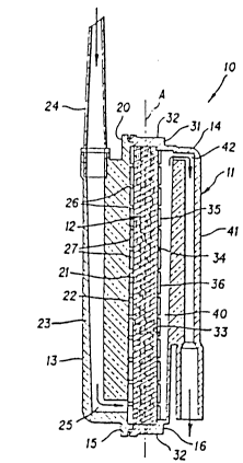
Figure 1 is a cross sectional view of an exemplary
depletion device employing the filter element provided
by the present invention.
Figure 2 is an elevation view of the inside
surface of the inlet section of the depletion device
shown in Figure 1.
Figure 3 is an elevation view of the inside
surface of the outlet section of the depletion device
shown in Figure 1.
Figure 4 is a cross sectional view of the outlet
section shown in Figure 3.
Material for Use in Construction
of LeucocYte Removal Devices
A variety of starting materials other than fibers
can be considered; for example, porous media could be
cast from resin solution to make porous membranes, or
sintered metal powder or fiber media could be used.
However, considerations of cost, convenience,
flexibility, and ease of fabrication and control, point
to fibers as a preferred starting material.
In order to achieve good priming with the fibrous
- 23 -
2016297
medium fully wetted and in the absence of surfactant
deliberately added to reduce the surface tension of the
blood product, it would appear at first glance from
elementary consideration of the physical chemistry
involved that blood component devices should be made of
materials which have CWST values in the range of 70 to
7S dynes/cm or higher. Practical considerations
dictate the use of commercially available fibers.
Synthetic resins from which fibers are prepared
commercially include polyvinylidene fluoride,
polyethylene, polypropylene, cellulose acetate, Nylon 6
and 66, polyester, polyacrylonitrile, and polyaramid.
An important characteristic of resins is their critical
surface tension (Zisman, "Contact angles, wettability
and adhesion", Adv. Chem. Ser. 43, 1-51, 1964). These
resins have critical surface tensions (~c) ranging from
25 to 45 dynes/cm. Experience has shown that the CWST
of filters in the pore sizes preferred in the products
of this invention can be expected to be less than about
10 dynes/cm higher than ~c For example, for
polytetrafluoroethylene, ~c is 18 and CWST is 27.5,
while for a polyester PBT fibrous mat, ~ is 45, and
CWST is 52. No commercially available synthetic fiber
has been found which has a CWST higher than about 52
dynes/cm.
Some natural fibers have CWST greater than 52, but
natural fibers smaller than about 15 ~m in diameter are
not generally commercially available. Synthetic fiber
webs in which the fibers are less than about 5 ~m in
diameter can be made by the melt blowing process, and
compared with natural fibers, such fibers require one
third or less the mass to provide equal fiber surface
area for adsorption of leucocytes, and consequently,
occupy less volume when fabricated into filters of a
- 24 -
201~297
given pore diameter. For this reason, natural fibers
are less suited for manufacturing leucocyte removal
devices with optimally low hold-up volume. For
example, a commercially available packed cotton fiber
device currently used for leucocyte depletion has a
priming volume of over 75 ml, which is more than twice
the volume of the preferred device described in this
application. Furthermore, the makers of this device
require saline to be passed before and after the PRC
has been passed, and the device is not suitable for
bedside use. Additionally, blood so processed must be
used within 24 hours.
The art of surface grafting has been the subject
of extensive research for 25 years or more. Numerous
publications in the scientific literature and a large
number of patents describe a variety of methods and
procedures for accomplishing surface modification by
this means. One such method employs monomers
comprising an acrylic moiety together with a second
group which can be selected to vary from hydrophilic
(e.g., -COOH or -OH) to hydrophobic (e.g., saturated
chains such as -CH2CH2CH3), and these have been used in
the process of this invention. Heat, UV, and other
reaction energizing methods can be used to initiate and
complete the reaction. However, cobalt source
radiation grafting has been selected as most convenient
and has been used in this invention to modify the CWST
of fibrous mats. By cut and try selection, mixtures of
monomers or single monomers can be found which will
produce a fibrous mat of polybutylene terephthalate in
which the CWST has been increased from 52 to any
desired value up to as high as is possible to be
measured by the method described above. The upper
limi-t is set by the paucity of liquids with surface
- 25 -
2016297
tensions at room temperature higher than about 110
dynes/cm.
During the development of this invention, devices
were prepared using media in which grafting was
accomplished by compounds containing an ethylenically
unsaturated group such as an acrylic moiety combined
with a hydroxyl group (for example, 2-hydroxyethyl
methacrylate, or "HEMA"). A second acrylic monomer,
such as methyl acrylate (MA) or methyl methacrylate
(MMA), which tend to cause the grafted porous webs to
have lower CWST, can be used in combination with HEMA,
and by varying the proportions, any CWST between 35 to
45 to over 110 dynes per cm can be obtained.
Liquids with surface tensions lower than the CWST
of the porous medium will wet the medium and, if the
medium has through pores, will flow through it readily.
Liquids with surface tensions higher than the CWST will
not flow at all at low differential pressures, but will
do so if the pressure is raised sufficiently. If the
surface tension of the liquid is only slightly above
the CWST, the required pressure will be small. If the
surface tension differential is high, the pressure
required to induce flow will be higher.
It has been discovered that, when a liquid is
forced under pressure to pass through a fibrous mat
which has a CWST more than 15 to 20 dynes/cm lower than
the liquid's surface tension, flow tends to occur in a
non-uniform fashion, such that some areas of the mat
remain dry. This is highly undesirable in a leucocyte
depletion device, first because the pressure drop is
higher causing earlier clogging, second because all the
flow passes through only a portion of the available
area, again increasing the probability of clogging, and
third because only a portion of the fiber surface area
- 26 -
~0162g7
available for adsorption of or retention by filtration
of leucocytes is used for that purpose and, as a
result, leucocyte removal is less efficient.
Solutions to the Problems of
Poor Wetting of Synthetic Fibers
Fiber surface characteristics of most or all of
the synthetic resins listed above, as well as of other
materials, can be modified by a number of methods, for
example, by chemical reaction including wet or dry
oxidation, by coating the surface by depositing a
polymer thereon, and by grafting reactions which are
activated by exposure to an energy source such as heat,
a Van der Graff generator, ultraviolet light, or to
various other forms of radiation, among which gamma-
radiation is particularly useful.
As examples of these various methods, stainless
steel fibers can be made water wettable, i.e., provided
with a ~c greater than about 72 dynes/cm by oxidation in
air at about 370C to produce a thin oxide surface
skin. Synthetic organic and glass fibers may be coated
by polymers which contain at one end a reactive (e.g.,
epoxide) moiety and at the other a hydrophilic group.
While the above methods and others known to those
familiar with surface modification techniques can be
used, radiation grafting, when carried out under
appropriate conditions, has the advantage that
considerable flexibility is available in the kinds of
surfaces that can be modified, in the wide range of
reactants available for modification, and in the
systems available for activating the required reaction.
In the subject invention gamma-radiation grafting has
been focused on because of the ability to prepare
- 27 -
2016297
synthetic organic fibrous media with CWST over the full
range of from below 50 to above 80 dynes/cm. The
products are very stable, have zero or near zero
aqueous extractables levels and, in addition, improved
adhesion between fibers is obtained when used in
preformed prefiltration or in adsorption/filtration
elements.
Selection of Fiber Diameter for
Use in LeucocYte DePletion Devices
As noted in the section headed "Characteristics
Desirable in a Leucocyte Depletion Device", adsorption
of leucocytes on fiber surfaces is widely accepted as
the mechanism of leucocyte removal. Since the surface
area of a given weight of fibers is inversely
proportional to the diameter of the fibers, and removal
of leucocytes by adsorption to the fiber surfaces is a
significant mechanism for leucocyte depletion, it is to
be expected that finer fibers will have higher capacity
and that the quantity, as measured by weight of fibers
necessary to achieve a desired efficiency, will be less
if the fibers used are smaller in diameter.
For this reason and because it is well known that
finer fibers quite generally contribute to higher
efficiency and longer life of filters, the trend has
been to use finer fibers for leucocyte depletion.
Historically, as the technology required to produce
smaller diameter fibers has advanced, they have soon
thereafter been packed into housings and/or proposed to
be used for leucocyte depletion.
- 28 -
2~162~7
Selection of Fiber for LeucocYte DePletion Devices
A number of commonly used fibers, including
polyesters, polyamides, and acrylics, lend themselves
to radiation grafting because they have adequate
resistance to degradation by gamma-radiation at the
levels required for grafting, and they contain groups
with which available monomers can react. Others, such
as polypropylene, are less readily adapted to
modification by grafting.
As noted above, fiber diameters should be as small
as possible. Synthetic fibers made by conventional
spinneret extrusion and drawing are not currently
available smaller than about 6 ~m in diameter.
Melt blowing, in which molten resin is attenuated
into fibers by a high velocity stream of gas and
collected as a non-woven web, came into production in
the 1960's and 1970's and has been gradually extended
over the years with respect to the lower limit of fiber
diameter with which webs could be made. Within recent
years, webs with fiber diameters less than 3 ~m have
been achieved, and more recently, webs of good quality
with average fiber diameter less than 2 ~m have been
made.
Some resins are better adapted to melt blowing of
fine fibers than are others. Resins which work well
include polypropylene, polymethylpentene, polyamides
such as Nylon 6 and Nylon 66, polyester PET
(polyethylene terephthalate), and polyester PBT
(polybutylene terephthalate). Others may exist that
have not yet been tested. of the above listed resins,
polyester PBT is a preferred material because it lends
itself to radiation grafting and to subsequent
conversion into preformed elements of controlled pore
size by hot pressing.
- 29 -
201t~297
Polyester PBT has been the principal resin used
for the development of the products of this invention
and is, except for a portion of a gel prefilter, the
resin used in the examples. It should be noted,
however, that other resins may be found which can be
fiberized and collected as mats or webs with fibers as
small as about 1.5 ~m in diameter or less, and that
such products, with their CWST adjusted if necessary to
the optimum range, may be well suited to the
fabrication of equally efficient but still smaller
leucocyte depletion devices. Similarly, glass and
other fibers, appropriately treated, may make suitable
devices with very low hold-up of blood.
Description of an Exemplary DePletion Device
As shown in Figures 1-4, an exemplary depletion
device 10 generally comprises a housing 11 and a
separation element or filter-adsorber assembly 12. The
housing 11 has an inlet 13 and an outlet 14 and defines
a fluid flowpath between the inlet 13 and the outlet
14. The filter-adsorber assembly 12 is disposed within
the housing 11 across the fluid flowpath and serves to
separate undesirable substances, such as gels, fat
globules, aggregates, and leucocytes, from a fluid,
such as a suspension of packed red cells, flowing
through the housing 11.
Housings can be designed to accept a variety of
shapes of filter-adsorber assemblies. One such is, for
example, a square. Those and other possible forms
would in principle all be functional, provided that
adequate flow area is provided.
A square filter-adsorber assembly would in theory
allow more economical use of material, but would be
less reliable if an interference fit seal were used in
- 30 -
2016~97
the manner described below for housings fitted with
disc shaped filter-adsorber assemblies. If sealing is
obtained by edge compression about the periphery,
significant effective area is lost at the seal. For
those reasons, cylindrical housings with disc shaped
filter-adsorber assemblies assembled with an
interference fit seal are preferred, although other
forms may be used. Circular housings with an effective
cross sectional area of about 62 cm2 have been used in
developing this invention.
Housings can be fabricated from any suitably
impervious material, including an impervious
thermoplastic material. For example, the housing may
preferably be fabricated from a transparent polymer,
such as an acrylic or polycarbonate resin, by injection
molding. Not only is such a housing easily and
economically fabricated, but it also allows observation
of the passage of the fluid through the housing. The
housings are designed to withstand normal abuse during
service, as well as internal pressures up to about 3
psi (0.2 Kg/cm2). This permits light construction,
which is a desirable feature of this invention made
possible by the use of preformed filter-adsorber
assemblies. The force required to compress the fibers
of an efficiently designed filter-adsorber assembly by
packing of fibers into a housing is as high as about 68
kilograms for a 62 cm2 disc, or about 1.1 kg/cm2,
requiring heavier, bulkier, and more costly housing
construction.
While the housing may be fashioned in a variety of
configurations, the housing 11 of the exemplary
separation device lO is preferably fashioned in two
sections, i.e., an inlet section 15 and an outlet
section 16. The inlet section 15 includes a circular
- 31 -
2016297
inlet plate 20, and the inside surface of the circular
inlet plate 20 defines a wall 21 which faces the
upstream surface of the filter-adsorber assembly 12.
The inlet 13 delivers the fluid to an inlet plenum
22 between the wall 21 and the upstream surface of the
filter-adsorber assembly 12. As provided by one aspect
of the invention, the inlet 13 delivers the fluid to
the inlet plenum 22 at or near the bottom of the
housing 11, as shown in Figures 1 and 2.
The inlet may be variously configured. However,
the inlet 13 of the exemplary separation device 10
includes a longitudinal inlet ridge 23. The inlet
ridge 23 extends along the outside surface of the
circular inlet plate 20 parallel to a diametrical axis
A of the housing 11, which, in use, is positioned with
the diametrical axis A oriented generally vertically.
The upper end of the inlet ridge 23 may be fashioned as
a socket for receiving a hollow spike 24 which is used
to pierce the bottom of a bag containing the fluid,
e.g., a blood bag. The inlet 13 further includes an
inlet passageway 25 which opens at the upper end of the
hollow spike 24, extends through the hollow spike 24
and the inlet ridge 23, and communicates with the inlet
plenum 22 at the bottom of the inlet section 15.
The wall 21 of the circular inlet plate 20
includes a plurality of generally concentric circular
ridges 26 which define concentric circular grooves 27.
The ridges 26 abut the upstream surface of the filter-
adsorber assembly 12. As shown in Figure 2, the ridges
26 terminate in the lower portion of the inlet section
15, defining a passageway or access 30. The access 30
extends between the inlet passageway 25 and each
circular groove 27, allowing fluid to flow from the
inlet passageway 25 to the circular grooves 27.
- 32 -
2016297
Collectively, the circular grooves 27 and the access 30
define the inlet plenum 22, which distributes the fluid
delivered by the inlet passageway 25 over the whole
upstream surface of the filter-adsorber assembly 12.
To prevent aggregates and other large obstructions from
blocking flow at or near the junction of the inlet
passageway 25 and the inlet plenum 22 and, at the same
time, to minimize hold-up volume in the housing 11, the
depth of the inlet plenum 22 is greatest at the bottom
of the housing 11 and decreases along the vertical axis
A to a minimum value at the horizontal centerline of
the housing 11.
The outlet section 16 of the housing 11 includes a
circular outlet plate 31 and a cylindrical collar 32
which extends from the periphery of the circular outlet
plate 31 to the periphery of the circular inlet plate
20. The cylindrical collar 32 is preferably integrally
formed with the circular outlet plate 31 and joined to
the circular inlet plate 20 in any suitable manner,
e.g., by an adhesive or by sonic welding.
The inside surface of the circular outlet plate 31
defines a wall 33 which faces the downstream surface of
the filter-adsorber assembly 12. The wall 33 includes
a plurality of generally concentric circular ridges 34
which define concentric circular grooves 35. The
ridges 34 abut the downstream surface of the filter-
adsorber assembly 12. The circular grooves 35
collectively define an outlet plenum 36 which collects
the fluid passing through the filter-adsorber assembly
12. The depth of the outlet plenum 36 is made as small
as possible to minimize hold-up volume within the
housing 11 without unduly restricting fluid flow.
As provided by another aspect of the invention,
the wall 33 further includes a passageway such as a
201S2~7
slot 40 which communicates with the outlet 14 at or
near the top of the outlet section 16. The slot 40,
which collects fluid from each of the circular grooves
35 and channels the fluid to the outlet 14, preferably
extends from the bottom to the top of the outlet
section 16 along the vertical axis A. In the exemplary
separation device 10, the width of the slot 40 remains
constant but the depth of the slot 40, which is greater
than the depth of the outlet plenum 36, increases from
the bottom to the top of the outlet section 16 along
the vertical axis A. Alternatively, the height may be
less than the diameter of the housing, the width may
vary, or the depth may remain constant. For example,
the slot may extend from the top of the housing along
the vertical axis A a distance in the range from about
80% of the inside diameter of the housing.
The outlet 14 may be variously configured.
However, the outlet 14 of the exemplary depletion
device 10 includes a longitudinal outlet ridge 41 which
extends along the outside surface of the outlet plate
31 parallel to the vertical axis A. The lower end of
the outlet ridge 41 may be fashioned as a tubing
connector or as a socket for receiving a tubing
connector or other apparatus. The outlet 14 further
includes an outlet passageway 42 which communicates
with the slot 40 at or near the top of the housing 11,
extends through the outlet ridge 41, and opens at the
lower end of the outlet ridge 41.
As blood starts to flow through the apparatus,
filling it from the bottom and emptying at the top, air
is displaced and flows towards and out of outlet
passageway 42. By careful design of the exemplary
apparatus, it has been possible to reduce, but not to
eliminate completely, the situation in which some
- 34 -
-- 2016297
liquid reaches the area adjacent to the outlet
passageway 42 before all of the air is cleared from the
inner parts of the housing assembly. In the absence of
slot 40, this lagging air flow would carry some red
cell-containing suspension into the outlet passageway
42. Slot 40 allows the blood so carried to flow into
the slot, where the air is harmlessly separated from
the liquid suspension. The air then rises harmlessly
to the outlet 14 ahead of the rising fluid level in the
slot 40 and is almost completely ejected before the
liquid level reaches the top of the outlet plenum 36
and outlet passageway 42. Thus, air is very
efficiently cleared from the housing 11 of the
exemplary depletion device 10 according to the
invention. For example, in a depletion device which
has an inside diameter of about 8.9 cm, an initial air
volume of 36 cc, and a slot about 8 cm high, about 0.73
cm wide, about 0.2 cm deep at the bottom, and 0.33 cm
deep at the top, the residual volume of air passing
through the outlet after 1 or 2 cc of blood has passed
through the outlet is estimated to be less than about
0.1 cc.
In order to understand the importance of the slot
and the flow passage configuration, the equivalent
operation of a conventional leucocyte depletion unit
will be described.
In conventional units, fluid enters at the top of
the housing and exits at the bottom. The housing of
such a unit is typically connected by plastic tubing
between a blood bag upstream from the conventional
housing and a transparent drip chamber downstream from
the conventional housing and thence to the patient.
During priming, the housing along with the drip chamber
is i-nverted and blood is forced through the
- 35 -
20162g7
conventional housing into the drip chamber. This has
the disadvantage that some pressure head is lost, but,
more seriously, fluid reaches the exit of the
conventional housing and enters the drip chamber while
as much as 1 to 2 cc or more of air is still trapped in
the conventional housing. Blood bank practice requires
that the volume of air delivered to the collection bag
be kept to the lowest possible value, even 1 or 2 cc
being undesirable.
The filter-adsorber assembly 12 preferably
comprises a number of individually preformed layers as
described below under the heading Fabrication of
Fibrous Elements. During the development stage,
housings were constructed for testing which
incorporated the basic internal configuration described
above, but in addition were variable with respect to
the thickness of the filter-adsorber assembly. In this
way, it was possible to test filter-adsorber assemblies
varying in total thickness. In each case, the distance
between the tips of the ridges 26, 34 of the inlet and
outlet sections was adjusted to be equal to the nominal
total thickness of the filter-adsorber assembly.
To provide an interference fit of the filter-
adsorber assembly 12 within the housing 11, the filter-
adsorber elements were cut from large precompressed
slabs to a diameter 0.1 to 0.5% larger than the inside
diameter of the cylindrical collar 32. The filter-
adsorber elements were cut in such a manner as to
maintain true right cylindrical form at their outer
edges. This, coupled with the slight oversizing,
provides good edge sealing, i.e., an interference fit,
between the outer edges of the filter-adsorber assembly
12, made up of the various filter-adsorber elements,
and the inner periphery of the housing 11.
- 36 -
2016297
-
Fabrication of a Gel Prefilter Element
A first element of those assembled into the above
described housing is referred to as a gel prefilter. A
proportion of PRC specimens contain gels, fat globules,
or microaggregates which can clog filter media. The
gels form a phase distinct from, and not miscible with,
the blood plasma in which they are suspended. The
state-of-the-art procedure for coping with clogging of
filters is enlargement of the pore openings of the
upstream layer or layers, continuously or in relatively
small steps, but this procedure is inefficient when
applied to the device of this invention, as a
significant number of graduated pore size layers are
required, and these tend to occupy a relatively large
volume, and for this reason would cause an excessive
volume of blood to be held up within the device.
Whereas the normal means calls for uniformly graduated
pore size, continuously or in relatively small steps,
the pore diameter of the preferred products of this
invention change abruptly, by a factor of about ten, in
the transition from the gel prefilter (first element)
to the immediately adjacent microaggregate filter
(second element), thereby accomplishing a substantial
reduction in the overall volume of the filter element.
Needle punched webs are made using staple fibers,
which for synthetic fibers are usually derived from
continuous filament by cutting or tearing the filament
into lengths of usually 3 to 6 cm. These straight
lengths are laid onto a moving belt after suspending
them in air, and the fibers are interlaced by
reciprocating multi-barbed needles.
The fibers assume the form of irregular loops,
- 37 -
~016297
-
circles, and spirals, interspersed with a variety of
other irregular shapes. Straight sections are few, and
fewer sections still are straight for more than a
fraction of a millimeter. A notable characteristic is
that at least about 90% of the fibers depart for at
least one portion of their length from the planar
structure which characterizes other non-wovens, i.e.,
significant portions of the fibers of needle punched
media are not parallel to the plane of the web. Gels
appear to penetrate easily into this type of web, but
to be effectively retained within the web, as may be
seen by post-test microscopic examination.
The structure of needle punched webs is in strong
contrast with respect to fiber orientation when
compared with non-wovens such as melt blown web, in
which the fibers are essentially parallel to the plane
of the web.
Needle punched webs are generally thicker as made
than is desired for gel removal, and for optimal use
are hot compressed to a controlled smaller thickness.
Fabric so made was discovered to be particularly
effective in retaining gels. Further, such fabrics can
be nearly filled with collected gel, yet allow free
flow of blood to the downstream component of the
system.
While the gel prefilter does not recover
microaggregates directly by filtration, the gels it
retains may contain microaggregates, and these are
efficiently retained along with the gels.
The "type A" gel prefilters used in the examples
of this invention comprise a needle punched web made
using polyester PET fibers of average diameter about 23
~m, bonded by polyethylene isoterephthalate. Nominal
weight is about .008 g/cm2. The fiber lubricant is
- 38 -
~16297
removed using a hot solution of trisodium phosphate and
detergent, and the web then thoroughly washed and dried
prior to use.
A needle punched web identical with the web
described above was used as one of the components of
the gel prefilter of U.S. Patent No. 4,925,572 (U.S.
Patent Application No. 07/259,773, filed October
19,1988). For use with PRC derived from freshly drawn
blood which has relatively fewer gels and
microaggregates, the same prefilter has been used, but
compressed to a smaller thickness, as described below.
Other gel prefilters that can be used in the
devices of this invention include melt blown fibrous
webs. Fibers in the gel prefilter typically have fiber
diameters of from 10 to 30 ~m, preferably about 20 ~m.
Fabrication of Preferred Microaqqreqate Filter
In U. S. Patent No. 4,925,572 (U.S. Patent
Application No. 07/259,773, filed october 19, 1988),
three layers of prefiltration are described. For use
with fresh PRC, fewer prefiltration layers can be used,
or indeed none at all need be used, with little or no
risk of clogging. Among the Examples provided by this
invention, we have used as the microaggregate filter a
6.5 mg/cm2 layer of web of fiber diameter 3.2 ~m
followed by a 6.9 mg/cm2 layer of web of fiber diameter
2.9 ~m in diameter. These are compressed to a voids
volume of 74% to 84%. The fibers of both these layers
are surface grafted to provide a CWST in the range of
60 to 70 dynes/cm.
- 39 -
2016297
Fabrication of an Adsorption/Filtration Element
Leukocytes are removed to only a small degree by
the gel prefilter and microaggregate filter. The
principal contributor to leukocyte removal is the
adsorption/filtration element, which comprises
preferably one or more hot compressed preforms of
multiple identical layers of relatively small fiber
diameter melt blown web.
Preforming and Assembly of the Elements
In a preferred filter of the invention, flow
through the above elements is in the order in which
they are listed, that is, gel prefilter, microaggregate
filter, and then the adsorption/filtration element.
The gel prefilter preferably comprises about two to
four layers, the microaggregate prefilter comprises
preferably one to four layers, ànd the
adsorption/filtration element generally comprises one
or more preforms, each comprising a number of layers.
In a more preferred embodiment, the
adsorption/filtration element comprises two sets of
multilayers, each comprising a different voids volume.
Multilayers may be preferred for the
adsorption/filtration element because the melt blowing
process is such that making a single layer of the
weight, thickness, fiber diameter, and uniformity
required is difficult.
These multiple layers can be used as individual
preformed layers assembled in the order noted, however,
it is sometimes more convenient to fabricate them as
subassemblies. In one preferred configuration of the
gel prefilter, two layers of needle punched medium and
- 40 -
2016297
one of melt blown medium are hot compressed together
into a single preform, while in another two or more
precompressed layers of melt blown web are used as
separate layers.
The values cited above and in the examples can be
varied within limits while meeting the objective of
this invention. To determine whether any particular
variation produces a fully equivalent product, tests
are required. Thus, it should be understood that,
while the precise materials, fiber diameters, weights,
densities, thicknesses, and number of layers can be
varied somewhat while achieving equivalent or possibly
even better results, that which is disclosed herein is
intended as a guide to the design of a device meeting
the stated objectives of this invention and that
devices made with such variations fall within the scope
of this invention.
With the exception of the gel prefilter, all of
the elements are preferably surface treated to a CWST
in excess of about 55 dynes/cm, but not in excess of 75
to 80 dynes/cm, and more preferably from 60 to 70
dynes/cm.
Hot compressed element preforms made using melt
blown fibrous mats which have been surface modified to
raise their CWST values by 5 or more dynes/cm are
palpably better with respect to firmness and resistance
to fraying when compared with discs made by hot
compression followed by radiation grafting. Grafting
prior to hot compression is for this reason preferred;
however, serviceable elements could be made by hot
compression followed by grafting.
While the examples provided by this invention have
used hot compression to form the integral elements
which together combine to provide prefiltration, gel
2016297
removal, and adsorption, it would be feasible to form
the integral elements by other means, such as resin
bonding, and a device utilizing this or similar
alternatives is within the scope of this invention.
Melt blown fibers have been preferred for use in
all but the first layer of these devices. Should finer
melt blown or other fine fibers, for example, fibers
made by mechanical fibrillation of larger diameter
fibers or by other means, become available in the
future, their use in elements for leucocyte depletion
devices would be within the scope of this invention.
Edge Sealing the Preformed Elements into the Housinq
The housing is preferred to be of generally disc
form, or more rigorously stated, in part have the form
of a right cylinder. The preformed elements are made
also in right cylindrical form, of dimension 0.1 to
0.5% larger in diameter than that of the inner surface
of the housing. When assembled, a good seal is
obtained, with no detectable bypassing during service.
In order to achieve good sealing, circular
elements must have a truly right cylindrical form.
That form is not achieved by all the means by which the
elements can be cut to circular form; for example, an
obvious means, stamping out a circle using a steel rule
die, does not provide an acceptable outer seal.
Instead, the disc must be cut to its finished diameter
using means which achieve the geometry of a true right
cylinder at the cut edges. This has been achieved in
the practice of this invention by construction of a
circular knife of the required diameter, which is
rotated to cut a true right cylinder while holding
gently but firmly in place the inner and outer surfaces
- 42 -
- - 2016297
of the precompressed slab from which the disc is cut.
Circular housings may be adapted to a procedure in
which a bag containing the collected PRC is attached to
the filter aseptically, followed by application of
pressure to the bag containing the PRC to force the PRC
through the filter into a second collection bag.
In an alternate procedure, the filter and a second
PRC collection bag are provided as part of the blood
collection set prior to attaching the set to the blood
donor. When this procedure is used, the blood is drawn
into the first collection bag, that bag along with the
filter and the auxiliary bags are placed into the
centrifuge bucket, following which the assembly is spun
to make the PRC. For use in this procedure it is
desirable for reasons of economy to use as small as
possible a bucket. In order to make this possible, it
may be preferred that the filter have a form other than
circular, for example, rectangular. Rectangular
filters can be sealed by an interference fit at their
outer edge into a rectangular housing, however, they
may in addition or alternately be preferred to have a
peripheral compression seal.
CWST of the Elements
The gel prefilter (first) element may have a low
CWST without harm, and indeed may function better in
that condition. The results of tests in which
sufficient PRC is run through a device to cause
clogging or near clogging, followed by dissection,
inspection, and testing of the pressure drops of the
individual layers, indicate that little if any
improvement can be accomplished by increasing the CWST
of this layer. The microaggregate filter element and
2016297
the adsorption/filtration element are preferably
modified to a CWST of between 55 to 80 dynes/cm, and
more preferably to between 60 and 70 dynes/cm, and
still more preferably to between 62 and 68 dynes/cm.
Red Cell RecoverY
No significant changes in hematocrit were detected
when the hematocrit values for the blood in the bag
were compared with the effluent from the devices
provided by this invention.
Some of the incoming blood or PRC is lost due to
hold-up within the depletion device. That loss is
reported as blood hold-up volume.
Characterization of Porous
Media by PhYsical Characteristics
Formulae have been proposed to predict pore
diameter. These formulae typically use fiber diameter,
for example as determined by BET testing; bulk
(apparent) density; and fiber density. One such, for
example, calculates the average distance between
fibers. However, the average distance between fibers
can not be a meaningful predictor of performance as in
any liquid flow path it is the largest pore or pores
encountered which control performances, and this is
particularly true of deformable "particles" such as
leucocytes. In a fibrous mat such as made by melt
blowing, the fibers are laid down in a random manner,
and the pore size distribution is quite wide. Other
means for forming fibrous mats, e.g., air laying, or
formation on a Fourdrinier screen, also produce wide
pore~size distributions. In these circumstances, the
20~6297
average distance between fibers is clearly a poor
predictor of performance. A variety of other formulae
have been proposed to allow calculation of pore
diameters from data on fiber diameter, fiber density
and bulk density, but applicants are unaware of any
formula that has proved useful for calculating a Priori
the effective pore diameter of filters for liquid
service.
Measurement of fiber surface area, for example by
gas adsorption - popularly referred to as "BET"
measurement - is a useful technique, as the surface
area is a direct indication of the extent of fiber
surface available to remove leucocytes by adsorption.
In addition, the surface area of melt blown PBT webs
can be used to calculate fiber diameter. Using PBT, of
density 1.38 g/cc as an example:
Total volume of fiber in gram = 1 cc, and this
1.38
volume is equal to the fiber cross sectional area
multiplied by its length, hence
rd2L = 1 ( 1 )
4 1.38
Surface area of the fiber is
~dL=Af (2)
Dividing (1) by (2) d
4 1.38
2016297
and d = 4 = 2.9 or (o.
1.38~ ~
where L = total length of fiber per gram,
d = average fiber diameter in centimeters,
and ~ = fiber surface area in cm2/g.
If the units of d are ~m, the units of ~ become
square meters/gram (M2/g), which will be used
hereinafter. For fibers other than PBT, their density
is substituted for that of PBT.
A second characteristic necessary to describe a
porous medium adequately to permit it to be reproduced
is its pore diameter (Dp). We have used a modified
OSU-F2 test for this purpose; this test and its mode of
use are described in the following section.
Other characteristics which describe a porous
medium include apparent (bulk) density (~) in
grams/cubic centimeter (g/cc), the fiber density (also
in g/cc), the thickness (t) of the elements of the
medium, specified in centimeters (cm), the cross
sectional area available for flow through the filtering
element in square centimeters (62 cm2 for all of the
examples), and the CWST in dynes/cm. Specifying these
parameters defines a filter or filter-adsorber element
of predictable behavior when used for leucocyte
depletion.
PROCESSING BLOOD USING THE PRODUCTS OF THIS INVENTION
In current blood banking practice, where platelet
concentrate (PC) is desired to be recovered, the
sequence of operation is:
- 46 -
201~2~7
a. Perform venipuncture and draw about 400 to
about 450 ml of blood into a sterile
collector bag in which an anti-coagulant has
been preplaced. This collection bag is
attached by tubing to two other bags via a
tee connection, these bags denoted
respectively as the platelet bag and the
plasma bag.
b. The collector bag is then placed in a
centrifuge and spun for about 3 minutes at
conditions which develops about 2000 G
(i.e., 2000 times the force due to gravity)
during which the red cells are concentrated,
forming the PRC at the bottom of the bag.
c. The collector bag is then placed in a device
which is denoted as an "expresser", but in
one commonly used version is denoted as a
"plasma extractor". The expresser squeezes
the bag, developing 7.03 x 10² to 1.05 x 10³
kg/m2 (1 to 1.5 psi) of internal pressure.
The operator opens a valve at the top of the
bag, allowing the supernatant fluid
containing the plasma and most of the
platelets to be decanted into the platelet
bag, and leaving in the collection bag 170
to 250 ml of PRC. It is this PRC which is,
in a separate step, processed by the filter
of this invention in order to reduce its
leucocyte content.
d. The remaining steps in the blood processing
as generally practiced are preparation of
plasma, or of platelet concentrate and
plasma. These are not described, as they
are not pertinent to this invention.
- 47 -
2016297
In alternate types of procedure, for example those
designated as Adsol or SAG-M, the procedure of steps 1,
2 and 3 is similar except that a harder (higher G) spin
may be used, and after decanting the supernatant liquid
in an expresser, the red cells are resuspended in a
physiological solution containing saline and an anti-
coagulant, forming the PRC.
In the practice of this invention, the collected
PRC prepared by these or similar processes is as the
next step passed through the filter of this invention.
This may be done in a separate step in which a filter
and second collecting bag are aseptically attached to
the PRC bag, and the PRC is forced, for example by a
pressure cuff developing a pressure of about 0.4 Kg/cm2,
to pass through the filter into the second collection
bag. Alternatively, the filter and the second
collection bag may be attached to the lower end of the
whole blood collection bag prior to collecting the
blood from the donor; then, after the supernatant fluid
in the blood collection bag has been removed by
centrifuging and decanting, and while the bag is still
in the expresser, the PRC is transferred through the
filter into the second collection bag using the
pressure provided by the expresser.
Examples
All tests run used blood drawn from human
volunteers and processed using either Adsol or CPDA-l
anticoagulant within 6 hours in accordance with the
standards of the American Association of Blood Banks to
provide one unit of PRC. Hematocrits of the PRC were
recorded and were with few exceptions in the range of
- 48 -
~016297
70 to 80%, while hematocrits of Adsol processed blood
were generally in he range of 55 to 65~. Leucocyte
counts of the PRC prior to processing were in the range
of 3500 to 17,000 per microliter (~L).
Priming time is defined as the time required to
fill the test housing with fluid.
Bag (i.e., influent) leucocyte counts were
determined using a Model ZM Coulter Counter. Leucocyte
counts are reported as number per microliter (~L). The
conventional centrifugal method was used to determine
hematocrits.
Use of automatic counters for the leucocyte
depleted filter effluents provides incorrect results
because automatic counters are designed to be operated
in the range of normal leucocyte content of whole blood
and of normal PRC. Thus, the normal operating range of
automatic counters is 103 to 107 times the levels
reached in the examples herein; as a consequence,
automatic counter data at these low levels is quite
useless.
A method to assay the degree of depletion of
leucocytes to the very low levels of this invention,
i.e., reduction of leucocyte count by a factor between
105 to 107 (99.999 to 99.99999~ efficiency) has become
available only recently. The method was developed, in
a cooperative project with Pall Corporation (Pall), by
the laboratory of the American Red Cross (ARC). Pall
supplied the necessary high efficiency filters, while
the ARC developed the assay method. This assay method,
subsequently routinely practiced in the Pall blood
laboratory, is described below.
Zeta potential was determined using a
conventional streaming potential apparatus.
^ The elements used in the examples were right
- 49 -
2016297
circular discs, about 88.9 mm in diameter at assembly.
The stacked layers of elements, with a total thickness
of t cm were assembled into a housing as described
above, with a clearance of about t cm between the faces
of the two plenums, i.e., between the tips of the
ridges 26 on the inlet plate 20 and the tips of the
ridges 34 on the outlet plate 31, as shown in Figure 1.
After piercing the blood bag, leucocyte content was
determined in the manner described in the preceding
part of this section.
Losses of red cells due to adsorption were, unless
noted, too small to be detected. For the examples of
this invention, losses due to hold-up within the filter
housing were about 30 cm3 of PRC.
Pore diameters of filter media were determined
using the modified OSU F2 method and are reported as
the diameter of hard particle at which about 99.9% of
the incident particles were removed. The F2 test used
in making pore size measurements is a modified version
of the F2 test developed in the 1970's at Oklahoma
State University (OSU). In the OSU test, a suspension
of an artificial contaminant in an appropriate test
fluid is passed through the test filter while
continuously sampling the fluid upstream and downstream
of the filter under test. The samples are analyzed by
automatic particle counters for their contents of five
or more preselected particle diameters and the ratio of
the upstream to downstream count is automatically
recorded. This ratio is known in the filter industry
as the beta ratio.
The beta ratio for each of the five or more
diameters tested is plotted as the ordinate against
particle diameter as the abscissa, usually on a graph
in which the ordinate is a logarithmic scale and the
- 50 -
~0162~7
abscissa is a log2 scale. A smooth curve is then drawn
between the points. The beta ratio for any diameter
within the range tested can then be read from this
curve. Efficiency at a particular particle diameter is
calculated from the beta ratio by the formula:
Efficiency, percent = 100(1-1/beta)
As an example, if beta = 1000, efficiency = 99.9%.
The removal rating cited in the examples presented
herein is the particle diameters at which beta = 1,000,
hence, the efficiency at the removal ratings cited is
99.9%.
In the modified F2 test, efficiencies in particles
in the range of from 1 to 20-25 ~m were determined
using as a test contaminant an aqueous suspension of AC
fine test dust, a natural silicious dust supplied by
the AC Spark Plug Company. Prior to use, a suspension
of the dust in water was vigorously mixed for three
weeks until the dispersion was stable. Prior to use
for measuring the pore size characteristics the
suspension passed through one of the gel prefilters of
this invention, thereby removing oversize particles
which would otherwise collect on the filter surface and
cause flow to stop. Pore diameter values below 1 um
were obtained by extrapolation per the following
tabulation:
Beta Value at lumPore Diameter (um)
1000-2000
1200-1800 0.9
1800-2500 0.8
2500-4000 0.7
- 51 -
20~L6297
Test flow rate was 100 liters per minute per square
foot of filter area and each pore size measurement as
reported is the average of four tests.
The needle punched web used in the examples was
scrubbed in order to remove the fiber lubricant, and
then dried.
Preform thickness was measured using a 7.7 cm
diameter anvil and with an applied pressure of 4.3
g/cm2 .
Unless otherwise noted all of the elements used in
the examples were right circular discs of diameter 88.9
mm at assembly. Properties of sub-sections of
composite discs are listed in the order in which the
blood flow through them.
Gel prefilter type A consisted of three layers.
The two upper layers consisted of 8 + 1 mg/cm2 of 23 ~m
average fiber diameter PET needle punched web, while
the third and last layer consisted of a 7.7 mg/cm2 20 ~m
average fiber diameter PBT melt blown web. The first
of the three layers was hot calendered to about 0.89 cm
thick by passing the web between a pair of moving belts
in an oven in order to heat the web to 165 to 170C,
following which it passed through calendering rolls.
The second and third layers were hot calendered in
assembly to about 0.10 cm thick. All of the above were
then hot calendered together to a thickness of about
0.13 cm. The resulting gel prefilter consisted of 3
integrated layers of approximate density, respectively,
0.14, 0.19, and 0.32 g/cm2, the last layer having a pore
diameter of 20 to 30 ~m.
Gel prefilter type B consisted of four adjacent
layers of about 20 ~m diameter fiber melt blown web
each separately hot calendered in the manner described
above to obtain the characteristics listed below:
- 52 -
2016297
Weight Thickness Voids
mq/cm cm Volume, %
3.1 .025 91
4.1 .025 88
5.7 .025 84
7.7 .025 78
Although the thickness of the separately measured
layers of the type B prefilter add up to about 0.1 cm,
the total thickness when the four were stacked on each
other was about .08 cm. The last layer had a pore
diameter of 20 to 30 ~m.
All type B gel prefilters were made using PBT
fibers with no surface modification.
The development procedure went through stages at
which first a laboratory (L) method was used to achieve
hot compression, and later a production (P) method was
used to achieve the same purpose. In the L method the
necessary microaggregate filter and adsorption filter
layers were assembled as a stack and the whole
compressed between TeflonTM lined aluminum alloy plates
at 165C for about 40 seconds. In the P method a
similar stack was passed between two moving TeflonTM
coated belts heated to 165 to 170C for a period of
about 30 seconds, following which they were passed
through a pair of calender rolls to achieve the desired
density or thickness.
The filter effluents were assayed to determine
leucocyte content using a "Ficoll" method,
alternatively referenced hereinafter as the "ARC"
method, in which the leucocytes are separated in
concentrate form enabling a high proportion of all of
the leucocytes present to be counted directly. This
- 53 -
20:~6297
method was developed in the laboratory of the American
Red Cross. Prior to this development no assay
procedure was available which was capable of counting
the very low levels of leucocytes, less than 102 to 105
per unit of PRC, which are obtained using the products
of this invention. The Ficoll Assay is described
below:
1.0 Purpose:
1.1 This test method is used to separate
leucocytes from filtered packed red cells
and determine the log efficiency of a
leucocyte depleting filter.
2.0 Materials and EquiPment:
- one unit of fresh PRC or of whole blood
- 5 mL disposable polypropylene test tubes
with caps
- 600 mL transfer bags
- filter
- pressure infusor
- hemastat clamps
- 500 mL graduated cylinder
- Ficoll solution (see section 4.1)
- 60 mL disposable polypropylene syringe
- plasma extractor
- Sorvall RC-3C General Purpose Refrigerated
Centrifuge
- blood bank pipets
- vacuum suction apparatus
- 1% ammonium oxalate solution (see section
4.3)
- aliquot mixer
- Neubauer hemacytometer
2Q16297
- plain hematocrit tubes
- Fluorescent microscope with phase contrast
and 20 and 40X objectives
- 250 mL disposable polypropylene centrifuge
tubes
- 15 mL disposable polypropylene centrifuge
tubes
- hematology control: normal level and low
abnormal level - Counter-CheckTM, Diagnostic
Technology, Inc.
- Acridine orange fluorescent stain (see
Section 4.4)
3.0 Procedure:
3.1 Mix the PRC unit and withdraw a sample in a 5 mL
tube for use to count influent leucocytes.
3.2 Connect the pre-weighed 600 mL transfer bag to the
filter outlet, and the filter inlet to the blood
bag.
3.3 Prime the filter with PRC using a pressure infusor
set at 300 mm Hg.
3.4 When flow has begun, lower pressure to 200 mm Hg
for remainder of filtration.
3.5 When filtration is complete, turn pressure off,
clamp the collection bag and remove it from the
filter.
3.6 Determine the effluent volume by weighing the
collection bag, subtracting the empty bag weight,
and dividing by 1.08 (the density of PRC). Record
this volume.
3.7 Adjust the volume to 300 cc by discarding the
excess, or by adding saline, then remove the
syringe and seal off the bag.
3.8 With a graduated cylinder, measure 300 cc of
Ficoll solution and add it to the collection bag
- 55 -
2016297
using a 60 mL syringe attached to the transfer bag
tubing.
3.9 Vigorously mix the Ficoll-blood solution and place
the bag in a plasma extractor.
3.10 Clamp the tubing and remove the syringe.
3.11 Apply the extractor clamp and let the blood settle
for 30 minutes.
3.12 Carefully express the upper layer into a 250 mL
centrifuge tube by opening the hemastat clamp. Do
not disturb the interface while expressing the
maximum amount of Ficoll.
3.13 Release the extractor clamp and repeat step 3.8
using a sufficient volume of Ficoll to fill the
600 ml bag, then repeat 3.9, 3.10, 3.11, and 3.12.
3.14 Centrifuge the tubes at 775 g for 15 minutes at
room temperature in the SorvallTM centrifuge.
3.15 When spin is complete, use a blood bank pipet
attached to a vacuum flask to extract and discard
the supernatant leaving a red pellet.
3.16 Resuspend the pellet in 250 mL of 1% ammonium
oxalate.
3.17 Allow the suspension to mix on an aliquot mixer
for 10 minutes to lyse the red blood cells.
3.18 Centrifuge the tube at 432 g for 10 minutes at
room temperature and discard the supernatant as
before.
3.19 Resuspend the pellet in 2-3 mL of ammonium oxalate
using a blood bank pipet to draw the pellet up for
mixing. Transfer this suspension to a 15 mL
polypropylene centrifuge tube combining all
pellets from one blood unit.
3.20 Fill the tube to the 15 mL mark with the 1%
ammonium oxalate solution, mix, and allow tube to
set for 10 minutes.
- 56 -
2016297
3.21 Spin the 15 mL tube in the centrifuge at 775g for
10 minutes at room temperature.
3.22 Decant the supernatant down to the 0.5 mL line on
the 15 mL tube. Carefully resuspend the pellet
using a pipetter. Add .05 mL of Acridine Orange
stain to the suspension, and weigh and record the
tube welght to determine the final volume of the
suspenslon.
3.23 Leucocyte Counts: All counts are performed
manually.
3.23.1 Control Counts:.23.1.1 Control counts are performed daily by making
a 1:100 dilution of each control in 1%
ammonium oxalate..23.1.1.1 Add 0.01 mL of the control to 0.9g mL of 1
ammonium oxalate using an adjustable
pipetter. Mix well and let the dilution sit
for at least 10 minutes to lyse the red
blood cells, then add .05 mL of Acridine
Orange..23.1.1.2 Charge each side of a hemacytometer using a
plain capillary tube, taking care not to
overload or underload the chamber..23.1.1.3 Allow the hemacytometer to sit in a moist
atmosphere (covered petri dish with
moistened filter paper in bottom half of the
dish) for ten minutes..23.1.1.4 Count the number of leucocytes in the nine
large squares on both sides of the
hemacytometer using the phase contrast UV
microscope..23.1.1.5 Record the counts from each side (each nine
squares) of the hemacytometer..23;1.1.6 To determine the number of leucocytes/~L,
2.0 ~ 6~97
use the following formula:
leucocytes/~L = total cells counted X dilution X 10
total squares counted
(if 18 large squares are counted, the total number
of cells counted x 56 = cells/~L)
3.23.2 Influent LeucocYte Counts:.23.2.1 Influent leucocyte counts are performed
using the same procedure as for the control
counts except that a total of 36 large
squares (two hemacytometers) are counted..23.2.2 To calculate the leucocytes/~L, use the same
formula as above. If 36 large squares are
counted, the total number of cells x 28 =
cells/~L.
3.23.2.3 The number of leucocytes/~L x 1000 equals
the number of leucocytes/mL..23.2.4 Multiply the number of leucocytes/mL by the
effluent volume to determine the total
leucocytes in the prefiltration sample
(note: the effluent volume is used in this
calculation, not the influent volume,
because log reduction is a direct
volume/volume comparison and does not take
into account the hold-up volume)..23.3. Effluent Leucocyte/Counts:.23.3.1 Effluent leucocyte counts are performed
using the undiluted final ammonium oxalate
suspension from step 3.24..23.3.1.1 Charge the hemacytometer and count as
before..23.3.1.2 If the number of cells counted on both
sides of the hemacytometer is 30 or less,
continue counting hemacytometers until 30
- 58 -
2016297
cells or 5 hemacytometers are counted..23.3.2 Determine the total leucocytes in the
postfiltration sample as follows:.23.3.2.1 Divide the effluent volume by the volume of
the final suspension to determine the
concentration..23.3.2.2 Use the following formula to calculate
leucocytes/~L:eucocytes/~L =
total cells counted x
total squares counted concentration x 10
.23.3.2.3 The number of leucocytes/~L x 1000 equals
the number of leucocytes/mL..23.3.2.4 Multiply the number of leucocytes/mL by the
effluent volume to get the number of
leucocytes in the postfiltration sample..23.3.2.5 The Ficoll procedure gives a nominal 53%
yield of leucocytes, thus the number from
the step above must be divided by 0.53 to
determine the total leucocytes in the
postfiltration sample.
3.24 Determine the log reduction by dividing the total
number of leucocytes in the prefiltration sample
by the total number of leucocytes in the
postfiltration sample and taking the log of this
quotient.
4.0 SuPPlementary Information:
4.1 Ficoll Formula:
100.0 g Ficoll* 400 DL
20.0 g Bovine Serum Albumin
2000.0 mL Stock EBSS (see below)
Mix the above ingredients in a 2L volumetric flask
and warm to 37C while stirring. Filter solution
.through a 1.2 ~m filter disc and then through a
- 59 -
2016297
0.45 ~m filter disc. Store at 4C.
* Ficoll 400 DL is a dialyzed, hydrophilized,
synthetic polymer of sucrose with a molecular weight of
approximate 400,000 available from Sigma Chemical Co.
4.2 Stock EBSS Formula:
200.0 mL Earle's Balanced Salt Solution
( lOx)
1800.0 mL deionized H20
40.0 mL Hepes Buffer Solution
Mix the above ingredients and store at 4C.
Warm to room temperature prior to use.
4.3 Ammonium Oxalate Formula:
10.0 g Ammonium Oxalate
0.10 g Thimerosal
0.43 g KH2P04
0.57 g Na2HP04
Mix the above ingredients in a lL volumetric flask
and dilute to lL with deionized water. Check the
pH and adjust to 6.8 if necessary. Store at 4C.
Warm to room temperature prior to use.
4.4 Acridine Orange Stain Formula:
4.4.1 Stock Acridine Orange (lOOOX solution):
- 6.69 mg Acridine Orange/ml of DI water
- Store in the dark at 2-5C
4.4.2 Working Acridine Orange (lOX solution):
- dilute Stock Acridine Orange with Stock EBSS
(see 4.2)
- good for 1 month if stored in the dark at
2-5C
The above described assay method has a nominal
recovery efficiency of 53~ of the leucocytes actually
- 60 -
2016297
present in the filtered PRC. The consistency of the
53% figure is about +15 to -25%; however, the log
reduction calculated is affected in only a minor way by
these deviations when a single test is run, and
consistency is improved by running four or more tests
on each filter tested.
The leucocyte removal efficiencies obtained for
the products of this invention using the ARC assay
procedure can be stated in several alternate ways.
Using as an example a unit of PRC containing 103
leucocytes in the total filtrate, and a value of 109
leucocytes contained in an equal volume of the PRC
prior to filtration, then the ratio of effluent
content/influent content is 103/109 = 10~, and
efficiency of leucocyte removal can be reported as:
(a) 100(1 - 10~) = 99.9999%
or (b) leucocyte content is reduced by a factor of
one million (l/10-6 = 106)
or (c) log reduction = -6
A convenient method is to use the term log
reduction and omit the minus sign, since minus is
inferred by the term "reduction". This nomenclature is
widely used, and we will use it when reporting
efficiencies hereinafter.
Examples 1-6 were performed in the ARC laboratory
using preformed filter elements prepared in the
inventors' laboratory by Pall Corporation personnel
using materials and procedures conceived and developed
by the inventors with no input from ARC. ARC's
contribution to this part of the development was
confined to developing and initially performing the
Ficoll test on filters developed and made by Pall, and
then reporting test results to the Applicant. Later
the Ficoll test was performed in Pall's laboratory
- 61 -
2016297
using Pall personnel.
This group of examples consisted of a single
preform made using 2.6~m fiber diameter melt blown PBT
web, hot compressed using the L (laboratory) technique
described above. No prefiltration was used. The tests
were run using 47.6 mm diameter discs, assembled into
Pall made housings of slightly smaller diameter. Each
test was run using one quarter of a unit of fresh PRC
of hematocrit 50 to 55%. Flow rate of 4 to 8 cc per
minute was obtained at a pressure of 40 inches of fluid
head. The resulting data for these examples is listed
in Table 1. When plotted, 5 of the 6 points fall on or
near to a straight line represented by the equation
Log reduction = 18.5p + 0.5 OR
~ =Log reduction ~ 0 5 (1)
where is the density in grams/cc, and the weight
of the adsorption/filtration element is
(0.5~ - .029)g/cm2 (2)
This equation provides guidance for selecting the
densities of PBT filters which have a desired average
log reduction between 4.5 to 7; however, filters with
log reduction in the upper part of the range, with
voids volumes less than 74% to 78%, may become clogged
prior to passage of a single unit of PRC, consequently
gel, microaggregate filters, or multilayers may be
required to prevent occasional clogging.
In example 7, ten discs with a pore diameter of
0.9 ~m duplicating those of Example 4, except 88.9 mm
in diameter, are assembled into an 88.6 mm diameter
housing with ridge to ridge depth of 0.439 cm. Priming
is accomplished in 50 to 100 seconds at a pressure of
- 62 -
201~297
0.4 Kg/cm2, and one full unit of fresh PRC of hematocrit
of 70 to 80% derived from blood collected into CPDA-l
anticoagulant is passed through each at a pressure of
0.27 Kg/cm2. The average time to pass one unit through
five of the ten discs ranges from 15 to 25 minutes.
The average log reduction is approximately 6.4. Flow
in the other five of the ten tests will fall within a 2
hour period to less than 0.7 cc/min, and these tests
are then discontinued.
In Example 8, ten integral preformed discs
duplicating those of Example 7 are assembled together
with a type A gel prefilter into an 88.6 mm diameter
housing with ridge to ridge depth of 0.569 cm, and one
unit of fresh PRC will be passed through each. The
total volume of one unit of PRC is passed at a pressure
of 0.27 Kg/cm2 in 15 to 25 minutes. The average log
reduction is approximately 6.4.
In Example 9, ten discs duplicating those of
Example 7 are assembled together with a type B gel
prefilter into an 88.6 mm diameter housing with ridge
to ridge depth of 0.519 cm, and one unit of fresh PRC
is passed through each in the same manner as in Example
7. The total volume of PRC is passed in 15 to 25
minutes. The average log reduction is approximately
6.4.
In Examples 8 and 9, the addition of the type A
and B prefilters will make clogging less likely than
would be the case if they were not used, as in Example
7.
Example 10 is prepared in the same way as Example
9, except that the adsorption/filtration element is
integral with a microaggregate filter, formed by
including two layers of media of larger fiber diameter
in the upstream side of the lay-up, followed by hot
- 63 -
2016297
compression to obtain a preformed integral element.
Specifically, the uppermost layer is made using 3.2 ~m
diameter fibers in the form of a mat of weight 0.0065
g/cm2, and the adjacent layer is made using 2.9 ~m
diameter fiber of weight 0.0069 g/cmZ, while the
balance, the adsorption/filtration element consists of
multiple layers, all of fiber diameter 2.6 ~m. Total
weight is 0.130 g/cm2 compressed to a voids volume of
76.5~ (density = 0.324 g/cc) and thickness of 0.439 cm.
The filter so made will possess capability for
microaggregate removal, and be more resistant to
clogging, as would otherwise be caused by the
occasional specimen of donated blood which contains an
unusually high content of microaggregates. The
properties of the filter of Example 10 with respect to
efficiency of leucocyte depletion and blood transit
time are not significantly altered from those of
Example 9, as the fiber surface area of Example 10 is
reduced from that of Example 9 by only about 1%. The
pore sizes obtained by F2 testing are essentially equal
for the two examples. Blood hold-up volume within the
voids of the gel prefilter, the microaggregate
prefilter, and the adsorption/filtration element are,
respectively, 3.9 cc, 1.6 cc, and 19.1 cc for a total
of 24.6 cc.
Examples 11 to 14, summarized in Table 2,
illustrate the use of equations (3) and (4) set forth
above to calculate the manner in which filter
assemblies may be made which have lower efficiencies,
but which during filtration pass PRC at a lower
pressure drop, making it possible to use gravity to
induce flow at a satisfactory rate when processing
blood with relatively high hematocrit. All contain
integral elements combining microaggregate elements
- 64 -
` - 2016297
with adsorption/filtration elements, with the fibers
surface modified to a CWST of 60 to 70 dynes/cm, and
preferably 62 to 68 dynes/cm. The device of Example 11
also incorporates a type B gel prefilter.
Other variations of density and thickness are
possible. All of the Examples 11-14 can be made with a
higher density (i.e. lower voids volume), while
retaining equal or better leucocyte removal efficiency.
Thus, Example 15 is made in the same manner as Example
11 except that the voids volume is changed to 76.5%.
The resulting log leucocyte reduction will be
intermediate between 6 and 6.5, and the blood hold-up
volume within the element is reduced by about 10%.
Similarly, Example 16 is the filter of Example 12 with
the voids volume decreased from 80.4 to 78.5%, with log
reduction between 5.5 and 6, and blood hold-up volume
reduced by about 10%, and Example 17 is the filter of
Example 13 with voids volume decreased from 82.4 to
80.4%, with log reduction between 5 and 5.5, and with
blood hold-up volume decreased by about 10%. Example
18 is the filter of Example 14 with voids volume
decreased from 84.3 to 82.4%, with log reduction
between 4.5 and 5, and with blood hold-up volume
decreased by about 11%.
The void volume of each of Examples 16, 17, and 18
could be further decreased, obtaining in this way equal
or higher efficiency along with still lower blood hold-
up volume compared with Examples 16, 17, and 18.
Example 19 is essentially the filter of Example 10
with voids volume decreased further. A type B
prefilter was used in this construction in order to
minimize the chances of clogging. Immediately
downstream of the prefilter was a section consisting of
nine layers, all of fiber diameter 2.6 ~m, with a CWST
- 65 -
2Q16297
of 66 dynes/cm and a total weight of 0.054 g/cm2,
compressed to a voids volume of 77% (density = 0.321
g/cc) and a thickness of 0.168 cm. Another section,
downstream of this one, consisted of 20 layers of the
same type of fibers, however, compressed to a lower
voids volume. The total weight of this section was
0.118 g/cm2, voids volume was 70% (density = 0.414
g/cc), and thickness was 0.285 cm. When tested with
blood in accordance with the previous examples, this
combination gave a leukocyte reduction of 6 log, and a
total filtration time of 32 minutes.
It was previously thought that the use of voids
volumes much below about 74% would pass PRC more slowly
and thus would tend to clog with higher frequency.
However, another surprising feature of this invention
is that voids volumes of 70% were found to be quite
suitable and efficient, and voids volumes as low as 60
may be useful for some special applications. When
lower voids volumes are utilized, it is found that it
is the combination of voids volumes with other factors,
i.e., the number of multilayers in each section, rather
than a particular voids volume by itself, that produces
an element that is suitable in carrying out the instant
invention.
While the examples all deal with PRC, generally
equivalent results will be obtained when anticoagulated
whole blood is used.
An extraordinary and very surprising feature of
this invention is the ability of red blood cells to
pass through a filter of pore diameter less than 1 ~m
without apparent injury and no apparent losses. The
ability to use such small pored filters was not
anticipated, and was seen only as a very unlikely
possibility meriting exploration in the absence of
- 66 -
20162~7
other approaches to the preferred goal of 100,000 to
1,000,000 fold reduction of leucocyte content in a
filter so small that red cell loss due to hold-up is
less than 10% of the average volume of a unit of PRC.
Red cell loss can be reduced by passing saline
post filtration into or through the filter. Passing a
volume about equal to that of the filter helps to
reclaim blood. It is generally a less desirable
procedure to flush the filter with larger volumes of
saline, as this undesirably increases blood volume and
may reduce efficiency by flushing out some white cells.
Use of saline requires manipulation which is relatively
costly in terms of labor, while at the same time
compromising sterility of the red cell concentrate.
For these reasons, small hold-up volume, with
correspondingly small red cell loss, is very desirable,
as it obviates the need for using saline.
The adsorption/filtration elements described in
the examples of this invention all comprise a non-woven
web of average fiber diameter 2.6 ~m. Of the various
media available to the inventors, this was selected
because it can be made in large quantity with very
reproducible properties. Should smaller fiber diameter
webs become available in the future, or if such exist
elsewhere at this time, these may be adapted by one
versed in the art of filter development to be used in
the manner described above possibly with advantage. A
product so developed would fall within the scope of
this invention.
Products resembling and similar in function to
those of this invention may be made using coarser
fibers. Such a product may perform similarly in a
general way to the product of this invention, and would
fall within the scope of this invention.
- 67 -
2016297
Similarly, other materials and methods of surface
modification may be used to achieve similar results,
but these also would be within the scope of this
invention.
- 68 -
20~6297
a~ ~ o
~ ~ .,,
a ~J ~ J~ 0 ~ O
.1 C O .
a
tG
~ O ~ ~ ~U~a~ ~ ~ -
O --I U V ~ I O ~ 1 L7
E ~
I,qh
u~ a~ ~
o ,~ o
oa~
C: ~ . , . . . o
h ~ ~ra~
a)
.rlN
a) ~ iq ~a~ t~ 0~~ q
P~ ,C ~ . . . , , ,,~
O ~ - ~1
Z
~ ,c
~ a t- u o a~oo r ~ o
~ o o,~ oq ~
c- a ~ ~d o
o~ 3 3 ~
a, I~ U '~
c a~- l 1 u~o ~ JJ
,~ E ~ ~ IJ~r ~ Ir
a~ ~ a~
o I ~ ~
o : : : ~ u
~D a~ ~-1 v o 1
C ~ N--I ~1 ~ 1~ ~
O ~ ~1 0 0 U
rl ~4-~
E I I a~
~ u
Ll
~1 0 1 ~ ~ o
-1 c ~ ~ ~ . . . . . .
~ , ,1~ U o a~a~ o ,1 a~ h
-- ~ O O U ~ ~1_I ~ ~ ~1 .C
~ ~ a~
c,
.,
,~ o
~
v~ ~ E~ r~ ~ o o o U
O o
~llq
oq O ~
o ~ o~ ~ O
Z o
c ~
~l ~ ~
P. ~ ~ a~
E o,~ D C ~
~ Z - c:
X C, H
- 68A -
~ 2016297
-
~ J u~ O u~
a~ O ~
C ~ `~
O ~ E ,1 ~ uJ er u~
o r c ~ :~,C ~ ~
rr ¢ o o ,1 ~,~
,- a)
E
a
JJ
c
., ~
~1 o ~ ~ o
'1 ~o ~ F
a,
r
S
J~ UJ
- ~ E u~
O ~1 000 ~ er
O ra~
,- c : :~
~O ~ ~J
o
,1 ~ u r o ~ ~D
un ~ Ua~ r
J U
~r ~a c ~
~ ~ a
o s ~ p
m
.) O 1 7
~ ~ ~t
~ u~ . o ~ ~ ~ a.
E Oa) r ~o ~
u
~J E _
~ ~ -
o q~ I r
~1
r~ ~4
.,1 ~ ~
3 r~ Eo~
~ C~ U~ oa~
uJ , ~_~ ~ o o
. ~,. . -
J~ _
,-~ v-
._~ u
a~ '
_I -
~
E O ~ ~ ~ ~r ;
x c
~q