Note: Descriptions are shown in the official language in which they were submitted.
` 201690{~ ~
TITLE OF INVENTION ~-
TINES STRUCTURE IN CLINICAL APPLICATOR
FIELD OF INVENTION
5The present invention is concerned with a novel
tines structure in clinical applicators for the
application of liquid antigen or vaccine to the
intradermal or subdermal region of skin.
10BACKGROUND TO THE INVENTION
The intradermal or subdermal application of liquid
antigen or vaccine using scarifying devices having a
plurality of tines or needles, usually to the forearm
for testing or vaccination, particularly in children, is
well known.
Not a great deal of attention has been paid to the
design of the tines and, as a result, inefficient use of
liquid antigen or vaccine results. The purpose of the
present invention is to provide a design of tine which
achieves the desired intradermal or subdermal
administration of the antigen or vaccine in a highly
efficient manner.
A search in the facilities of the United States
Patent and Trademark Office has located the following -
U.S. Patents as the closest prior art.
U.S. 3,074,403 Cooper et al
U.S. 3,322,121 Banker ~ ~-
U.S. 3,688,764 Reed
U.S. 3,905,371 Stickl et al
U.S. 4,109,655 Chacornac - -~
As will be seen from the description below, none of
these prior patents describe the novel tine structure of
the present invention.
35SUMMARY OF INVENTION
In accordance with one aspect of the present -~
invention, there is provided a novel tine structure for ~` ` `~i
201~9~0
a clinical applicator. The tine comprises a body
member of pyramid shape having a channel formed in one
face thereof extending from the tip to the base of the
body member. The walls of the channel are at right
angles to each other for the length of the channel and
at right angles to the base.
In accordance with another aspect of the present
invention, there is provided an improvement in a
clinical applicator having a plurality of tines for
intradermal or subdermal administration of an active
substance in a liquid vehicle. The improvement lies in
an array of eight individual ones of the tines defined
above. The tines are arranged in the array having their
apices arranged in a circle and being oriented in two
group of four tines in which the channel of each member
forms one corner of a square.
In one embodiment, a further tine may be provided
comprising a body of pyramid shape and located with its
apex at the center formed by the apices of the array of
eight individual tines.
The tine structure of the present invention
contrasts markedly with that illustrated in the prior
art described above. For example, U.S. Patent no.
3,074,403 describes a device intended for the delivery
of dried materials, the tines being dipped in liquid and
then dried. In contrast, in the present invention, the
tines are designed for the delivery of liquid product.
The capillary action which is attributed to the
design in U.S. Patent no. 3,074,403 arises from an edge
30l space between individual tine members, which litsellf
results from stamping out metal, while the capillary
action in the present invention arises from the channel
in the one-piece molded tine element.
Further, the structure of the tines in the device
of U.S. Patent no. 3,074,403 results, in use, in two
single slit punctures into the skin carrying dried
X01690~
antigen, whereas in the present invention, the action of
the tines is to form a channel in the punctured skin to
facilitate the flow of liquid antigen into the skin.
U.S. Patent no. 4,109,655 describes a scarifying
5 device which employs tines having a star-shaped cross-
section. In the tine structure of the present
invention, a groove is formed by a channel which runs
from the tip of the tine to the base where the channel
surfaces are perpendicular to the base, which enables
10 antigen to be transferred along the tine and into the
skin through an opening which has equal proportion from
base to tip. In contrast, in U.S. Patent no. 4,109,655,
channel surfaces meet the base at an angle, so that, a
channel in the skin, if made initially, would not
15 enlarge and would allow minimal or no flow of antigen to
the skin.
The frictional forces for retention of liquid
applied to the tines are greater in the present
invention. In the prior art, the angle of tine inner
20 surface is 120 and, as a consequence, the tine would
lose liquid material on it if force towards the tip is
applied.
We have previously filed U.S. patent application
Serial No. 302,928 filed January 30, 1989, the
25 disclosure of which is incorporated herein by reference,
relating to a novel structure for a clinical applicator,
which provides for a visual indication of the location
of administration of the active substance. The tines
structure defined herein has particular application to a
30 clinical applicator of the type described in our prior
application, but may be used with any convenient
applicator for liquid antigen or vaccine.
The antigens or vaccines which may be administered
using the device of the present invention may be one of
35 a variety of materials known to be administered using
similar multi-tine devices, including tuberculin tests,
: ,...
` " ;' ~ ` '`:
- , : -,
:. 20169(~
tetanus sensitivity, diagnosis of histoplasmosis,
blastomycosis, coccidimycosis, cryptococcisis,
sporotrichosis, allergen sensitivity and smallpox
vaccination.
BRIEF DESCRIPTION OF DRAWINGS
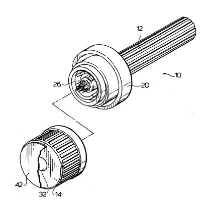
Figure 1 is a perspective view of one form of
clinical applicator or scarifying device, with the cap
member removed from the inoculator member, employing an
arrangement of tines in accordance with one embodiment
of the invention;
Figure 2 is a sectional view of the clinical
applicator of Figure 1; :
Figure 3 is a perspective view of the clinical -
applicator of Figure 1, with the cap member assembled
with the inoculator member;
Figure 4 is a sectional view of the clinical
- , ~.
applicator of Figure 3;
Figure 5 is a sectional view of the inoculator
member during administration of liquid antigen or
vaccine to skin: -
Figure 6 is a perspective view of the inoculator ;~
member and skin showing the visual indication of the
location of administration of the active substance;
Figure 7 is a close-up perspective view of the
arrangement of tines;
Figure 8 is a perspective view of the tines of one
embodiment of the invention; :. :
Figure 9 is a plan view of the tines of Figure 8; : .
30l and j !
Figures 10A and 10B are close-up perspective views : ~:
of the structure of two different tines used in the
arrangement of Figure 8.
.. . .
DESCRIPTION OF PREFERRED EMBODIMENT ~:.
Referring to the drawings, a scarifying device 10
' '
9~
comprises an inoculator member 12 and a cap member 14.
The inoculator member 12, which is a one-piece molded
part, comprises an elongate handle 16 in the form of a
hollow tube and a head 18 having a cylindrical outer
channel structure 20 joined to the handle 16 by a
radially-directed flange or wall 22.
As more particularly described in the
aforementioned U.S. patent application Serial No.
302,928, a series of projections 24 extend from the
wall 22 in the shape and form of an identifying location
indicating marking. A plurality of parallel needles or
tines 26 protrude from the handle 16 beyond the
projections 24 to effect intradermal or subdermal
application of an active substance.
The cap member 14, also a one-piece molded part,
comprises an outer wall 28, a parallel inner wall 30
defining a central openlng 32 and a radially-directed
wall 34. To enable the cap member 14 to be releasably
mounted to the inoculator member 12, the outer wall 28
is provided with an integral protrusion 36 at its upper
extremity, which snap-locks into a corresponding recess
formed in the channel member 20 when the outer wall 28
is received in the channel.
At the same time, the inner wall 30 bears against a
central cylindrical protrusion 38 on which the tines 26
are mounted. In this way, the opening 32 into which the
tines 26 protrude is sealed off.
The cap 14 is assembled with the handle 12. With
the assembly in a vertical orientation, an active liquid
30l material 40 is placed in the opening 32 and ~a foil
covering 42 is adhered over the open end of the opening
32 to hermetically seal the active substance in the
cavity into which project the needles 26.
The cap member 14 also includes a ring 44 of
absorbent material which is seated against the inner
surface of the wall 34 and which has a marking fluid
~' '~' '''.'''
~ '': :,..',
.,: : ',:
9~o
absorbed therein, such as a dye or ink which, when dry,
resists removal by washing. When the cap member 14 is
assembled to the handle member 12, the projections 24
engage the absorbent pad 46, as seen in Figure 4.
When it is desired to make an application of the
active material 40 to a patient's skin, the inoculator
member 12 is removed from the cap member and the head 18
is brought into contact with the skin 46 of the patient.
The needles 26, which are wetted with the active
material 40, penetrate the skin 46 and make an
intradermal or subdermal application of the active
material, depending on the length of the needles 26. At
the same time, the projections 24, which are wetted with
marking fluid from contact with the absorbing surface,
engage the outer surface of the skin, as seen in Figure
5, and form a pattern on the skin corresponding to the
pattern of the projections 24, as seen in Figure 6.
The pattern 48 of marking fluid, a "happy face" in
the illustrated embodiment, thereby provides a visual
indication of the location of the intradermal or
subdermal application but does not, in any way,
interfere with the application site. For adult use, the
visual image may take the form of a simple ring.
Turning now to consideration of Figures 7 to 10,
wherein there is illustrated the tines structure 26 in
more detail. As may be seen, the tines structure
comprises a central tine 50 which is in the shape of a
pyramid and a circular array of eight tines 52 which are
each in the shape of a channelled pyramid.
Each of the tines 52 has a groove 54 found therein
which runs from the tip of the tines to the base and has
surfaces which are perpendicular to the support surface
56 and to each other. Each of the complete faces is in
the form of an isosceles triangle having an apex angle
of about 5 to about 20.
The tines 52 are arranged with their apices joined
: ' '
Z016900
7 -
by a circle, the center of which is the apex of the tine
50. The tines 52 are arranged in two groups of four,
each made up of an alternate one of the tines 52, in
which the channels 54 define the corners of a square.
When the tines penetrate the skin, the channels 54
enable liquid antigen or vaccine to be transferred along
the tine and into the skin through an opening which has
an equal proportion from base to tip.
The 90 angle between the walls of the channel 54
inhibits skin elasticity from closing the channel,
enabling efficient administration of the liquid to be
effected. ~-
The individual tines, 50, 52 may be metal but
preferably are of plastic material integrally molded
with the remainder of the innocular member 12.
SUMMARY OF DISCLOSURE
In summary of this disclosure, the present
invention provides a novel form of tines structure which ~ -
enables efficient administration of liquid antigen or
vaccine to be effective. Modifications are possible
within the scope of this invention.
~`~ ~'''`; ''''' '``''
~ -: .. ..
-: -., .,:: ..-.
',',~,,', .~"',,,,,,',,,
: . ~:
" :~, . - .
: ~ ,
~ ~.
