Note: Descriptions are shown in the official language in which they were submitted.
201 9873
74761-12
BACKGROUND OF THE INVENTION
This invention relates to diagnostic assay modules for
analytical applications and, more particularly, it concerns assay
modules including a resilient assay element wherein the
interaction of a component in a sample fluid with one or more
reagents present in the assay element causes a detectable change
corresponding to the presence of the æample component.
In commonly assigned United States Patent No. 5,051,237
which issued on September 24, 1991, there is disclosed a liquid
transport device which is particularly suited for use in
immunoassay applications. In such applications a surface of an
assay element is retained in contact or virtual contact with a
relieved surface on the underside of a member which includes an
aperture for allowing a sample fluid to be applied and distributed
uniformly over the surface of the assay element. The relieved
surface is defined by a plurality of projections arranged
throughout the fluid flow zone to control the flow of fluid
between opposed surfaces of the fluid flow zone In a preferred
embodiment the projections are arranged in orthogonal rows and
columns to provide a uniform distribution of a sample fluid across
the surface of an assay element. The positioning of the assay
element in contact or virtual contact with the projections
controls the volume of fluid in the fluid flow zone.
In addition to ensuring a uniform spread of the fluid
sample across the surface of the assay element, the disclosure in
the above-mentioned U.S. patent recognizes the importance of
retaining the assay element in a desired orientation to provide,
among other purposes, for optical precision where the change
resulting from interaction of a sample analyte with the assay
element reagent(s) is read out by means of an optical apparatus.
- 20 1 9873
74761-12
Retention of the assay element in such an orientation can be
achieved by a æupport member arranged to engage the surface of the
assay element in a manner to sandwich the element between a
surface on the support member and the relieved surface. The
support member is provided with a transparent window through which
the change in the assay element may be read by the optical system.
This arrangement is not completely satisfactory in all instances
since the electromagnetic radiation used to read the change in the
assay element must pass through the material from which the
support member is made. Where relatively small changes are being
read it may be more desirable not to interpose anything between
the assay element and the optical system.
In commonly assigned United States patent No. 4,977,325
which is~ued on December 11, 1990, a highly efficient dual channel
fluorometer is disclosed in which enhanced optical efficiency
enables the use of a low co~t tungsten halogen illumination source
in combination with solid state photodetectors to detect the low
levels of sample emitted light encountered in fluoroanalysis. The
optical ~ystem is embodied in an optics head designed to be
positioned under a sample receiving vessel of the general type
represented by the physical embodiment disclosed in the
aforementioned U.S. Patent No. 5,051,237.
It is apparent from the combined disclosures of the
aforementioned U.S. patents that the attainment of reliable
results in analytical procedures requires the assay module in
which the assay element is contained to accommodate the
requirements for obtaining a uniform spread of the sample fluid
across the surface of the assay element, for volume control of the
fluid in a fluid flow zone where applicable and retention of the
assay element under conditions which optimize the optical system
_ ` 201 9873
74761-12
by which the optical signal is read from the assay element. In
this context, inclusion of a transparent window in the path of
light directed to and reflected from the surface of the assay
element represents an efficiency loss in the optical system of a
magnitude which may have a substantial effect on the accuracy of
the overall diagnostic equipment.
SUMMARY OF THE PRESENT INVENTION
In accordance with the present inventionr there is
provided an assay module which has a structure adapted for
manufacture on a volume
201 9873
productlon basls and by whlch the very strlngent tolerance
requlrements for retentlon and optlcal presentatlon of the
assay element ln the conduct of analytlcal procedures are
achleved.
Accordlng to the present lnventlon, there ls
provlded a dlagnostlc assay module for analytlcal dlagnostlc
procedures ln whlch an optlcal slgnal developed by lnteractlon
between a component ln a sample fluld and one or more reagents
ln a reslllent sheet-llke assay element ls read by optlcal
means, sald assay module comprlslng: a sheet-llke assay
element; a flrst member lncludlng wall means to deflne an open
slde and lnner and outer surface formatlons, sald wall means
lncludlng an openlng to permlt sample fluld to be lntroduced
therethrough; a second member for closlng sald open slde of
sald flrst member, sald second member having an open optlcal
read aperture posltloned to be ln reglstratlon wlth at least a
substantlal portlon of sald assay element when sald assay
element and sald flrst and second members are assembled; and
means for flexlng the assay element to malntaln lt ln a
preclsely controlled orlentatlon.
By means of the lnventlon, the assay element can be
optlcally located for readlng by an optlcal system through an
unobstructed openlng ln the assay module thereby obvlatlng the
need to lnterpose a support layer between the optlcal system
and the assay element.
In a preferred embodiment of the lnventlon, one of
the members whlch form the assay module also deflnes a fluld
transport surface whlch, together wlth the upper surface of
76207-3
;~
20 1 9873
.,
the assay element deflnes a fluld transport zone to provlde
for the unlform spreadlng of a sample fluld across the surface
of the element. The spreadlng of the sample fluld ls achleved
by a plurallty of pro~ectlons arranged throughout the lntended
fluid flow zone. As wlll be dlscussed ln detall herelnafter
ln con~unctlon wlth the detalled descrlptlon of the preferred
embodlments of the lnventlon, lt ls very lmportant to control
the volume of fluld whlch ls present ln the fluld flow zone
above the assay element. In thls embodlment of the lnventlon
the assay element ls retalned ln contact or vlrtual contact
wlth the plurallty of pro~ectlons whlch
- 4a -
76207-3
. .
~w
20138~
control the fluid flow thus providing precise
control of the volume of fluid above the assay
element as well as optically locating the assay
element.
The two complementing members, which
together with the assay element comprise the assay
module of the invention, may be of various
dimen&~ions and may be provided initially as two
separate parts or they may be joined such as by
being hinged together so as to allow the assay
module to be assembled after the assay element is
inserted in place.
In a preferred embodiment, one of the
molded parts of the assay module, hereinafter
referred to the first, or top, member is of an
oblong configuration and has one open side defined
by a continuous peripheral lip on mutually opposed
side and end walls which, in turn, join commonly
with a closing wall. The closing wall is provided
on its interior with a planar fluid transport
surface of rectangular configuration and displaced
toward the open side of the member from the
remainder of the closure wall. A pair of inclined
ramp formations in the side walls extend in parallel
spaced relation from the side edges of the fluid
transport zone and lie closer to the open side of
the member than the fluid transport zone. The other
of the two molded parts, hereinafter referred to as
the second, or bottom, member is generally shaped as
a plate member having a peripheral configuration to
fit the open side of the first member. The second
member is formed along opposite sides of its inner
surface with ramp formations which complement the
ramp surfaces on the side walls of the first member.
2 ~ 7 ~3
A relatively large rectangular opening is provided
between the ramp formations on the second member.
During assembly of the assay module, the
assay element is positioned so that the side edges
of the assay element overlie the respective ramp
formations on the first and second members. When
the two parts are closed against the assay element,
the latter is flexed in a manner to be biased under
the resiliency of the element into contact or
virtual contact with the projections of the fluid
transport surface and across the entire area of the
intended fluid flow zone. The first and second
members are then secured such as by adhesives,
ultrasonic welding or the like.
The exterior of the top member of the
assay module is shaped to define a relatively deep
central well having a floor through which an
aperture passes and opens to the fluid transport
surface which carries the projections. The
provision of the relatively deep well facilitates
the introduction of sample fluid to the surface of
the assay element at this time being flexed against
the fluid transport surface. In addition, a pair of
relatively deep cylindrical wells are provided in
the exterior of the top member on opposite sides of
the central well so that other fluids may be stored
therein if desired.
A principal object of the invention is,
therefore, the provision of an improved assay module
for analytical applications. Another object is the
provision of such a module in a structure capable of
volume production while maintainin~ very close
tolerances of the type required in immunoassay
procedures. A still further object of the invention
is to provide an assay module which facilitates the
8 7 3
introduction of sample fluid to the surface of the
assay element. Other objects and further scope of
applicability of the present invention will become
apparent from the detailed description to follow
taken in conjunction with the accompanying drawings
in which like parts are designated by like reference
numerals.
BRIEF DESCRIPTION OF THE DRAWINGS
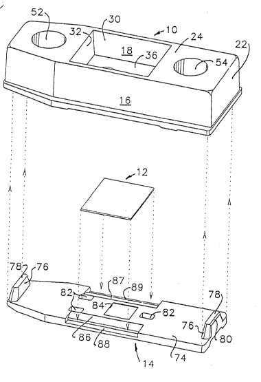
Fig. 1 is a exploded perspective view
illustrating the respective parts of a preferred
assay module according to the invention;
Fig. 2 is a bottom plan view of one of the
parts shown in Fig. 1;
Fig. 3 is a cross section on line 3-3 of
Fig. 2;
Fig. 4 is a cross section on line 4-4 of
Fig. 3;
Fig. 5 is a bottom plan view of the other
of the two major components shown in Fig. 1 of the
drawings;
Fig. 6 is a section on line 6-6 of Fig. 5;
Fig. 7 is an end elevation of the part
shown in Figs. 5 and 6;
Fig. 8 is an enlarged cross section on
line 8-8 of Fig. 6;
Fig. 9 is an enlarged cross section
illustrating representative layers of a preferred
embodiment of an assay element which may be
incorporated in the assay module of the invention;
Fig. 10 is an enlarged cross section in
the plane of Fig. 4 but with the components of the
invention assembled;
Fig. 11 is an enlarged fragmentary plan
view of the area represented by the sight circle 10
in Fig. 2;
2019~73
Fig. 12 is a fragmentary cross section on
line 12-12 of Fig. 11; and
Fig. 13 is a generally schematic view
illustrating the invention in relation to an optical
read system.
DETAILED DESCRIPTION OF THE PREFERRED EMBODIMENTS
In Fig. 1 of the drawings, the principal
parts of the assay module of the present invention
are shown generally and prior to assembly as
including a first, or top member 10, an assay
element 12 and a second, or bottom, member 14.
Although each of the parts 10, 12 and 14 will be
described in considerably more detail below with
reference to drawing figures illustrating such
detail, it may be appreciated from Fig. 1 that the
first member 10 and second member 14 are self-
contained units capable of formation by injection
molding techniques and may be closed one against the
other to retain the assay element 12 in a precisely
- 20 positioned location. Although the first member 10
has considerably higher sidewalls than the second
member 14 in this preferred embodiment, the first
and second members may be provided with various
sidewall dimensions. For cxample, the first member
may have relatively small sidewalls, particularly
where fluid storage wells are not required.
Further, although the first and second members are
shown as two separate parts, as noted previously,
they may be connected such as by being hinged
together along one of their peripheral dimensions
thus permitting them to be folded together and
secured after the assay element is arranged in
place.
In Figs. 2-4 of the drawings, structural
features of the first member 10 are detailed in
` 2019873
bottom plan, longitudinal and transverse cross
sectional views, respectively. In these figures it
may be seen that the first member 10 is formed with
mutually opposed side walls 16 and 18, end walls 20
and 22 and a shaped closure wall 24. The projecting
edges of the side and end walls 16-22 define a
peripheral flange-like lip 26 having a continuous
chamfer 28 joining with the respective side and end
walls.
In Figs. 1, 3 and 4 of the drawings, it
may be seen that the closure wall 24 is shaped to
define with the side walls 16 and 18 a centrally
located, generally rectangular well 30 delimited at
opposite ends by mutually facing linear wall
portions 32 and 34 extending between the side walls
16 and 18. The well 30 opens to the exterior of the
first member 10 and is closed between the side walls
16 and 18 and wall portions 32 and 34 by a floor 36.
Viewed from the opposite side of the well 30 or from
the interior of the first member 10, the wall 36 is
thickened centrally to establish a rectangular fluid
transport surface 38 displaced from the interior
surface of the wall 36 toward the open side of the
first member circumscribed by the lip 26. A sample
fill opening 4~ opens through the floor 36 of the
well 30 to the plane of the rectangular surface 38.
Also as may be seen in Figs. 2-4, a plurality,
specifically 3 post-like projections 42 extend from
the inner surface of the well floor wall 36. The
projections 42 are located to be spaced from the
ends of.the rectangular surface 38 and oriented such
that one such post lies off one end of the
rectangular surface 38 whereas the other two posts
42 lie off the opposite end of the rectangular
surface 38. A pair of inclined ramp surfaces 44 and
_g _
20 1 9873
74761-12
46 are defined on step-like formations 48 and 50, respectively
lying at the intersection of the inner surface of the ramp floor
wall 36 and the interior of the side walls 16 and 18. As æhown in
Fig. 2, the ramp surfaces 44 and 46 are in spaced parallel
relation with the side edges of the rectangular surface 38 and
extend symmetrically beyond opposite endæ of the surface 38. As
may be æeen most clearly in Fig. 4, the ramp surfaces 44 and 46
are displaced generally toward the lip 26 from the rectangular
surface 38 and are inclined æo as to diverge outwardly from the
plane of the surface 38.
With reference to Figs. 1 and 3, the first member 10 is
formed with a pair of cylindrical wells 52 and 54 spaced from
opposite ends of the central rectangular well 30. Theæe
cylindrical wellæ open through the outer surface of the covering
wall 24 and are cloæed at their inner ends by dome-like formations
56 and 58, respectively. The cylindrical wells 52 and 54 may be
uæed in practice to store other fluids such aæ diluents or
reagents used in the analytical procedures in which the assay
module of the invention is used.
The rectangular fluid transport æurface 38, as
described, projects from the interior of the well floor wall 36 to
present a well defined rectangular planar æurface at the open side
of the first member 10. The surface 38 operateæ to asæure the
spread of liquid between it and a parallel opposed surface aæ
described in the above-mentioned U.S. Patent No. 5,051,237. To
thiæ end, and aæ shown in Figæ. 11 and 12 of the drawingæ, the
planar surface 38 is relieved by a uniform pattern of projections
60. The fluid tranæport surface including the
20 1 9873
projections, toqether with the surface of the assay
element which is retained in contact or virtual
contact with the projections, defines a fluid flow
zone wherein the surface 38 and the surface of the
assay element are spaced apart a capillary distance
to permit capillary flow of fluid between them. The
height of the projections is generally from about 50
to about 150 microns and preferably from about 80 to
about 120 microns. The illustrations of Figs. 11
and 12 are greatly enlarged relative to the already
enlarged illustration of Figs. 1-4, for example, to
provide an appreciation of the configuration and
dimensioning of the projections 60. In particular,
it will be noted that in a preferred embodiment of
the present invention, the projections 60 are shaped
as truncated rectangular py-amids which terminate at
their outer ends in a square surface, the side
dimensions of which are designated by the letter
"t". In practice the, preferred size of the
dimension t is approximately 0.05 millimeter. The
surfaces of the projections diverge from their outer
end toward the plane of the rectangular surface 38
at an angle of approximately 45 and are of a height
"_" equal to approximately 0.10 millimeter. The
pyramidal projections 60 are spaced on centers "S"
both in rows extending parallel to the length of the
shelf 38 and in columns extending across the width
of the surface 38. The preferred size of the
spacing dimension S is 0.38 millimeter.
As may be seen in Figs. 1 and 2, the
interior corners at the juncture of the end wall 22
with the side walls 16 and 18 are filled by bosses
62 and 64 which extend to flat end surfaces 66 and
68, respectively, spaced slightly inward of the
peripheral lip 26 circumscribing the open side of
~O1~J73
the first member lo. A spacer pin 70 is located
centrally of the surface 68 and projects slightly
therefrom in a manner to establish the assembled
position of the second member 14 in a manner to be
described. Identical pin-like projections 70 are
provided on each of the shelf-like formations 48 and
50 as well as on a bridge 72 extending from the
inner wall surface of the cylindrical well 52 to the
end wall 20.
Structural details of the second member 14
may be understood and appreciated by reference to
Figs. 1 and 5-8 of the drawings. As shown, the
second member 14 is generally of plate-like
configuration and of a peripheral contour to
complement the open side of the first member 10 as
defined by the inner chamfered surface 28 on the
peripheral lip 26. The inner side of the bottom
member is shown most completely in Fig. l to include
a planar inner surface 74. A pair of locating lugs
76 projéct from the surface 74 at opposite ends of
the member 14. As may be seen in the drawings, the
lugs 76 taper so as to converge from the surface 74
to a truncated flat 78 defining the inner ends of
each lug. A chamfer 80 joins the outer end surfaces
of each lug 76 with the respective truncated flats
78 thereof. A central working portion of the bottom
member 14 and particularly of the inner planar
surface 74 thereof is defined longitudinally between
three through-holes 82 located to fit over and
receive the posts 42 projecting from the inner
surface of the well floor wall 36 of the top member
lO. Also a relatively large square read aperture 84
opens through the planar surface 74 and is offset
slightly from the longitudinal center of the member
14.
-12-
20~ 73
The inner surface of the bottom member 14
is recessed along opposite sides of the central
working area to establish a pair of parallel and
generally elongated ramp surfaces 86 and 87 which
diverge from the planar surface 74 at an angle a
(see Fig. 8 ~ and end at step-like planes 88 and 89,
respectively. The ramp surfaces 86 and 87 on the
bottom member 14 complement the ramp surfaces 4 4 and
46 in the top member 10. In this respect, the angle
of inclination a is the same for both the ramps 86,
87 and the ramps 44, 46. A preferred angle a for
the inclination of the surfaces from the plane of
the rectangular fluid transport surface 38 in the
top member 10 and from the plane of the surface 74
on the bottom member 14 is preferably on the order
of lS. As will be appreciated from the description
to follow concerning the function of these ramps,
the specific angle at which they are inclined may
vary substantially from 15.
~ 20 The outer surface of the bottom member 14
may be appreciated from Figs. 5, 7 and 8 of the
drawings. As shown particularly in Figs. 5 and 8,
the read opening 84 which opens through the inner
planar surface 74, is delimited by a very narrow lip
as a result of flaring the periphery of the opening
toward the outer surface of the bottom member 14. A
continuation of the outwardly flared opening 84
results in a pair of longitudinal rails 90 extending
over the length of the member 14. The rails
function not only to strengthen the bottom member 14
but also to allow an adequate amount of material to
define the recesses on which the ramps 86, 87 and
steps 88, 89 are located.
The assay element 12 may comprise any
analytical assay element. Further, the assay
-13-
-- 201~87~ -
element may be a single layer or a multilayer
element. Assay elements which are based on
immunological interactions are preferred. A typical
thin film assay element has a thickness of about 0.1
mm and comprises one or more reagent layers residing
on a support layer which is transparent to permit
reading of the element from below. The assay
element may also include various other layers such
as are known in the art including,-for example, a
light-blocking layer to permit the signal generating
species in one layer to be read out without
interference from material present in another layer,
a registration layer for holding a signal generating
species formed in, or released from, another layer,
etc. For the purpose of further illustrating the
invention a particularly preferred multilayer assay
element 12 is shown in Fig. 9. Specifically, one
surface of the assay element 12 is defined by the
outer surface of a transparent support layer 92
whereas the other surface of the assay element is
defined by the outer surface of a layer 94 which may
be a reagent layer, a protein filter layer, an anti-
abrasion layer or the like. A reagent layer 96 and
a light blocking layer 98 lie between the outer
layers 92 and 94. Although the chemical and/or
immunological properties of the assay element 12 in
the use of the present invention for analytical
procedures is important, the physical properties of
the element are of greater significance to an
understanding of the present invention. In
particular, the sheet-like assay element 12 is not
only very thin, as indicated, but because the
support layer 92 is typically a polymeric material,
it is resilient in the sense that if it is distorted
out of its initial planar condition, it will exhibit
2 0 ~
a bias to its original position. Also the assay
element is of generally rectangular configuration as
shown in Fig. 1, and is of a size to extend onto and
lie against the ramps 44 and 46 in the first member
10 as well as to lie between the posts 42 on
opposite ends of the rectangular fluid transport
surface 38.
In a particularly preferred embodiment
reagent layer 96 comprises an immunocomplex of a
fluorescent labeled antigen and an antibody directed
against the antigen. In this embodiment the
antibody is immobilized in reagent layer 96 such as
by being covalently bound to the surface of support
layer 92 or to a matrix material or by being
physically held by the matrix material. In practice
a sample fluid is introduced through the opening 40
in the first member 10 and is spread uniformly
across the surface of the assay element 12
corresponding to the fluid flow zone defined by
rectangular fluid transport surface 38. A
substantially uniform concentration of any analyte
present in the sample fluid is distributed across
the assay element and the fluid diffuses throughout
layers 94, 96 and 98 as well as filling the fluid
flow zone between the surface of layer 94 of the
assay element and the rectangular fluid transport
surface 38 of first member 10. An equilibrium is
established. When present, the sample analyte, in
this illustrative discussion an antigen of interest,
will compete with the fluorescent-labeled antigen
(the same antigen as the sample antigen or an
analogue thereof) for the available binding sites on
the immobilized antibody. The fluorescent-labeled
antigen initially complexed to the antibody in
reagent layer 96 will be dissociated therefrom and
20 1 ~873
replaced by the sample antigen in a ratio
approximately equal to the relative amounts of
sample antigen and fluorescent-labeled antigen.
Thus, depending upon the amount of antigen present
in the sample fluid, some percentage of the
fluorescent-labeled antigen will bind to those
immobilized antibodies which are not bound to the
sample antigen. The remainder of the labeled
antigen will be distributed throughout the remainder
of the assay element, i.e., throughout layer 94 and
98, and the fluid flow zone between the surface of
layer 94 and the opposed rectangular fluid transport
surface 38 of the first member ~0. The amount of
labeled antigen bound to the immobilized antibodies
in reagent layer 96 at any time is inversely
proportional to the amount of sample antigen. A
quantitative determination is obtained by
irradiating the reagent layer 96 through the
aperture 84 of the bottom member 14 with appropriate
excitation energy. Since the reagent layer 96 which
includes the immobilized antibody is preferably very
thin in comparison to the combined thickness of
layers 94 and 98 and the fluid flow zone, preferably
a ratio of from about 1:20 to about 1:100 or more,
and because light-blocking layer 98 prevents any of
the excitation energy from entering layer 94 or the
fluid flow zone, the optical readout system will
measure the amount of labeled antigen which is bound
to the immobilized antibody and a very small
percentage of the free labeled antigen which is
distributed throughout the remainder of the assay
element and the fluid flow zone.
It will be appreciated by those skilled in
the art that in this preferred embodiment it is very
important to control the volume of fluid in the
-16-
2 ~ 3
fluid flow zone. This is accomplished according to
the invention by retaining the surface of the assay
element in contact or in virtual contact with the
projections 60 carried on fluid transport surface 38
by means of the flexure developed between the
complementary ramp surfaces of the respective first
and second members of the assay module.
Assembly of the first member 10, assay
element 12 and the second member 14 is effected
simply by placing the assay element 12 into the
first member 10 so that the layer 94 faces the
rectangular surface 38. This placement is
facilitated by a combination of the three posts 42
and the formations 48 and 50 which serve to guide
the element to a preliminary position in which the
side edges Oc the element 12 rest on the ramps 44
and 46 and so that the top face of the element, that
is, the outer surface of the layer 94, properly
overlies the rectangular surface 38 to space the top
surface initially from the surface 38. The second
member 14 is then advanced into the open side of the
first member 10 as depicted by phantom line
illustration in Fig. 10 and also by the exploded
perspective illustration in Fig. 1 of the drawings.
Final guiding of the second member into position is
aided by the tapered lugs 76 as well as by the
continuous internal chamfer 28 on the peripheral lip
26 of the first member lO.
During final movement of the bottom member
14 into the top member 10, a combination of the
ramps 86, 87 on the bottom member and the
complementing inclination of the ramps 44, 46 in the
top member result in a flexure of the assay element
12 so that the layer 94 in the illustrated
embodiment of the assay element, which represents
2019~73
the top surface of the assay element, is biased by
the inherent resiliency of the assay element into
uniform continuous contact or virtual contact with
the projections 60 on rectangular surface 38. The
final position of the second member 14 is
established by engagement of inner surfaces thereon
with the projecting locator pins 70 spaced about the
inner periphery of the first member 10. Once in
place, the bottom member 14 is secured by
appropriate means such as adhesives, ultrasonic
fusion, or the like.
The relationship of the three parts 10, 12
and 14 after assembly is shown most clearly in Fig.
10 of the drawings. In particular, it will be noted
that the surface of the assay element 12 is
constrained to be in contact or virtual contact with
the truncated ends of the individual pyramid
projections 60 on rectangular surface 38. This
orientation of the assay element surface is,
moreover, assured by flexure of the assay element 12
as a result of coaction between the respective ramps
44, 46 in the first member 10 and the ramps 86, 87
on the bottom member 14.
It is to be noted that the optical read
opening 84 in the secondary member 14 is displaced
so that it does not overlie any portion of the fill
opening 40 in the first member 10. Because the size
of the read opening 84, a substantial portion of the
assay element 12 is unsupported by structure over a
relatively large area of the fluid flow zone defined
by rectangular surface 38. As noted above, the area
of assay element 12 left out of contact by structure
in the area of the opening 84 must lie in a planar
configuration to be retained in contact or virtual
contact with projections 60 on rectangular surface
-18-
201 9873
74761-12
38 for the purpoæe of controlling the volume of fluid in the fluid
flow zone between the outer æurface of layer 94 and rectangular
surface 38 and for optical precision. Becauæe of the manner in
which the assay element is stressed to conform with the planar
orientation of rectangular surface 38 in accordance with the
invention this condition is assured.
In Fig. 13, the assembled aæsay module including the
firæt member 10, the assay element 12 and the æecond member 14 are
shown poæitioned relative to an optical æystem of the type
diæclosed in the above-mentioned U.S. Patent No. 4,977,325 which
issued on December 11, 1990. In light of the detailed disclosure
of the optical system in this U.S. patent the components thereof
need only be summarized in this instance aæ including a light
source 100 from which broad band illumination iæ collimated along
one channel 102 including a filter 104 to impinge againæt the
transport support layer surface of the assay element 12, light
energy of a specific narrow frequency range. Light emanating from
the assay element 12 as a result of the interaction between a
sample analyte and the reagent layers in the aæsay element is
directed through a second æeparate channel 106 including a filter
108 to paæs light of another specific wavelength to a sensor 110
to develop a voltage æignal corresponding to the amount of analyte
in the sample. As pointed out in the aforementioned U.S. patent,
the efficiency of the optical system is important particularly
from the standpoint of being able to uæe a relatively inexpensive
- tungsten halogen lamp for the illumination source 100 and a
photodetector for the
2019873
sensor 110. Thus, not only is the planar
orientation of the assay element 12 critical to
efficiency of the optical reading system shown, but
also the absence of any media underlying the bottom
surface of the element 12 due to the read opening 84
in the second member 14 is important to optical
efficiency of the system.
Thus, it will be appreciated that as a
result of the present invention, a highly effective
assay module is provided by which the stated
objectives, among others, are completely fulfilled.
Also, it will be understood by those skilled in the
art from the preceding description and accompanying
drawings that variations may be made in the
disclosed preferred embodiments without departure
from the invention. It is expressly intended,
therefore, that the description and illustration is
of preferred embodiments only, not limiting, and
that the true spirit and scope of the present
invention will be determined by reference to the
appended claims.
-20-