Note: Descriptions are shown in the official language in which they were submitted.
203-231 (1106)
L ~
~~ APPARATUS AND METHOD FOR APPLYING SURGICAL 2 0 213 ~
CLIPS IN LAPAROSCOPIC OR ENDOSCOPIC PROCEDURES
BACKGROUND OF THE lNV~NLlON
1. Field of the Invention
This invention relates to an apparatus and method for applying
surgical clips, especially hemostatic clips, to body tissue such as blood
vessels. More particularly, this invention relates to a surgical clip
applier which can be used in laparoscopic or endoscopic procedures, and a
method for using same.
2. Background of the Related Art
In surgical operations it is often necessary to apply
hemostatic clips to blood vessels, and apparatus for applying clips are
known in the art. See, for example, U.S. Patent No. 4,616,650, and
4,624,254, both of which are hereby incorporated by reference, which
disclose a surgical clip applying apparatus having a pair of ring-like
handles. The handles are squeezed to force jaws to move distally
relative to the apparatus where they are forced together by a pair of
inclined surfaces. A surgical clip between the jaws is thereby squeezed
closed.
In laparoscopic procedures surgery is performed through a small
incision; in endoscopic procedures surgery is performed through narrow
endoscopic tubes inserted through small entrance wounds in the skin.
Laparoscopic and endoscopic procedures generally require that any
instrumentation inserted into the body be sealed, i.e., provisions must
` 2021362
be made to ensure that gases do not enter or exit the body~through the
laparoscopic or endoscopic incision as, for example, in surgical
procedures in which the surgical region is insufflated. Moreover,
laparoscopic and endoscopic procedures often require the surgeon to act
on organs, tissues, and vessels far removed from the incision, thereby
requiring that any instruments to be used in such procedures be both long
and narrow. Up to now there have been no instruments for placing
surgical clips in laparoscopic or endoscopic procedures.
Because endoscopic procedures are more common than laparoscopic
procedures, the present invention shall be discussed in terms of
endoscopic procedures and apparatus. However, use herein of terms such
as Nendoscopic", "endoscopically" and "endoscopic portion", among others,
refer generally to instruments having elongated and relatively narrow
operating portions for inserting into a cannula or a small wound in the
skin and should not be construed to limit the present invention to an
apparatus for applying surgical clips only in conjunction with an
endoscopic tube. To the contrary, it is believed that the present
invention may find use in any procedure where access is limited to a
small incision, including, but not limited to laparoscopic procedures.
3. Objects of the Invention
Accordingly, it is one object of the present invention to
provide a surgical clip applier.
It is another object of the present invention to provide a
surgical clip applier which can be used endoscopically.
It is a further object of the present invention to provide a
surgical clip applier which is adapted to prevent gases from
communicating between the interior and exterior of the body during an
endoscopic procedure.
i` . 202~6~
It is yet another object of the present invention to provide a
surgical clip applier which is at least partially disposable.
These and further ob;ects and advantages are achieved by
providing a surgical clip applier insertable through a small incision or
narrow tube for applying surgical clips to blood vessels or other body
tissue.
SUMMARY OF THE INV~NLlON
In accordance with the present invention, an apparatus is
disclosed for applying surgical clips to body tissue which comprises
frame means, endoscopic means connected to the frame means of generally
elongated configuration and extending distally from the frame means and
including means for storing a plurality of surgical clips, means for
selectively advancing the clips to the distal portion of the endoscopic
means for positioning adjacent the body tissue to be clipped; and means
for at least partially closing each clip at least sufficient to grip the
body tissue after the clip has been advanced distally to the
predetermined portion of the endoscopic means.
Preferably, the apparatus for applying surgical clips to body
tissue comprises a frame, and an elongated endoscopic section connected
at the proximal end thereof to the frame and extending distally from the
frame. The endoscopic section includes means for storing a plurality of
surgical clips and a pair of jaws positioned at the distal portion of the
endoscopic section and adapted for reception of the clips. Means is
provided for sequentially advancing the clips distally for positioning
within the pair of jaws to be positioned adjacent the body tissue to be
clipped; and means is provided for sequentially at least partially
closing the jaws about the clips to close the clips at least partially
about the body tissue.
2021362
In one embodiment, a completely disposable apparatus is
disclosed for applying surgical clips to body tissue which comprises a
frame configured and dimensioned for manual gripping, an elongated
endoscopic section connected at the proximal end thereof to the frame and
extending distally therefrom, the endoscopic section including means for
storing a plurality of surgical clips in generally aligned relation
facing the distal portion thereof, jaw means positioned at the distal end
thereof and adapted for sequential reception of the clips, means for
sequentially advancing the clips distally as to be positioned between the
jaw means for positioning adjacent the body tissue to be clipped, and
means for sequentially at least partially closing the jaw means about
each clip after the clip is advanced therebetween while simultaneously
repositioning the clip advancing means for distal advancement of the next
clip .
In another embodiment of the present invention the endoscopic
portion is formed as a disposable unit detachable from a reusable frame
and handle portion.
Preferably, an instrument body is provided and an actuating
handle mounted to the instrument body, with first transmission means for
linearly transferring motion from the actuating handle to the clip
advancing means and means to close the jaw means. Second transmission
means is provided for linearly transferring motion from the actuating
handle to the jaw closing means, and means is provided for locking the
handle such that after actuating the handle to close the jaws the handle
cannot be actuated unless the locking means is released. The endoscopic
section is rotatable independent of the handle, with means being provided
to selectively lock the endoscopic section at a predetermined angular
orientation relative to the handle. Means is provided to release the
~ 2021 362
lock means of the endoscopic section so as to permit rotation thereof
relative to the handle.
Handle locking means comprises a first resilient catch movable
in response to actuation of the handle from an unlocked position to a
locked position wherein the first transmission means is advanced and
locked. Release means is adapted to release the first resilient catch,
the first resilient catch being returnable to the unlocked position in
response to actuation of the release means. A second resilient catch is
movable in response to actuation of the handle from an unlocked position
to a locked position wherein it engages and locks the second transmission
means. The second resilient catch is resiliently returnable to the
unlocked position in response to the release of the resilient catch. The
first transmission means comprises means responsive to actuation of the
release means to release the second transmission means. The jaw means
preferably comprises a pair of jaws positioned in spaced relation and
configured and dimensioned for reception of a surgical clip
therebetween. The jaws are resiliently movable toward and away from each
other in response to distal movement of a c. ing means from a proximal
position to a distal position. The c~ ing means comprises a channel
member slidably mounted within the endoscopic section and longitudinally
movable in response to actuation of the handle. The channel member
having at least two distal c ing surfaces for biasing the jaws into the
closed position. Means for storing surgical clips comprises a track for
holding a longit-~dinAl array of surgical clips, and resilient means
located proximal to the array of surgical clips for biasing the surgical
clips toward the distal direction. A clip track is positioned between
the jaw means and the clip follower. Means for advancing the surgical
clips comprises a pusher bar for advancing the distal-most clip in the
2021362
area of the pair of jaws, the pusher bar being longitudinally slidable in
response to actuation of the handle, and escapement means located at the
distal end of the array of clips for preventing more than one clip at a
time from being advanced into the jaw means. The escapement means
comprises a plurality of projections upstAndine from the clip track and
extending into the clip path.
The first transmission means comprises a pusher bar, and a
proximal pusher tube connected to the proximal end of the pusher bar.
The pusher bar is movable between a first position wherein the distal end
of the pusher bar is located proximally of the surgical clip to be
advanced, and a second position wherein the distal end of the pusher bar
advances the surgical clip to the jaw means. The first pusher tube
includes mounting means for rotatably connecting the pusher bar thereto.
The mounting means of the pusher tube comprises a generally circular
shaped projection dimensioned for reception and engagement of at least
one cooperating projection on the pusher bar.
The second transmission means comprises a channel member
positioned within the endoscopic section, a proximal channel tube
connected to the proximal end portion of the channel member, and a
channel tube adapted for rotatably connecting the channel member
thereto. The channel member is connected to the c. Ine means for
closing the jaw means.
The jaw means preferably comprises a jaw blade fixed to the
endoscopic section and having a pair of distal spaced jaws which are
resiliently movable between a closed position for closing a surgical clip
and an open position for reception of the surgical clip. The c~ ine
means is comprised of a channel member having c~ ine surfaces movable
from a first position proximal of the jaws, and a second distal positiDn
-6-
. 2021362
wherein the camming surfaces of the channel member move the jaws to the
closed position. The channel member is connected at its proximal end to
the channel tube.
The rotatable mounting means of the channel tube comprises a
circumferential projection dimensioned for engaging at least one
cooperating notch in the c~ ~ng means. The endoscopic section is
rotatable about a longitudinal axis extending relative to the frame
between a plurality of click-stop settings. Further, the endoscopic
section is preferably adapted to provide a gaseous seal means,
optionally, in the form of silicone grease.
According to the method of the present invention the endoscopic
portion of the apparatus is inserted into the body through a small
incision or, more likely, through an endoscopic tube. The blood vessel
or other tissue to be clipped is engaged by the jaws of the apparatus. A
clip is positioned between the jaws and the jaws are closed, thereby
applying the clip to the blood vessel.
The present invention advantageously permits a surgeon to
perform internal clip application without full access to the operation
site, i.e., without providing a large opening in the body to allow access
to the operation site. The frame and handle portion of the apparatus are
manipulated outside of the patient's body. Additionally the endoscopic
portion may be rotated so as to facilitate positioning of the clip.
The ability to apply surgical clips through a small incision or
tube dramatically reduces blood loss, tissue trauma, and patient recovery
time, thereby contributing to improved health care practices.
` 2Q21362
BRIEF DESCRIPTION OF THE DRAWINGS
Preferred embodiments of the invention are described
hereinbelow with reference to the drawings wherein:
Fig. 1 is a perspective view of a completely disposable
apparatus for placing clips in laparoscopic or endoscopic procedures
constructed according to the present invention;
Fig. 2 is a perspective view with parts separated for purposes
of illustration of the endoscopic section of the apparatus of Fig. l;
Fig. 3 is a perspective view with parts separated for purposes
of illustration of the handle section of the apparatus of Fig. 1 utilized
for activating the endoscopic section;
Fig. 4 is a plan view from above, of the distal portion of the
endoscopic section of the apparatus of Fig. l;
Fig. 5 is a cross-sectional view taken along lines 5-5 of Fig.
4 illustrating the clip pusher in position to push the clip next in line
to a position between the jaws of the clamp of the apparatus;
Fig. 5a is a cross-sectional view of the distal portion of the
endoscopic section of the apparatus of Fig. 1, illustrating the position
of the clip pusher after the last clip has been advanced into the jaws of
the endoscopic section;
Fig. 5b is a cross-sectional view taken along lines 5b-5b of
Fig. 5a;
Fig. 6 is a plan view from above similar to Fig. 4 of the
distal portion of the endoscopic section of the apparatus with the clip
pusher in the distal position with the clip shown in Fig. 5 now
positioned between the ~aws of the clamp;
Fig. 7 is a cross-sectional view taken along lines 7-7 of Fig.
6;
-8-
2021362
,
Fig. 8 is a schematic view of the jaws positioned around the
cystic duct;
Fig. 9 is a cross-sectional view taken along lines 9-9 of Fig.
6;
Fig. 10 is a cross-sectional view taken along lines 10-10 of
Fig. 6;
Fig. 11 is a cross-sectional view taken along lines 11-11 of
Fig. 6;
Fig. 12 is a plan view from above of the distal portion of the
endoscopic section illustrating a clip positioned within the clamp jaws
after clamping is completed about an artery;
Fig. 13 is a side view thereof;
Fig. 14 is a cross-sectional view taken along lines 14-14 of
Fig. 12;
Fig. 15 is a cross-sectional view taken along lines 15-15 of
Fig. 12;
Fig. 16 is an elevational cross-sectional view of the handle of
the apparatus of Fig. 1 illustrating the channel tube in the normal "at
rest" position and the pusher tube in the loaded position corresponding
to the position of the clip pusher in the proximal position about to push
the next clip distally into the jaws;
Fig. 17 is an elevational cross-sectional view of the handle of
the apparatus of Fig. 1 with the pusher tube extended to its distal-most
position corresponding to the position of the clip pusher after
positioning a clip between the jaws, and the channel tube in its proximal
position corresponding to open jaws at the distal-most position of the
endoscopic section;
202I362
Fig. 18 is an elevational cross-sectional view of the handle
section of the apparatus of Fig. 1 illustrating the pusher tube in its
proximal position corresponding to the clip pusher in position behind the
next clip to be advanced and the channel tube in its distal-most position
after having closed the jaws of the clamp;
Fig. 19 is an elevational cross-sectional view of the handle of
Fig. 1 illustrating the channel tube in locked position after the last
clip has been pushed into the jaws of the endoscopic section and the
pusher bar has engaged a distal slot in the clip follower, locking the
pusher tube in the position shown;
Fig. 20 is a cross-sectional view taken along lines 20-20 of
Fig. 17 and illustrating the channel tube and pusher bar;
Fig. 21 is a view taken along lines 21-21 of Fig. 17
illustrating the pusher tube and the channel tube in cross-section;
Fig. 22 is a cross-sectional view taken along lines 22-22 of
Fig. 17 illustrating the pusher tube mainspring and the connection
between the pusher tube and the pusher bar;
Fig. 23 is a cross-sectional view taken along lines 23-23 of
Fig. 17 illustrating the pusher tube and the pusher tube link pin;
Fig. 24 is a cross-sectional view taken along lines 24-24 of
Fig. 17, illustrating the pusher tube and the mainspring;
Fig. 25 is a cross-sectional view taken along lines 25-25 of
Fig. 17 illustrating release of the channel tube latch plate by the
pusher tube; and
Fig. 26 is a cross-sectional view taken along lines 26-26 of
Fig. 17 illustrating the lost motion spring which permits partial
clamping of the surgical clips.
Fig. 27 illustrates a cutaway perspective view of a second
embodiment of the present invention;
-10-
` ` 2021 362
Fig. 28 illustrates a perspective view of the frame portions of
the second embodiment;
Fig. 29 illustrates an exploded perspective view of the
actuating mechanism of the second embodiment;
Fig. 30 illustrates a perspective view of the handle;
Fig. 31 illustrates a side view of the right toggle lever;
Fig. 32 illustrates a perspective view of the left toggle lever;
Fig. 33 illustrates a side view of the pusher bar front link;
Fig. 34 illustrates a side view of the pusher bar rear link;
Fig. 35 illustrates a perspective view of the rear channel;
Fig. 36 illustrates a perspective view of the rear pusher bar;
Fig. 37 illustrates a perspective view of the pusher bar stop
spring;
Fig. 38 illustrates a front view of the pusher slide;
Figs. 39a and 39b illustrate a front view of the channel lock
pin in the unlocked and locked positions, respectively;
Fig. 40 illustrates the channel lock pin in perspective view;
Fig. 41 illustrates an exploded perspective view of the
transmission mechAnl~ of the second embodiment;
Fig. 42 illustrates a sectional top view of the transmission
coupling;
Fig. 43 illustrates a sectional top view of the collar fitting;
Fig. 44 illustrates an exploded perspective view of the
endoscopic portion of the second embodiment;
Figs. 45a and 45b illustrate in perspective view the closing of
the jaw blade to apply a surgical clip;
Fig. 46 illustrates a cutaway perspective view of an
alternative embodiment;
2021362
Fig. 47 illustrates the frame of an alternative embodiment;
Fig. 48 illustrates an exploded perspective view of the
actuating and transmission system of an alternative embodiment;
Fig. 49 illustrates the handle of an alternative embodiment;
Figs. 50a and 50b illustrate in sectional side view and bottom
view respectively, the pusher tube of an alternative embodiment;
Figs. 51 and 51a illustrate the channel tube of an alternative
embodiment in perspective and side sectional views, respectively.
Fig. 52 illustrates the leaf spring of an alternative
embodiment;
Fig. 53 illustrates the release button of an alternative
embodiment;
Fig. 54 illustrates a sectional side view of the outer tube of
an alternative embodiment;
Fig. 55 illustrates in exploded view the endoscopic portion of
an alternative embodiment;
Fig. 56 illustrates a sectional top view of the collet and
sleeve of an alternative embodiment;
Fig. 57 illustrates a sectional side view of the upper portion
of the cartridge of an alternative embodiment;
Fig. 58 illustrates a sectional side view of the lower portion
of the cartridge of an alternative embodiment;
Figs. 59a and 59b illustrate the proximal section of the
endoscopic portion of an alternative embodiment;
Figs. 60a and 60b illustrate the endoscopic portion of the
apparatus used in conjunction with the cannula of a trocar in top and
side views, respectively;
Fig. 61 illustrates the sealing block in perspective view.
~ `~ I 2021362
DETAILED DESCRIPTION OF THE lNv~:NllON
The surgical apparatus described herein is adapted to apply
surgical clips to blood vessels and the like, in endoscopic or
laparoscopic procedures.
The apparatus or instrument generally comprises a frame which
is of a size convenient for being held in the hand and which houses the
non-endoscopic body of the apparatus. An endoscopic portion defining a
longitudinAl axis extends distally from the frame and is rotatable around
the longitudinal axis relative to the frame. The endoscopic portion is a
long tube-like portion having a relatively narrow outer diameter (e.g.,
about 10 millimeters) for insertion into an endoscopic tube such as a
trocar cannula, or a small incision.
The endoscopic portion comprises means for holding a
longitudinal array of surgical clips, such as a track, with a spring
means to bias the clips forward in the distal direction. The clips are
generally U-shaped pieces of integral construction and comprise two
spaced apart legs connected by a bridge portion. The endoscopic portion
has a clip closing means comprising a pair of flexible opposing jaws
which are cammed together into closure by a distally-moving channel, and
means such as a spring mounted pusher bar for advancing the surgical
clips one at a time to the jaws.
The apparatus further has actuating means such as a pivoting
handle and connecting links and levers, and transmission means for
transferring the pivotal movement of the actuating means linearly along
the instrument axis to both the clip closing and clip advancing means.
The apparatus also includes tubular members with circumferential coupling
means such as circumferential notches or projections, which allow a
connection of the endoscopic portion to the frame such that linear
-13-
2021362
actuation movement may be transmitted thereto while allowing the
endoscopic portion to rotate around the instrument axis.
The apparatus further comprises a locking means such that once
the jaw means have been actuated and opened, the apparatus cannot be
reactuated until the locking means is released, usually by a release
button. The locking means comprises a resilient catch (such as a catch
in conjunction with a spring) which is movable in response to actuation
of the apparatus, i.e., application of a clip, from an unlocked position
to a locked position. In the locked position, the locking means is
engaged, thereby preventing linear transfer of movement to the clip
closing means by the transmission means. Preferably two spring catches
are used, one for locking the transmission means for actuating the clip
advancing mechanism (e.g., pusher bar), and a second catch for locking
the transmission means for actuating the jaw closing means (e.g., the
channel). Pressing the release button releases both the first and second
catches. The first catch is released directly thereby allowing the first
transmission means to slide forward, and the second catch is released in
response to the forward movement of the first transmission means.
As mentioned above, endoscopic instrumentation is usually
required to have a gaseous seal to prevent communication of gases through
the endoscopic incision. This gaseous seal may be accomplished in the
apparatus of the present invention by providing close tolerances between
the outer diameter of the endoscopic portion and the inner diameter of
the trocar c~nntll~ through which it is inserted, and by providing close
tolerances for the internal moving parts of the endoscopic portion. Thus
adapted, the instrument of the present invention will provide a suitable
gaseous seal.
2~21362
The instrument of the present invention has four basic actions
or functions. First, the endoscopic portion is introduced into the body
and positioned with the jaws eng~ging the blood vessel to be clipped.
This may involve rotation of the endoscopic portion relative to the body,
either by rotating the apparatus as a whole, or by rotating the
endoscopic portion relative to the frame, or by a combination of both
actions.
The second action is unlocking the instrument and positioning a
clip between the jaws.
Third, the instrument has a means for applying a surgical clip
to a blood vessel or other tissue. This may be accomplished by a
camming and clamping action. With a surgical clip in position between
the jaws of the instrument and the jaws and clip surrounding a blood
vessel, a channel member is moved distally which cams the jaws closed and
thereby clamps the surgical clip onto the blood vessel.
The fourth action is that of locking the instrument after a
clip has been applied and the jaws opened so that the jaws cannot
inadvertently be closed again without further action, e.g., pressing a
button to feed a new clip.
After the clipping operation has been completed the instrument
may be removed from the body. In one embodiment of the present invention
the entire instrument may be discarded. In another embodiment the
endoscopic portion may be detached and discarded, and the frame and
handle portion may be retained for a subsequent reuse with a replacement
of the endoscopic portion.
Referring initially to Fig. 1, the apparatus for applying clips
in endoscopic and laparoscopic procedures is disclosed in perspective
view. The apparatus is preferably constructed as a disposable item of
2Q21362
,
several materials as will be described. Essentially, however, two basic
materials are used, i.e., a polycarbonate material such as LEXAN brand
plastic material by General Electric Company and stainless steel. Other
suitable materials are contemplated.
Briefly, the apparatus 10 includes two main sections. A handle
section 12 and an endoscopic section 14 which is distal of the handle
section. A clip pushing system which will be described hereinbelow is
operative by single finger operative trigger 16 and a clip clamping
mechanism is operative by squeezing handle 18 toward hand grip 20 using
multiple fingers of the operator.
Referring now to Figs. 2 and 4-14 in conjunction with Fig. 1,
the endoscopic section 14 of the apparatus 10 will now be described. The
endoscopic section 14 is preferably housed in a non-removable cartridge
formed of upper half section 15 and lower half section 16. Each half
section is formed of a material capable of withstanding the stresses
applied by the inner working compartments without deformation or
compromise of precision. A polycarbonate material such as LEXAN brand
material marketed by General Electric Corporation has been found to
satisfy the requisite strength and deformation requirements. Other
suitable materials may be used. If desired, the cartridge can be
constructed to be removable from the handle.
The lower housing half section 16 includes upstRn~ing tabs and
the upper housing half section 15 includes correspondingly positioned
slots (not shown) which are dimensioned to receive tabs such that the two
half sections may be attached by ultrasonic welding techniques.
Preferably, the slots are dimensioned to receive the tabs in interference
relation to assist securement of the half portions together.
Alternatively, the half sections may be adhesively attached. Further,
2Q2 ~ 362
upper half section 15 includes longitudinally extending slots which
receive correspondingly dimensioned ribs in the collar of the handle
section (to be described) to facilitate rotation of the endoscopic
section with the collar.
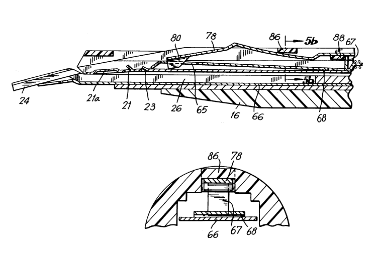
Referring once again to Fig. 2, a plurality of U-shaped clips
22 are positioned within the housing for movement in the distal direction
in preparation for the clamping procedures. The clips are preferably of
titanium for use in clipping blood vessels such as arteries. This
material avoids the "starburst" effect and facilitates enhanced CT
imaging. The clips 22 are aligned in a row as shown, with the leg
portions facing distally. A jaw blade 26 is positioned at the distal end
and includes a pair of jaws 24 for reception of each clip whereby the
jaws are brought together to close the clip about the artery.
The basic objective is to bias the clips toward the distal
direction and to sequentially advance each clip into the jaws after the
jaws have been positioned about an artery. Thereafter, the jaws are
closed and both legs of the "U" shaped clip are brought together to just
sufficiently close the artery as shown in Fig. 12.
The jaw blade 26 is fabricated of a material having sufficient
resilience such that clamping of the distal pair of jaws 24 toward each
other to close a clip therebetween will be followed by return of the jaws
to their original position upon release of the clamping forces.
Stainless steel has been found to be a preferred material capable not
only of withstanding the requisite number of clamping cycles without
ad~erse affect, but also of being suitably sterilized. Furthermore, jaw
blade 26 includes three square shaped apertures 28 dimensioned to receive
three correspondingly shaped pins 30 molded into the lower body half
section 16 of the housing to position the jaw blade 26 with respect to
the body.
` ~1)2~36~
Referring further to Fig. 2, crimping channel 32 is dimensioned
and positioned for slidable movement within the body of the housing and
defines elongated slot 34 having a wider portion 36 at the distal end for
reception of square pins 30. The width of the slot 34 in distal portion
36 of crimping channel 32 is just sufficient to receive the pins 30 to
maintain relative alignment between the jaw blade 26 and the pins 30. A
channel bracket 38, also preferably of stainless steel, is positioned
atop the jaw blade and defines two downwardly extending side walls 40, 42
positioned to be welded to the distal portions of correspondingly
positioned and dimensioned upwardly extending side walls 48, 50 of
crimping channel 32. This channel bracket 38 is positioned just distally
of upst~n~ine tabs 44, 46. It will be appreciated that the crimping
channel 32 forms with channel bracket 38, a rectangular slidable housing
surrounding the jaws 24 of jaw blade 26. Moreover, since the jaw members
24 are formed of outwardly tapered side walls 50, 52, movement of the
crimping channel 32 in the distal direction will cause inward movement of
the jaw members, while movement of the crimping channel in the proximal
direction will result in corresponding proximal movement of channel
bracket 38 thereby relieving the jaw members 24 of the crimping forces
and permitting the jaw members to open.
Referring now to Figs. 2 and 15, jaw members 24 include
generally longitudinal grooves 54, 56 dimensioned to receive a clip 22
therebetween for clipping a body portion. Tissue stop plate 60 shown in
Fig. 2, is positioned between jaw blade 26 and crimping channel 32 and
includes aperture 62 at the proximal end portion for reception of an
appropriate pin (not shown) which extends through the jaw blade 26 and
tissue stop plate 60 to maintain alignment of the jaw blade 26 and the
tissue stop plate 60 when these components are welded together. At the
-18-
2~213S2
distal portion of the tissue stop plate a tab 64 is oriented at
approximately the same downward angle as the jaws 24 for alignment
therewith and includes an arcuate cut-out portion as shown, dimensioned
to snugly receive an artery for locating and positioning the artery in
the precise area within the jaw blades 8S required for applying a clip to
the artery with predetermined precision. The tissue stop plate is
preferably fabricated of a thin stainless steel sheet material.
Referring further to Fig. 2, cover plate 66 is appropriately
dimensioned to rest atop the clip clamping mechanism described
hereinabove, and supports the row of clips 22. Proximally of clips 22 is
positioned a clip follower 68 which is "U" shaped at the distal end to
snugly engage and advance the clips under the action of clip feed spring
72 connected thereto at the distal end and to a pin 74 at the proximal
end. Pin 74 is in turn connected to cover plate anchor tab 66a while
clip pusher bar 78 is positioned for slidable movement thereon between a
proximal position and a distal-most portion. When the next clip 22 is
engaged by the distal nose 80 of clip pusher 78, distal movement of the
clip pusher 78 advances the clip into the slots 54, 56 of jaws 24 of the
jaw blade 26.
Referring again to Fig. 2, upper housing half section 15
includes a longitudinal slot 82 having bridge connections 84, 86, 88 as
shown. In position, the clip pusher bar 78 is snaked over bridge 88 and
under distal bridges 86 and 84 such that bridge 88 will act as a stop
mech~ni~ to prevent the advancement of clip follower 68 when upst~n~ng
tab 67 engages bridge 88 as shown in Fig. 5a. This occurs when the last
clip 22 has been advanced and crimped thereby permitting the clip
follower to advance to its distal-most position under action of spring 72.
Thus by sliding clip pusher bar 78 between the proximal and
-19-
2021362
distal positions, the clip pusher bar may be alternately positioned with
nose 80 behind each successive clip, and thereafter advancing the clip
into the jaws 24 of jaw blade 26 by a pusher mechAnis~ in handle section
12 which will be described. The connection between the mechanism in the
handle 12 is made with the proximal end portion 90 of clip pusher 78
which extends into the handle section. Further, the connection between
the appropriate link of handle 12 with the crimping mechAni~ of jaw
blades 24 is made with the proximal end portion 92 of crimping channel 32
as will be described. The precise action of the handle 12 and its inner
mechanism is such that proximal force applied to trigger 16 causes clip
pusher 78 to push the next clip 22 into the jaws 24 while simultaneously
releasing the crimping channel 32 to the "ready" position for crimping
the clip. Next, the operator squeezes handle 18 toward hand grip 20
which causes crimping channel 32 to move distally to crimp the clip
positioned within jaws 24, while simultaneously moving clip pusher 78
proximally in position to push the next clip 22 into the jaws 24. These
movements are alternately repeated until the last clip 22 is spent.
- Referring to Fig. 3 the handle section 12 of the apparatus is
illustrated with the transmission mechAnis for manually activating the
endoscopic section described previously, i.e., advancing clips distally
and crimping the clips about an artery. The parts of the handle section
12 are separated for convenience of illustration. The handle section 12
includes left body 100 and right body 102. The body parts are fastened
together by fasteners such as screws or rivets extending through
appropriate bosses. Alternatively, the body parts may be ultrasonically
welded or adhesively attached together along their seams or by bosses and
transverse rods or pins in engaged relation. The body parts 100, 102 are
preferably fabricated of a hard plastic material such as LEXAN Brand
-20-
20213G2
polycarbonate material marketed by General Electric Co. Other rigid
-
materials are contemplated. Materials capable of being molded into shape
while being able to sustain the forces applied by the transmission
mechanism are preferred.
The clip loading and crimping system is divided into two
separate systems as described in connection with the endoscopic section.
As noted, a first system pushes the clip next in line from a row of clips
to a position within a pair of clamping jaws 24 as described in
connection with the endoscopic section of the apparatus. The second
system closes the pair of jaws 24 around the clip to cause the clip to
grip the intended artery, tissue, or other blood vessel, while
simultaneously repositioning the clip pusher mechanism to push the clip
next in line into position between the jaws. This procedure is repeated
alternately and sequentially until all clips are spent.
Referring now to Figs. 16-19, in conjunction with Fig. 3, the
clip pusher and clamping loading mechanism will now be described. Handle
18 is pivotally mounted via aperture 108 on pin 104 extending
transversely of the body parts. The handle lB includes a rearward
extension 112 which defines arcuate slot 126 through which pin 110
extends. Pin 113 extends through aperture 114 and functions as a pivot
for left channel link 116 and right channel link 118 which extend in a
generally forward direction. Rearwardly directed left pusher link 120
and right pusher link 122 are mounted for pivotal motion on pin 110
extending through arcuate slot 126.
At the opposite ends left channel link 116 and right channel
link 118 are pivotally mounted to channel tube 124 by pivot pins 127, 129
formed integral therewith and pusher links 120, 122 are connected to
transverse pins 94, 96 arranged for slidable movement within the forward
-21-
2~21~62
cut-out portion 132 of pusher tube 134 for engagement with shoulders 136,
138 of the pusher tube when the links are moved in the proximal
direction. Main spring 140 connects channel tube 124 with pusher tube
134 via pins 119, 131, such that the spring is loaded when the tubes are
separated by squeezing handle 18 toward handle grip 20 causing distal
movement of channel tube 124 and proximal movement of pusher tube 134.
Referring now once again to Figs. 3 and 16 in conjunction with
Fig. 2, it can be seen that pusher tube 134 is connected to clip pusher
bar 78 by proximal end tabs 90 which are inserted by squeeze and release
action into the distal opening 133 of pusher tube 134 with annular steel
pad 142 positioned as an interface between the plastic pusher tube and
the steel pusher bar. Similarly, the crimping channel 32 is connected to
the channel tube 124 by insertion of the proximal legs 92 into the distal
opening 123 of channel tube 124 with annular steel pad 121 positioned as
an interface between the plastic channel tube 124 and the steel legs 92.
With the connections described, the crimping channel and clip pusher are
free to rotate independently of the channel tube and pusher tube as
permitted by the rotation of the proximal legs 90 and 92, within the
distal opening 133 of pusher tube 134 and opening 123 of channel tube 124.
Referring once again to Fig. 3, latch plate 150 is pivotally
mounted and biased upward toward apertured plate 146 in lower wall of
channel tube 124 by spring 148 such that tongue 156 enters the aperture
of plate 146 when the channel tube 124 is moved to its proximal
position. This prevents unwanted forward movement of the channel tube
124 prior to advancing a clip in position within jaw members 24 of jaw
plate 26. Release of tongue 156 is accomplished by engagement of the
latch plate 150 by pin 158 extending downwardly from pusher tube 134 when
pusher tube moves distally under action of mainspring 140 as will be
-22-
2021362
.
developed further. Similarly, pusher release leaf spring 160 is
positioned for entry of tab 162 into a slot 161 in the bottom wall of
pusher tube 134 when the tube is moved proximally by pusher links 120,
122 against the force of mainspring 140, permitting the leaf spring 160
to retain the pusher tube in position against the force of the mainspring
140. Release of the pusher release leaf spring 160 is accomplished by
proximal movement of release lever 164 via finger activated pusher
release button 16 supported at the proximal end by lever support block
168 which slidably moves against the lower wall of pusher tube 134.
In operation, squeezing the handle 18 toward hand grip 20
causes pusher links 120, 122 to pivot and move proximally, resulting in
proximal movement of pusher tube 134 by engagement of pins 94, 96 with
shoulders 136, 138 of pusher tube 134. Proximal movement of pusher tube
134 continues with pusher release spring 160 continuously biased upwardly
until tab 162 enters the slot 161 in the bottom wall of pusher tube 134
thereby ret~inine pusher tube 134 in position against the bias of
mainspring 140. Simultaneously, this action withdraws clip pusher 78 to
a position just proximal of the next clip 22 in preparation for pushing
the clip distally between the jaw members 24 of jaw blade 26. Retention
of the pusher tube in this proximal position by release spring 160 also
retains the clip pusher 78 in the corresponding position until the clip
next in line is to be pushed into the jaws 24. When this is desired,
proximal v~ -..t of pusher release button 16 causes proximal movement of
release lever 164 and engagement of proximal tip 169 with pusher release
spring 160 causing downward movement of the spring and corresponding
release of the pusher tube 134. This action causes distal movement of
pusher tube 134 and clip pusher 78 with corresponding distal engagement
of nose 80 with the next clip 22 thereby positioning the clip into the
slots 54, 56 of jaws 24.
-23-
` ` 2021362
Once clip 22 is positioned within jaws 24 of jaw blade 26
squeezing handle 18 proximally toward hand grip 20 causes distal and
pivotal movement of channel links 116, 118, resulting in distal movement
of channel tube 124 and corresponding distal movement of crimping channel
32. This action causes channel bracket 38 together with crimping channel
32 to engage and squeeze the jaw members 24 of jaw blade 26 thereby
crimping the clip 22 positioned therebetween. At the same time, the
proximal movement of pusher tube 134 resets clip pusher 78 to a position
just proximal of the next clip in re~diness for the next clipping
operation. Reentry of the tab 162 of pusher release spring 160 into slot
161 of pusher tube 134 retains the pusher 78 in position behind the next
clip 22.
Referring further to Fig. 3 in conjunction with Fig. 2, the
feature relating to the rotatable endoscopic section will be described.
Rotating collar 170 is constructed of the same material as the handle,
i.e. preferably a polycarbonate material such as LEXAN brand material.
This collar 170 includes a distal cylindrical nose section 172 and a
proximal barrel section 174. The proximal face of the barrel section 174
includes a plurality of proximally extending teeth 176 positioned
circumfer-entially about the proximal face of the barrel section and the
cylindrical nose section includes an inwardly extending rib 178 at the
distal end. In the assembled condition, the cylindrical nose section
rests within the cylindrical distal opening 182 of the distal end of the
handle and nose piece 184 is fitted over the distal cylindrical end 183
of the handle as shown in Figs. 16-18. Bearing washer 186 and spring
washers 188, 190 are positioned between shoulder 192 of collar 170 and
shoulder 194 formed in the handle body to bias the rotatable collar in
the proximal direction causing tooth 180 on the handle body to engage ~he
-24-
2021362
teeth 176 of the collar 170 to thereby fix the rotatable orientation of
the collar. When the surgeon desires to change the angular orientation
of the endoscopic section, the collar 170 is merely pushed distally to
disengage tooth 180 to free the collar and permit rotation relative to
the handle body. Such rotation of the collar is clearly permitted by the
fact that the cylindrical nose section of the collar is fit snugly within
the corresponding cylindrical distal section 182 of the handle. Except
when the tooth 180 of the handle body is engaged with teeth 176 of collar
170, the collar is otherwise free to rotate within the handle.
Referring now to Fig. 2 in conjunction with Figs. 1 and 3, the
distal cylindrical section 172 of collar 170 includes a distal
cylindrical opening dimensioned to receive the endoscopic cartridge
formed of upper half 15 and lower half 16, with distally positioned tooth
178 of collar 170 positioned within longitudinally extending groove of
upper cartridge half 15 to cause the cartridge to rotate with the collar
170. Similarly, the proximal legs 90 of clip pusher bar 78 are permitted
to rotate within the distal end portion 133 of pusher tube 134 and the
proximal legs 92 of the crimping channel 32 are permitted to rotate
within the distal end portion 123 of channel tube 124. Thus, the entire
endoscopic section may be selectively rotated by the surgeon by simply
pushing collar 170 in the distal direction sufficient to disengage tooth
180 on the handle body and by rotating the collar 170 until the
endoscopic section reaches the desired angular orientation. Thereafter,
by merely releasing the collar the bias of spring washers 190, 188,
causes the collar to move proximally, such that tooth 180 on the handle
body engages the appropriate teeth 176 on the collar 170 to lock the
position of the collar and the endoscopic section. This feature
represents a significant advance in endoscopic surgery when it is fully
-25-
2021362
.
appreciated that the orientation of human tissue or arteries to be
clamped vary widely and that selectivity of orientation of the clip is a
necessity. Without the above-described feature, the entire apparatus
must otherwise be rotated until the proper orientation of the endoscopic
section is reached. Such rotation of the entire apparatus during a
delicate surgical operation would be prohibitive.
Referring now to Figs. 12, 13, 14 and 15, the jaws of the
clamping section of the apparatus are illustrated. Fig. 12 illustrates
the jaws 24 of the apparatus in position after having applied a clip 22
about an artery 98 or other blood vessel to stop the blood flow as
illustrated graphically in Fig. 15. As shown in Fig. 15, the jaw members
24 include longitudinally extending grooves 54, 56 which receive clip 22
as the clip is advanced distally by pusher bar 78. It can be seen that
at the time the jaw members 24 are clamped together, the nose 80 of
pusher bar 78 has been withdrawn proximally to a position proximal of the
next clip 22 and is not permitted to advance in the distal direction
until the surgeon pulls pusher release button 16 in the proximal
direction to release the clip pusher mechanism described previously. Tab
23 prevents the next clip from moving proximally with the pusher bar when
the pusher bar returns to a position proximal of the next clip for the
sequence. Also, prior to release of the pusher bar for distal movement,
fingers 21 upst~n~ing from track 66 prevent distal -,v~ t of the next
clip preventing the clip from falling out through the jaws. In addition,
it is significant to note that once the ~aw members 24 are released from
their clamped condition shown in Fig. 12, by release of handle 18,
clamping of the jaw members 24 may not be repeated until the pusher
release button 16 has been depressed to deliver the next clip between the
jaws 24. Such clamping action is prevented by the position of tongue 156
` ~021362
within the aperture of plate 146 in the bottom wall of channel tube 124
under the upward bias of spring 148. This position prevents distal
movement of the channel tube 124 until the tongue 156 is released from
the aperture of plate 146 by engagement of downwardly extending finger
158 of pusher tube 134 with latch plate 150 when pusher tube 134 is
caused to advance distally by releasing pusher release button 16.
The release action on tongue 156 is shown more clearly in Fig.
17 which illustrates the handle with the pusher tube 134 in the
distal-most position after pusher release button 16 has been depressed to
advance the next clip into the jaw members 24 of jaw blade 26. It can be
seen clearly in Fig. 17 that finger 158 has engaged latch plate 150
pivoting the latch plate downwardly in the counter clockwise direction
against the upward bias of latch spring 148. It will similarly be
appreciated that the proximal movement of pusher tube 134 during the
squeezing action of handle 18 and jaw members 24 will continue until the
tab 162 of the upwardly biased pusher release spring 160 engages the slot
161 in the bottom wall of the proximal section of pusher tube 134 thereby
causing the pusher tube to be locked in position corresponding to the
nose 80 of pusher bar 78 being positioned just proximal of the next clip
22 for the next clip advancing step as described hereinabove. It can be
appreciated readily that this safety feature avoids the possibility of
squeezing the jaw members 24 about an artery or other tissue with no clip
positioned therebetween. Thus, the only time in the sequence of
operation that the jaws can be squeezed is after the advancement of a
clip 22 therebetween.
Referring now to Figs. 4-11, the inner mechanism and function
of the distal portion of the endoscopic section are illustrated. In Fig.
4 a plan view from above, is shown of the distal portion of the
-27-
2021 362
endoscopic section, illustrating the jaw members 24 and the nose 80 of
pusher bar 78 in position to advance the clip 22 into the jaw members.
At this time, the row of clips 22 are advanced to their distal-most
positions under bias action of clip feed spring 72 between anchor shaft
74 on cover plate pin anchor tab 66a and pin 71 on clip follower 68 shown
in Fig. 2. Fig. S is a cross-sectional view taken along lines 5-5 of
Fig. 4 illustrating the clip 22 and the nose 80 of pusher bar 78 in
position just proximally thereof. The view of the nose 80 of pusher bar
78 shown in dotted lines is intended to illustrate the proximal-most
position of the nose 80 of pusher bar 78 as represented by the last
portion of the squeezing motion of handle 18 toward hand grip 20 thus
establishing with certainty, that the nose 80 of clip pusher 78 is in
fact positioned proximally of the next clip 22 after the handle 18 is
released and the nose 80 of pusher bar 78 is permitted to move distally a
small distance as shown behind clip 22 as represented by relaxation of
the combined tolerance build-up of the components interacting with each
other. Escapement means in the form of upst~ndine tabs 21 in cover plate
66 prevent the next clip 22 from distal movement before it has been
advanced distally by the pusher bar 78. Arch 21a assists proper
orientation of the clip entering the jaws. Tab 23 prevents proximal
movement of clip 22 once it has been advanced distally by nose 80, i.e.
the proximal return JV~ t of nose 80 does not move clip proximally (by
friction) along with the nose.
Fig. 5a illustrates still another significant feature of the
present invention which prevents further distal advancement of the clip
pusher 78 after the last clip 22 has been advanced distally into the jaws
24 and clamped about an artery. In particular, the proximal portion of
clip follower 68 includes upst~ding tab 67 which is positioned and
-28-
~021362
dimensioned to engage bridge 88 on upper cartridge half 15 when clip
follower 68 assumes the distal-most position shown in Fig. 5a under bias
of spring 72. This position is assumed by clip follower 68 after the
last clip has been advanced distally into the jaws 24. Thus, the
engagement of upstanding tab 67 with bridge 88 prevents further distal
movement of the clip follower at this stage. Furthermore, as shown in
Fig. 5a, the distal position of clip follower 68 results in slot 65 now
assuming lB distal-most position such that nose 80 of clip pusher bar 78
may drop into slot 65 thus preventing further distal movement of the
pusher bar 78 after the last clip has been spent. This is a further
safety feature of the invention in that the apparatus is inactivated
after the last clip is spent, thus avoiding the possibility of the
surgeon clamping the jaws 24 about an artery with no clip in position.
Fig. 5b is a cross-sectional view taken along lines 5a-5a of Fig. 5
illustrating the clip follower 68 and the clip cover plate 66 in the
position shown in Fig. 5a.
Referring now to Fig. 6, a plan view from above similar to Fig.
4 is shown of the distal portion of the endoscopic section with clip 22
shown in Fig. 5 now advanced distally to a position within the jaws 24 by
nose 80 of clip pusher 78. Fig. 7 is a cross-sectional view taken along
lines 7-7 of Fig. 6 illustrating the clip 22 and clip pusher bar 78 in
the distally advanced position after advancing clip 22 into the jaws 24.
Fig. 8 is a schematic view of the distal end of the completely
disposable instrument of the present invention positioned in the body
cavity. The jaws of the intrument are positioned around cystic duct 202
where two clips have already been closed and a third is being positioned
for closure.
-29-
2021362
,
Fig. 9 is a cross-sectional view taken along lines 9-9 of Fig.
6, illustrating the crimping channel 32, the tissue stop 64, clip 22,
pusher bar 78 and jaws 24, and cover plate (or clip track) 66.
Fig. 10 is a similar cross-sectional view taken along lines
10-10 of Fig. 6 illustrating the clip advancing components. Fig. 11 is a
cross-sectional view similar to Fig. 10 illustrating the clip advancing
mech~ni~ distal of the cross-section shown in Fig. 10.
Referring now to Figs. 16-19, the inner clip advancing and jaw
squeezing mechanism is shown in various stages of the operation. Fig. 16
is an elevational cross-sectional view of the handle 18 of the apparatus,
illustrating the pusher tube 134 in the proximal-most position
corresponding to the position of the pusher bar 78 shown in Fig. 5, i.e.
with the nose 80 just proximal of the next clip 22 in readiness to
activate the clip distally into the jaws 24. Additionally, with pusher
tube in the proximal position, downwardly extending finger 158 has moved
out of engagement with latch 150 thereby permitting tongue 156 to enter
the aperture of channel latch plate 146 thus preventing any distal
movement of channel tube 124. This condition locks handle 18 in the
distal position whereby squeezing the handle toward hand grip 20 is
prevented.
Referring now to Fig. 17, there is shown a cross-sectional view
of the handle 18 of the apparatus with the pusher tube in the distal-most
position corresponding to the position of pusher bar 78 as shown in Fig.
7, i.e. with the clip 22 advanced distally into the jaws 24 of jaw blade
26. As can be seen further in Fig. 17, the distal position of pusher
tube 134 has now resulted in release of tongue 156 of latch plate 150
from the aperture of channel latch plate 146 in the bottom wall of
channel tube 124 thereby permitting advancement of channel tube 124 ~nd
-30-
2021362
crimping channel 32 distally to squeeze jaws 24 in conjunction with
channel bracket 38.
Referring now to Fig. 18, a cross-sectional view of the handle
18 is shown after the crimping action has taken place on clip 22
positioned within jaws 24 shown in Figs. 16 and 7. The position of the
components shown in Fig. 18 corresponds to the position of the jaws shown
in Figs. 12-15, i.e., in the clamped position about clip 22. In the
cross-section shown in Fig. 18, the pusher tube 134 in the proximal-most
position and the channel tube is in the distal-most position such that
crimping channel 32 and channel bracket 40 are in the distal-most
position shown in Figs. 12-15.
Referring to Fig. 19 a cross-sectional view of the handle 18 is
shown after the last clip 22 has been spent, i.e. corresponding to the
position of the clip follower 68 shown in Fig. 5a. As noted hereinabove,
the clip follower 68 of the endoscopic section is prevented from moving
further distally by interaction with bridge 88 formed in upper cartridge
half section 15. Additionally as noted, it can be seen in Fig. 5a that
clip follower 68 defines slot 65 at the distal portion which is bounded
on the distal end by a bridge 63 which is positioned to engage the nose
80 of pusher bar 78 when clip follower 68 has advanced to the distal-most
position shown in Fig. 5a, i.e. after the last clip has been spent. In
this position, the clip follower is now sufficiently distal to engage the
nose 80 of clip pusher bar 78 which is biased downwardly by the
configuration of pusher bar 78 and by the resilient properties of the
material from which the pusher bar is fabricated, i.e. stainless steel.
This engagement with bridge 63 prevents further distal movement of clip
pusher bar 78 and correspondingly of pusher tube 134. By preventing
pusher tube 134 from distal movement with channel tube 124 locked in its
2021362
proximal position by tongue 156 of latch plate 150, further squeezing
-
action of handle 18 toward hand grip 20 is also prevented. This locking
action correspondingly prevents distal movement of crimping channel 32.
As shown in Fig. 19, pusher release button is depressed but full distal
movement of pusher tube 134 is prevented by the engagement of nose 80 of
pusher bar 78 with bridge 63 of clip follower. Only a small distal
movement is permitted as seen by the position of slot 161 in pusher tube
134 relative to the position of pusher release spring 160. This locked
position of pusher tube 134 also serves to prevent downwardly depending
finger 158 of pusher tube 134 from distal movement sufficient to release
tongue 156 of latch plate 150 from channel tube 124 as shown. Thus, the
crimping mech~ni is inactivated for safety purposes.
This feature is extremely significant in disabling the
apparatus from squeezing jaws 24 of the jaw blade 26 on an artery alone,
i.e. with no clip positioned therebetween. Further, all movement of the
clip advance mechanism is now prevented after the last clip has been
spent. At this stage, the entire instrument is considered disposable and
may be disposed of in accordance with correct approved disposal
procedures.
Referring once again to Fig. 3 in conjunction with Figs. 16-19,
the lost motion spring 210 is shown having transverse arms 212 and tab
214. Spring 210 provides bias force on pusher links 120, 122 such that
squeezing action on handle 18 ~ eS proximal movement of pusher tube
134. Thus, partially closing the jaws 24 of jaw blade 26 will cause
pusher tube 134 to move sufficiently proximal to make certain that pusher
bar 78 has moved proximally of the next clip 22. Without such JV~_ -nt
it may be possible for the surgeon to squeeze the jaws, not fully
appreciating that the pusher bar 78 has not moved to a position proximal
-32-
2021362
.
of the next clip 22. This proximal movement of the pusher bar
transmission is thus assisted by lost motion spring 210 which -~i izes
the repositioning movement of the pusher bar 78 behind the next clip
whether the jaws are squeezed fully or partially. In particular, the
proximal bias provided by spring 210 on pusher links 120, 122 ~xi izes
the movement of pusher tube 134 in relation to the movement of handle 18
by maint~ining pusher links 120, 122 in their proximal-most positions
prior to squeezing the handle 18. This ~i proximal movement of
pusher links 120, 122 in turn results in proximal movement of pusher tube
sufficient to engage tongue 162 of release spring 160 thus -kjng certain
that pusher bar 178 is repositionèd sufficiently proximally to advance
the next clip 22 into the jaw members 24.
Fig. 27 shows a cutaway perspective view of a second embodiment
of the present invention 100' which generally comprises an actuating body
lOOa' supporting a non-detachable endoscopic portion lOOb'. Included are
means for actuating the instrument, transmission means, means for
applying a surgical clip to a blood vessel or the like, means for locking
the instrument, and means for unlocking the instrument and repositioning
another clip. Clip applier 100' is intended to be fully disposable.
More particularly, referring now to Figs. 27 and 28, frame 102'
comprises a left portion 102L' and a right portion 102R'. These portions
are optimally fastened together by means of fastening screws, although
rivets, welds, adhesives, or other means of joining the frame portions
may be used. Frame 102' is elongated and has an interior surface
defining a distal opening 102c', a proximal end 102h', an interior distal
chamber 102a', a circumferential groove 102b', an upper guideway 102e' to
receive pins 107' and 103' (see below), a mounting slot 102f' (to receive
spring 116'), elongated access aperture 102g', and pin mounting holes
2021362
.
102i', j', k', m' and n' for receiving pins 101', 119', 105', 118' and
101', respectively. The frame is of overall size and shape convenient
for being held in the hand.
Referring additionally now to Figs. 29, 30, 31, 32, 33 and 34,
handle 112' is pivotally mounted to the frame 102' by means of handle pin
101' which is disposed through holes 102n' and 102i' in the frame, and
hole 112b' in the distal portion of the handle 112'. Handle 112' also
has an elongated cavity 112a' for receiving the toggle levers 110' and
113'. Hole 112c' receives lever pin 145'. Handle 112' serves as a means
to activate the instrument when said handle is pivoted clockwise by the
user of the instrument.
Toggle levers 113' and 110' are T-shaped levers which are
pivotally mounted to the handle 112'. Lever 113' has a proximal aperture
113b' for receiving pin 145', a distal aperture 113d' for receiving pin
101', said pins being respectively disposed through holes 112c' and 112b'
in the handle 112'. Lever 113' also has an elongated slot 113a' for
receiving pin 111', and a lower slot 113c' for receiving pin 103'.
Toggle lever 110' is similar to toggle lever lI3' except that
the lower portion is offset to the left by bend llOe'. Slot llOc' in the
lower portion is for receiving pin 103'. Slot llOa' in the upper portion
receives pin 111', and apertures llOb' and llOd' receive pins 145' and
101', respectively. The lower legs of the toggle levers 113' and 110'
transmit motion of handle 112' to the rear channel 121' via pin 103'.
Pusher bar rear link 109' is a flat elongated piece having an
aperture lO9a' for receiving pin 111', an aperture lO9b' for receiving
pin 107', and a curved notch lO9c' for accommodating pin 145' when the
handle 112' is pushed down into a closed position. Pusher bar rear link
109' transfers movement from the handle 112' to the rear pusher bar 108'.
-34-
~0~1362
Pusher bar front link 114' is an elongated flat piece having a
bend 114c', aperture 114a' for receiving pin 111', and aperture 114b' for
receiving pin 119'.
Referring additionally now to Figs. 35 and 36, rear channel
121' is longitudinally movable and has a distal aperture 121a' to receive
screw 122' for attachment to the rear channel tube 123' (see Fig. 41.).
Rear channel 121' also has a proximal aperture 121b' to receive pin
103'. Slot 121g' receives pin 119' and aperture 121c' receives channel
lock pin 105'. Rear channel 121' has an overhang 121d', and a lower
offset flap 121e' with notch 121f' for holding the distal end of spring
106'. Rear channel 121' receives pivotal movement from toggle levers
110' and 113', and transfers motion linearly to the rear channel tube
123'.
Rear pusher bar 108' is longitudinally movable and has a distal
aperture 108a' to receive pin 124' for mounting the distal end to the
rear pusher bar tube 125' (see below). Rear pusher bar 108' also
comprises upper slot 108b' for receiving pin 119', lower slot 108d' for
receiving the channel lock pin 105', cr ing surface 108c', and offset
flap 108e' with proximal notch 108f' for receiving the proximal end of
spring 106'. Rear pusher bar 108' further possesses a stopping edge
108g' which provides a catch means for engaging the locking flap of the
pusher bar stop spring 116' (see below). Rear pusher bar 108' provides
means for transferring pivotal movement from link 109' linearly to rear
pusher bar tube 125'.
Referring additionally now to Figs. 37 and 38, resilient stop
spring 116' has an elongated distal end with flap 116a' for mounting into
slot 102f' in the right frame 102R'. Spring 116' has a bend 116e', and a
proximal end divided into a pusher bar stop latch 116c' which provides
` 202136~
means for locking the rear pusher bar 108' and a c~~ ine surface 116b'
which is angled by bend 116f', as illustrated. Slide member 115' has a
cam member 115a', an anchor post 115b' for engaging the distal hook end
of spring 117', an upper base 115c', a connecting portion 115d', a lower
base, 115e', and a curved pushing surface 115f'. Slide member 115' rides
along the longitudinal access aperture 102g' and is mounted in the frame
102' such that the upper base is in the enclosed interior space of the
frame 102', and the lower base 115e' and pushing surface 115f' project
outside of the frame. The cam member 115a' is engagable with the camming
surface 116b' of the spring 116'. Upon moving distally the cam member
115a' pushes spring 116' downward, thereby pivoting the stop latch 116c'
into a non-engagable position below the stopping edge 108g' of the rear
pusher 108'.
Spring 117' is connected by a distal hook to the anchor post
115b' and by a proximal hook to the pin 118', which is received into
aperture 102m' in right frame portion 102R'.
Referring additionally now to Figs. 39a, 39b and 40, channel
lock pin 105' provides means for locking the rear channel 121' and
comprises a cylindrical portion 105a' with c~ ine surface 105b', and an
axial shaft portion having ends 105c' and 105d'. When in the non-locking
position channel lock pin 105' is located as illustrated in Fig. 39a.
End 105d' of the shaft is disposed axially through spring 104' and into
hole 102p' in the left frame portion 102L'. End 105c' is disposed
through slot 108d' of the rear pusher bar 108'. When channel 105' is in
the locking position as shown in Fig. 39b and 105c' is disposed through
aperture 121c' in the rear channel 121', and hole 102k' in the right
frame portion 102R'.
Fig. 41 illustrates the first transmission means ~for
transmitting linear movement to the clip advancing means) comprising the
-36-
202 1 362
rear pusher bar 108', rear pusher bar tube 125', and front pusher bar
tube 126', and the second transmission means comprising the rear channel
121', rear channel tube 123', and front channel tube 129'. The rear
channel 121' is connected to rear channel tube 123' by means of screw
122' disposed through aperture 121a'. As can be seen additionally from
Fig. 42, rear channel tube 123' has a proximal projection 123b' having an
aperture 123a' for receiving screw 122'. Rear channel tube 123' provides
means for transferring linear movement from the rear channel 121' to the
front channel tube 129' and is generally cylindrical in shape having a
hollow bore and an external circumferential notch 123c' for enabling the
front channel tube to rotate.
Rear channel tube 123' is slidably mounted within the bore of
front channel tube 129' which provides means for transferring linear
motion from rear channel tube 123' to the front channel 133'. Front
channel tube 129' has a distal projection 129b' having an aperture 129a'
for receiving screw 130' which is the mounting means for the front
channel 133'. Front channel tube is slidably mounted within the distal
cylindrical chamber 102a' of frame 102'. Aperture 129a' in the front
channel tube 129' receives pin 128' which projects into groove 123c' in
the rear channel tube 123'.
Rear pusher bar 108' is connected to the proximal projection
125b' of the rear pusher bar tube 125' by means of pin 124' disposed
through apertures 108a' in the rear pusher bar 108' and 125a' in the rear
pusher bar tube 125'. Rear pusher bar tube 125' provides means for
transferring linear movement from the rear pusher bar 108' to the front
pusher bar tube 126' while permitting the front pusher bar tube 126' to
rotate. Rear pusher bar tube 125' is generally of cylindrical shape and
has an external circumferential notch 125c' for engaging proximal thrust
202 1 362
collar 126b' in the front pusher bar tube 126'. Front pusher bar tube
126' has a distal projection 126c' having an aperture 126a' for receiving
screw 127'. Screw 127' is for mountin~ the proximal end of front pusher
bar 143' and is disposed through aperture 143a' in the front pusher bar.
Front pusher bar tube 126' transfers linear movement to pusher
bar 143' from rear pusher bar tube 125'. Cooperating thrust collar 126b'
and cylindrical notch 125c' provide means for allowing rotation of front
pusher bar tube 126' relative to rear pusher bar tube 125'.
Rear pusher bar 108', rear pusher bar tube 125', front pusher
bar tube 126', rear channel 121', front channel tube 129', and rear
channel tube 123' are all slidable in the longitudinal direction. When
the instrument 100' is actuated rear pusher bar 108', pusher bar tube
125' and front pusher bar tube 126' move proximally as indicated by arrow
~pn ~ and the rear channel 121', rear channel tube 123', and front channel
tube 129' move distally as indicated by arrow "D". In addition to
longitudinal ve~-nt~ front pusher bar tube 126' and front channel tube
129' are rotatable around the instrument axis.
Referring additionally now to Fig. 43, collar 132' is generally
cylindrical in shape having a radial aperture 132a', for receiving pin
131'; a circumferential detent 132b' for mounting into circumferential
groove 102b' in the frame; a rectangular slot 132c' for receiving front
channel 133', jaw blade 135', and front pusher bar 143'; and a distal
portion 132d' which is located exterior to the frame. Collar 132' is
rotatable but does not move longitnd~nA]ly. The proximal end of front
channel 133' is mounted to the front channel tube 129' by means of screw
130' disposed through apertures 133a' and 129a'. Front channel 133' has
a slot 133b' to allow longitudinal movement without interference from pin
131'. When the instrument 100' is actuated, by pressing handle 112',
202i36~
front channel 133' moves distally. Slot 133b' must therefore extend
longitudinal for a distance sufficient to permit full distal movement of
the front channel 133'.
Front channel 133', which provides means for closing jaws
135b', also has upper and lower guide rails 133c' which project
transversely from the top and bottom of front channel 133', and which
extend longitudinally. Guide rails 133c' serve as means to retain and
align front pusher bar 143' and jaw blade 135' as well as means to close
jaws 135b'.
Front pusher bar 143' is connected to the front pusher bar tube
126' by means of screw 127' disposed through apertures 143a' and 126a'.
Front pusher bar 143' has a bend 143b' to widen the distance between it
and the front channel 133' so as to accommodate jaw blade 135' disposed
therebetween. Front pusher bar 143' has a slot 143c' to allow
longitudin~l movement without interference from pin 131'. When the
instrument is actuated by pressing handle 112', front pusher bar 133'
moves proximally. Slot 143c' must therefore extend longitudinally for a
distance sufficient to permit full proximal movement of the front pusher
bar 143'.
Jaw blade 135' has an aperture 135a' for receiving pin 131' and
has a proximal end disposed within cylindrical collar 132'. Jaw blade
135' provides clip closing means.
As can be seen from Figs. 41, 42 and 43, when collar 132' is
rotated for example by manually turning the distal portion 132d', jaw
blade 135', front channel 133', and front pusher bar 143' are likewise
rotated, as well as front tube channel 129' and front pusher bar tube
126'. Unlike front pusher bar 143' and front channel 133', jaw blade
135' does not also move longitudinally.
-39-
2021362
Referring now to Fig. 44, the endoscopic portion of the
-
instrument comprises a cover tube 144' enclosing front pusher bar 143',
spring anchor shaft 142', spring 141', rear pusher clip 140', front
pusher clip 139', (optional) clips 138', clip carrier 137', safety stop
136', jaw blade 135', tissue stop 134', front channel 133', and sealing
block 401'.
Cover 144' is an elongated tube fixed at its proximal end to
collar 132'. Front pusher bar 143' is an elongated piece longitudinally
disposed within cover 144'. In addition to features discussed above,
front pusher bar 143' comprises an elongated longitudinal slot 143d', and
inclined pusher tip 143e'. Carrier 137' is longitudinally positioned
along the side of pusher 143' and provides a means for carrying surgical
clips 138' which are disposed within the longitudinal guide rail 137a'.
At its proximal end carrier 137' has a mounting post 137b' for spring
anchor shaft 142'. The proximal end of spring 141' is mounted on anchor
shaft 142', and the distal end of spring 141' is mounted to the proximal
end of rear pusher clip 140'. Rear pusher clip 140' has a bar 140a'
which rides in slot 143d' of the front pusher bar. The distal end of the
rear pusher clip 140' contacts the proximal end of the front pusher clip
139', which engages and pushes the clips 138' distally.
Safety stop 136' is an escapement with prong members 136a'
which project into the path of the clips 138' to limit the distal loading
of the clips to one clip at a time. Safety stop 136' is attached to the
distal end of carrier 137'.
Jaw blade 135' is disposed within the front channel 133' and
has a pronged distal end with jaws 135b' which are flexibly movable
toward each other. When the instrument is actuated the jaws are forced
together by the c ing action of the guide rails 133c' of the distally
-40-
2 0 2 1 3 6 ~
moving front channel 133'. Tissue stop 134', which is fixed to the side
-
of the jaw blade 135' prevents the blood vessel or other tissue from
proximally moving beyond the jaws 135b'.
Figs. 45a and 45b illustrate a clip located between jaws
135b'. Grooves 135c' facilitate the proper alignment and positioning of
the clip. Jaws 135b' also have c~ ~ng surfaces 135d' which are
contacted by the distally moving guide rails 133c' thereby forcing the
jaws (and the clip 138') into closed position. The jaws blade 135' does
not move longitudinally.
Uhen handle 112' is pressed, it pivots clockwise thereby
pivoting levers 110' and 113', and link 109' (which pivots
counterclockwise) and link 114' (which pivots clockwise). Link 109'
pushes the rear pusher bar 108' longitudinally in the proximal direction,
and link 114' pushes the rear channel 121' longitudinally in the distal
direction. Rear pusher bar 108' pulls the front pusher bar in the
proximal direction via rear and front pusher bar tubes 125' and 126'
respectively. The front and rear pusher bar tubes 126' and 125' are
coupled so as to permit rotation of the front pusher bar tube 126'
relative to the rear pusher bar tube 125'. The rear channel 121' pushes
channel 133' distally via rear channel tube 123' and front channel tube
129'. The front and rear channel tubes 129' and 123' are coupled so as
to permit rotation of the front channel tube 129' relative to the rear
channel tube 123'. When channel 133' advances distally, it cams the jaws
of jaw blade 135' into the closed position, thereby closing a surgical
clip .
As the rear pusher bar 108' is being pushed proximally to the
rear of the instrument, offset flap 108e' rides over the stop latch
portion 116c' of spring 116', and when the offset flap passes proximally
-41-
2021362
beyond the latch 116c', said latch 116c' then springs upward and engages
edge 108g' of the rear pusher bar 108'. The pusher bar is then locked
and prevented from going distally forward. Furthermore, when the rear
pusher bar is moved to a proximal position, channel lock pin is permitted
to extend further towards 102R'. When the handle 112' is subsequently
released, the rear channel 121' is allowed to return proximally to its
original prefiring position. When it does so, aperture 121c' becomes
aligned with aperture 102k' in the right frame 102R', and the lock pin
slides therethrough, locking the actuating mechanism so that the handle
cannot be pressed again. The reason for this safety locking mechanism is
that, having already closed a clip 138', jaws 135b' are not yet chambered
with another clip. As a precautionary measure, lock pin 105' prevents
the operating surgeon from mistakenly closing the jaws when they are
empty.
To release the lock mechanism, the surgeon pushes the exposed
pushing surface 115f' of the slide member 115' forward in the distal
direction. Cam member 115a' then presses down the c~ 1ng surface 116b'
of spring 116'. This pushes down latch 116' and disengages the rear
pusher bar 108', which slides forward. When rear pusher bar 108' slides
forward the inclined c~ ing surface 108c' wedges the lock pin 105' out
of engagement with apertures 102k' and 121c', thereby unlocking the
actuation mechanism. Furthermore, as rear pusher bar 108' springs
forward the front pusher bar 143' also moves forward and pushes another
clip 138' into position between jaws 135b'. The instrument 100' may then
be reactuated to apply another clip.
A feature of this invention is that the transmission system
transmits linear, longitudinal motion to the endoscopic portion of the
instrument, while allowing the endoscopic portion of the instrument to
-42-
2021 362
rotate relative to the frame and actua~hg ~ pnlsm. This is
accomplished by means of the couplings between front and rear pusher bar
tubes 126' and 125', between front channel tube and rear channel tube
129' and 123', and between the collar 132' and the frame 102'.
In the above described embodiment the endoscopic portion is not
detachable. However, a further alternative embodiment of the present
invention comprises a disposable and replaceable endoscopic portion which
is detachable from a reusable frame.
Fig. 46 shows a cutaway perspective view of the alternative
embodiment of the present invention 200' which generally comprises a
reusable actuating body 200b' supporting replaceable and disposable
endoscopic portion 200a'. Included are means for actuating the
instrument, transmission means, means for applying a surgical clip to a
blood vessel or the like, means for locking the instrument, and means for
repositioning another clip and unlocking the mech~n1 ! _ .
More particularly, referring to Figs. 46, 47 and 48, frame 201'
comprises a left frame portion 201L' and right frame portion 201R'.
These portions may be either cast or ~chin~d pieces of polymeric resin
or metal. The left and right portions may optionally be fastened
together by means of fastening screws although rivets, welds, adhesives,
or other means of joining the frame portions may be used. Frame 201' is
elongated and is of overall size and shape convenient for being held in
the hand. Frame 201' has an interior surface defining a distal opening
201h', a proximal opening 201c', an interior transmission guideway 201g',
apertures 201a' and 201b' for receiving pins 224' and 204' respectively,
backstop notch 201d' for spring 205', bottom notch 201e', mounting notch
for spring 225', and distal opening detents 201i'.
-43-
2021362
, . ,
Referring additionally to Figs. 49, 50a, 50b, 51, 51a, 52 and
53, handle 222' is an elongated piece which is pivotably mounted to the
distal end of frame 201' by means of pin 224' which is disposed through
aperture 222a' in the handle and 201a' in the frame. Aperture 222b 'is
adapted to receive link pivot pin 223'. Elongated cavity 222c' is
adapted to receive pusher links 216' and 216a', and channel links 218'
and 219'.
Channel links 218a' and 218b' preferably are curved elongated
pieces having upper apertures for receiving pin 223', and lower apertures
for receiving pins 217' and 220' respectively. Channel links 218a' and
218b' provide means for transferring movement from the handle 222' to the
channel tube 212', and act as a load limiter as well.
Pusher links 216a' and 216b' are elongated pieces having
apertures for receiving pin 223' and apertures for receiving pins 215'
and 242' respectively. Pusher links 216a' and 216b' provide means for
transferring movement from the handle 222' to the pusher tube 209'.
Pins 215', 217', 220' and 242' each comprise a disc like center
portion, and two shafts projecting axially therefrom. The inwardly
projecting shaft is adapted to be received into the respective aperture
of the channel or pusher link, and the oppositely projecting shaft is
adapted to be received into the axial bore of the respective roller
bearings 241', 240', 221', and 243'. The inwardly projecting shafts of
pins 217' and 220' are received into apertures in the channel tube 212,
whereas the inwardly pro;ecting shafts of pins 215' and 242' are disposed
through the side slots of the pusher tube 209'.
The roller bearings 240', 221', 241', 243' are adapted to slide
along the guideways 201g' in frame 201'.
-44-
gO21362
Pusher tube 209' provides a first transmission means for
transmitting linear movement to the pusher bar 229' and comprises an
elongated tubular piece located within the transmission guideway 201g'.
At its proximal end pusher tube 209' has apertures 209a' for receiving
cross pin 208', and proximal terminal notch 209b'. Pusher tube 209' also
comprises a bottom slot 209c' for enepging release button 206', an
aperture 209d' for receiving pusher tube c~ ing pin 210', side slots
209e' for receiving pins 215' and 242', and an interior circumferential
projection 209f' at the distal end of the pusher tube for engPging the
proximal end of the pusher 229' (see below).
Channel tube 212' provides a second transmission means for
transmitting linear movement to channel 238' and comprises a proximal
aperture 212a' for receiving pin 211', apertures 212b' for receiving pins
217' and 220', bottom aperture 212c' for engaging spring clip 225', and
proximal slot 212d' for receiving camming pin 210'. Pusher tube 209' is
slidably mounted within channel tube 212' and the two are biased into
alignment by means of spring 207' which is axially disposed within pusher
tube 209' and which is attached at its proximal end to pin 208' and at
its distal end to pin 211'. Channel tube 212' further has an interior
circumferential projection 212e' at the distal end of for engaging the
proximal end of channel 233'.
Leaf spring or spring clip 225' is a catch means for locking
the channel tube in the distal position after the instrument has been
actuated. Spring clip 225' has a distal end with angled portion 225a'
for being mounted in notch 201f' of the frame. At its distal end,
resilient spring clip 225' has a catch 225b' which is engagable with
aperture 212c' of the channel tube 212'. Spring clip 225' has a c~ Ine
surface 225c' which is contacted by pusher tube c~ ing pin 210' when the
-45-
2021~62
pusher tube moves in the distal direction, thereby depressing the spring
clip 225' and disengaging and unlocking the channel tube 212'.
Release button 206' is pivotally mounted at the proximal end of
frame 201' by means of pivot pin 204' which is disposed through aperture
206a' and 201b'. Release button 206' has a proximal end 206b' which
projects through aperture 201c' in the frame, and a catch 206c' at its
distal end for engaging aperture 209c' of the pusher tube 209'.
Referring additionally now to Fig. 54, outer tube 213' is
adapted to be received into distal access opening 201h' of the frame.
Outer tube 213' has a proximal opening 213a', a circumferential outer
notch 213b' which is adapted to receive detent 201i' in the distal
opening of the frame, an aperture 213c' for receiving pin 214', and a
circumferential inner notch 213d' at the distal end of the outer tube for
engAging and interlocking with the collet 227'.
Pin 214' is received into aperture 213c' of the outer tube and
projects a small distance beyond the outer surface thereof. It provides
a contact surface to engage the interior notches of the sleeve 226' as
discussed below.
The above described portion of the alternative embodiment is
the reusable portion 200b'. The replaceable endoscopic portion 200a'
described below is adapted to be received into the distal end of the
reusable portion 200b' and to project outwardly therefrom. The
endoscopic portion 200a' is rotatable relative to the reusable portion
200b'.
Referring additionally now to ~igs. 55 and 56, the replaceable
portion 200a' includes a coupling which comprises sleeve 226' and collet
227'. The coupling is adapted to connect the endoscopic portion to the
non-endoscopic instrument body portion while permitting rotation of the
-46-
2021362
endoscopic portion. Sleeve 226' is a tubular piece having several
longitudinal notches 226a' on the interior surface of the proximal end
for engaging the protruding portion of pin 214'. The pin 214' and
notches 226a' cooperate so as to form click-stop settings wherein the
sleeve can rotate to any of several positions and be temporarily seated
in the chosen position. Sleeve 226' has slot 226e' and detent 226b'
which are adapted to cooperate with the collet 227' to retain the collet
227' within the sleeve 226' once the collet 227' has been inserted.
Detent 226b' serves as a backstop by abutting the distal surface of
projection 227e' in the collet. Key 226d' on the inside surface of
sleeve 226' is adapted to fit into longitudin~l spline 227d' in the
outside surface of collet 227'. C ine surface 226c' is adapted to push
against surface 227c' in the outer surface of the collet 227'.
The proximal portion of collet 227' comprises several
proximally extending prongs 227a', each prong having on its outer surface
a projection 227b' for ene~eine and locking into circumferential notch
213d' on the interior surface of the outer tube 213'. Once inserted into
sleeve 226', collet 227' locks into place by means of projecting surface
227e' which abuts detent 226b' in the sleeve. Projections 227f' lock
into cooperating notches in the cartridge halves 228', 229'. When
camming surfaces 227c' on the exterior surface of the prongs 227b' are
contacted by c~ ine surface 227c' in the sleeve as for example when the
sleeve 226' and collet 227' are pushed together, the prongs 227b' are
biased closer to the axial center of the collet 227', thereby enabling
them to disengage from the outer tube.
The endoscopic portion is axially disposed through the collet
and sleeve, and connects to the actuating and transmission mechanisms
described above. As can be seen from Fig. 55, the endoscopic portion
-47-
20213~2
comprises upper and lower cartridge portions 228' and 239', and disposed
within the cartridge portions are pusher bar 229', clip pusher 232', clip
feed spring 231', spring anchor shaft 230', bottom clip track 234',
capture plate` 235', jaw blade 236', tissue stop 237', channel 238', clip
233' and sealing block 401'.
Referring additionally now to Figs. 57 and 58, upper cartridge
portion 228' comprises an elongated relatively small diameter piece
which, in addition to lower cartridge portion 239', houses the endoscopic
portion of the transmission and clip applying mechAnis ^. The cartridge
may be constructed of polymeric material suitable for surgical procedures
and has a width of about 0.3 inches for practical use in endoscopic or
laparoscopic surgical procedures. Upper cartridge portion 228' comprises
a proximal end 228a' having a projection 228b' for engaging corresponding
notch 239b' in the lower cartridge portion, notches 228e' for engaging
corresponding projections 227f' in the collet, stopping surface 228f' for
limiting the distal or forward movement of the pusher bar 229' by
engaging projections 229f', stopping surface 228g' for limiting the
rearward or proximal movement of the pusher bar 229' by eneAeine bend
229b', rectangular projections 228d' for alternating alignment with
projections 239c' in the lower cartridge portion 239', and guide way
228c' for aligning the pusher bar 229'.
Lower cartridge portion 239' comprises a proximal portion 239a'
having a notch 239b' for receiving projection 228b', projections 239c',
and projections 239d' for engaging the corresponding apertures in the jaw
blade 236'.
Channel 238' is an elongated piece having a distal end with
c ine portions 238a' for contacting the jaws 236b' and pushing them
into a closed position. Slots 238b' are for receiving capture pl~te
-48-
` 20213~2
235'. Channel 238' has an aperture 238c' for receiving pro;ections 239d'
_
of the bottom portion of the cartridge 239', bend 238e', and proximal
prongs 238d' having notches 238e' for engaging projection 212e' in the
channel tube. Prongs 238' are resiliently flexible towards each other.
Tissue stop 237' has a proximal end 237a' to protect the blood vessel or
other tissue from entering too far between the jaws 236b'.
Jaw blade 236' has a proximal end with apertures 236a' for
engaging projections 239d' in the cartridge thereby aligning and firmly
seating the jaw blade 236'. Jaws 236b' at the distal end have c~ ing
surfaces 236c' which are contacted by the c~ ing surfaces 238a' of the
channel. When the channel moves in the distal direction the jaw blades
236b' are cammed into the closed position, thereby closing a clip
positioned within said jaws.
Bottom track 234' for clips 233' is an elongated flat piece
having a distal end with projections 234a' which serve as an escapement
means to limit the feeding of clips 233' to one clip at a time. Bottom
track 234' has a mounting post 234b' for the spring anchor 230', and flat
side projections 234a' for fitting and alignment in the cartridge 228'.
Clip pusher 232' has a distal end with prongs 232b' adapted to
fit around any distally push clips 233' forward. Clip pusher 232a' has a
proximal post 232a' for mounting spring 231'.
Spring 231' is axially mounted at its proximal end to anchor
230', and at its distal end to clip pusher 232'. Spring 231' resiliently
moves the clip pusher forward in the distal direction.
Pusher bar 229c' provides a means for advancing the clips into
the jaws. Distal end 229c' pushes the clips forward one at a time from
track 234' to a position between the jaws.
Pusher bar 229' has a bend 229b' which abuts stopping surface
-49-
202~362
228g' to limit the proximal movement of he pusher bar, projections 229f'
which abut stopping surface 228f' for limiting distal movement of the
pusher bar 229', and a proximal end with resilient prongs 229a', each
prong having a notch 229d' for e~ging projection 209f' in the pusher
tube. The resiliency of the prongs allow them to bend inward
sufficiently to enable them to snap into place when the endoscopic
portion of the instrument is inserted into the reusable body.
Referring additionally now to Fig. S9a, which shows a top
sectional view of the proximal coupling end of the endoscopic portion
200a', and to Fig. S9b which shows the same portion in side view, the
cartridge portions 228' and 239' which house the endoscopic portion of
the transmission means, are disposed axially through the collet 227' and
sleeve 226'. As can be seen from the illustration pusher bar 229' and
channel 238' are sandwiched between the cartridge portions, but have
proximal ends 229a' and 238d' which, being slightly wider than the
cartridge section 239a' and 228a', protrude slightly from the sides.
Inclined proximal edges of the pusher bar (229e') and the channel (238f')
allow the respective prongs to be biased inward while the endoscopic
portion 200a' is being inserted into the reusable body 200b'. The
proximal portions 229a' and 238a' will then snap into place as channel
tube projection 212e' engages notches 238e' and pusher tube projection
209f' engages notches 229d', the prongs 229a' and 238d' thereupon
resiliently expanding to their original position. The proximal movement
of pusher tube 209' will draw the pusher bar 229' rearward in the
proximal direction, and the distal movement of channel tube 212' will
push the channel 238' forward.
Initially, the clip applier of the present invention is in the
locked position, i.e., there is no clip loaded between the jaws. The
-50-
2021362
,
jaws are biased open and are free to cam between the open and closed
positions. This facilitates insertion of the endoscopic portion into an
endoscopic tube or into the body since the jaws can cam partially closed,
thereby avoiding interference with the positioning of the instrument.
When the clip applier has been properly positioned, the user
may release the first and second transmission means from the locked
position by pressing the release button (206'). This disengages the
first transmission means (pusher bar 209') which slides forward. The
first transmission means comprises means to release the catch from the
second transmission means. For example, in the first described
embodiment the rear pusher bar has an inclined cr ing surface 108c' to
push aside channel lock pin 105' which locks the second transmission
means. In the alternative embodiment pusher tube 209' (the first
transmission means) has a depending pin 210' which, upon being carried
forward, depresses leaf spring 225', thereby releasing the catch 225b'
from the channel tube 212' (second transmission means).
The first transmission means of both embodiments transfers
motion to the pusher bar (143' and 229') which has a distal pusher end
located behind the distal most clip in the array. When the pusher bar
(i.e., the clip advancing means) moves forward the distal most clip is
advanced to the jaws.
The second transmission means, upon being released, returns
backward, thereby pulling the channel (133' in the first described
embodiment; 238' in the alternative embodiment). The instrument is now
ready to be actuated for clip application.
The endoscopic portion of the instrument 200a' may be rotated
relative to the reusable body 200b' by manually turning sleeve 226'.
As in the previously described first embodiment, the clip
2021362
applier of the alternative embodiment is actuated by pressing the
handle. When handle 201' is pressed pusher links 216a' and 216b' move
pusher tube 209' proximally to the rear of the instrument thereby
repositioning the pusher bar behind another clip, and channel links 218a'
and 218b' move the channel tube 212' forward thereby causing the jaws to
be closed for applying the surgical clip. The distally moving channel
tube 212' pushes channel 233' forward, thereby c~ in~ the jaw blade 236'
into a closed position for applying the surgical clip. The pusher tube
209' is locked in the proximal position when catch 206c' of the release
button 206' engages aperture 209c' in the pusher tube. The channel tube
212' is locked in the distal position when catch 226b' of the leaf spring
engages aperture 212c' of the channel tube.
The instrument remains locked until the release button 206 is
pressed, thereby unlocking the instrument as explained above.
Figs. 60a and 60b illustrate the apparatus of the present
invention in conjunction with the cannula 300', or endoscopic guide tube,
of a trocar to apply a surgical clip to blood vessel 302'. (Trocars
generally comprise a cutting tip, cannula, and valve means for sealing
the cannula). The trocar is used to penetrate the skin 301' of a patient
and is inserted into the patient's body. Upon withdrawal of the cutting
tip (not shown) of the trocar, the endoscopic portion of the apparatus
200a' is inserted axially through the cannula 300' and maneuvered to the
operating site where ~aws 236b' engage blood vessel 302'. The endoscopic
portion is in sealing engagement with the valve means to prevent entry or
egress of gases.
Optimally, the gaseous sealing means comprises sealing block
401', as illustrated in Fig. 51. Sealing block 401' comprises U-shaped
upper and lower portions 401a' and 401b', respectively, which are
-52-
- 2021362
positioned together so as to define an axially extending opening 401c'
_
through which components such as channel 238' and pusher 229' may be
disposed. The inner surface of opening 401c' is in close contiguity with
the surface of the components disposed therethrough. A layer of silicone
grease 402' may be employed to prevent gases from leaking between the
surfaces. The sealing block 401' may be located within the cover tube
144' or the cartridge portions 228' and 239' of the embodiments disclosed
herein. See, for example, Figs. 44 and 55.
While the above description contains many specifics, these
specifics should not be construed as limitations on the scope of the
invention, but merely as exemplifications of preferred embodiments
thereof. Those skilled in the art will envision many other possible
variations that are within the scope and spirit of the invention as
defined by the claims appended hereto.
-53-