Note: Descriptions are shown in the official language in which they were submitted.
2 ~
SCATTERED TOTAL INTERNAL REFLECTANCE APPARATUS
This application is a continuation-in-part of U.S. Serial
No. 149,243 filed January 27, 1988 which in turn was a
continuation-in-part of U.S. Serial No. 879,236 filed June
26, 1986, each of which applications was assigned to the
assignee of the present invention and which are hereby
incorporated by reference into the present application.
Field of the Invention
This invention relates to apparatus and methods fox
scanning, detecting and manipulating light including a
scattered total internal reflectance immunoassay ystem.
Back~round of the Invçntion
Many human disease states are identified on the basis of
immunoassay techniques which rely upon the specificity
between immunoglobulins, whether monoclonal or polyclonal,
and their respective binding partners, which may be
haptens, antigens, or other analytes, all of which may
hereafter be collectively and interchangeably referred to
herein as "ligands" and "ligand binding partners."
Furthermore, "ligand" also means any molecule having an
affi~ity to bind or complex with a "ligand binding
part~er", including chelators, immunobinders, nucleic acid
ætrands, bioreceptors, and hydrophobic binders. Over the
past fifteen or so years, there has been a substantial
amount of ef~ort involved in the development of
immunoassay techniques utilizing the so-called sandwich
and competitive techniques. The sandwich technique
involves the immobilization oF an antigen by one antibody
and then subsequent labaling by attachment of a second
antibody having associated therewith a detectable label.
ORD-82
,. . ~
Reverse immunoassays for the detection cf antibody are
similar but instead put antigen on the surface for
reaction with the sample antibody. Competitive techniques
are useful for antigens having only a single epitopic site
for reaction with an antibody. Accordingly, and as the
name implies, such techniques rely upon the competition of
the antigen with another labeled antigen for a binding
site on an immobilized antibody. The substitutions
necessary for antibody detection tests are obvious and
need not be covered here in any great detail.
Of great importance in the laboratory is the development
of highly sensitive techniques which can be run in either
batch random access, panel, or stat modes. Preferably,
such techniques will be homogeneous in nature, i.e., and
as used herein, they will be conducted solely within one
container without any accompanying requirement to
physically separate out components following reactions
during the assay~
It is one object of the present invention to provide a new
immunoassay system which is highly sensitive and which is
homogeneous in nature.
U.S. Patent 3,939,350 to Kronick and the Kronick citations
therein referenced describe an immunoassay system which
allows for the measurement of biochemical analytes by
fluorescence in a liquid sample. Kronick employs a
physical phenomenon known under the name of total internal
reflectance. This optical phenomenon occurs wherein
light, when directed through a high refractive index
material toward the interface of that material with a
second material having a lower refractive index at greater
than a critical angle, all light is reflected from that
interface save for a microscopic evanescent wave which
~RD-82
~2~2~
.
- 3 -
, propagates into the second material for only a short
distance. The second material may, for instance, be water
or another aqueous medium in which an assay is being
conducted. Kronick noted that when he brought materials
; 5 which had been fluorescently labeled down to the interface
and within the field of the evanescent wave, he could
energize the fluorescent molecules and detect fluorescence
which then emanated into the overlying solution. The
Kronick system, however, looks at fluorescence which
cannot be readily modified by alteration of the
fluorescent labels in order to suit the system under
study. Due to the nature of the specificity of the
fluorescent label with respect to the wavelength of the
escitation frequency, one is limited to a discrete light
source providing the critical excitation frequency. To
date, most investigators favor the He-Ne laser light
source due to its reliability and low cost as well as the
low cost of associated optics. Such a light source,
however, brings concomitant difficulties in tailoring
fluorescent molecules to be excited by the He-Ne laser
outputO The organic, inorganic, and bio-organic
techniques required are especially difficult to contrsl in
the immunoassay arena. Further, Kronick's reliance on
fluorescence is accompanied by additional disadvantages
associated with bleaching of the fluorescent molecules and
generally critical matching of fluorescent molecule
e~citation wavelength with laser output wavelength
necessary to obtain good quantum efficiency.
It is an sbject of the present invention to provide a new
immunoassay system which avoids the disadvantages
associated with fluorescent labels and the criticality
associated with matching an e~citation source.
ORD-82
_ 4 _ ~ 'S~2
It is another object of the present invention to employ
the principles of total internal reflection but with far
greater flexibility regarding the choice of illumination
sources.
U.S. Patent 4,181,441 to Noller describes a system similar
to that of Kronick. Noller, however, taught that the
assay should be conducted by measurement of light
absurption in a liquid sample which could then be
correlated to the presence of biochemical analytes,
Although the Noller system employs diffexent physical
principles than the Kronick system, light absorption
measurements are similarly subject to poorer
signal-to-noise ratios due to small differences in large
light signals thereby making such a system inherently less
sensitive than desired.
It is another object of the present invention to avoid
employing light absorption measurements while still
gaining the advantages to be provided by the total
internal reflectance phenomenon~
U.S. Patent 4,521,522 to Lundstrom teaches yet another
immunoassay based upon reflectance and the use of
Brewster's angle. This system relies upon a different
optical phenomenon wherein directing a light beam,
polarized in the plane of incidence, upon an interface,
for example that formed between plastic and liquid,
results in the transmission of a strong light beam into
the liquid when such light strikes the interface at the
Brewster angle. At the Brewster angle, substantially no
light is reflected.
The Brewster angle is a function of the refractive indices
of the two materials as well as the direction of
ORD-R2
2~2~2~
polarization. Lundstrom noted that upon the growth of a
hiochemical layer at the interface, the Brewster angle
condition would be disrupted resulting in increasing light
reflectance, particularly at angles less than the Brewster
angle. Unfortunately, the Lundstrom assay only works
effectively with a wash step since the transmission of the
beam into the liquid also results in the generation of
light scatter and thus a spurious signal.
It is another object of the present invention to utilize
light scatter but to avoid light scatter generated by the
transmission of light into the liquid which occurs
naturally when light is directed at an interface at the
Brewster angle. Accordingly, it is yet another object of
the present invention to avoid employing a Brewster angle
condition.
It is another object of the present invention to provide
an apparatus and method for scanning, detecting and
manipulating light.
Summary of th~ Invention
In accordance with various aspects and the principles of
the present invention, there is provided an immunoassay
system which utilizes scattered total internal reflectance
~STIR) as a measure of the presence of particular ligands
to b~ determined in an aqueous solution. The invention
relies in part upon the identificatisn of the critical
ang~e associated with total internal reflectance. The
angle is largely a function of the refractive index of the
material through which an incident light wave is directed,
e.g. plastic, and the relatively lower refractive inde~ of
the material in which the immunoassay is being conducted,
e.g. an aqueous solution. It is measured from a line
ORD-82
` ~0222~.~
-- 6 --
perpendicular to the interface between the two materials,
and thus at its ma~imum, 90, will lie in the plane of the
interface.
Light direct~d through the plastic toward the interface
formed by the aqueous sample and plastic materials at the
critical angle will result in total internal reflectance
of the light within the plastic. It is recognized that no
materials in the real world are perfect and accordingly,
it is preferred that the incident light be directed toward
the interface at an angle several degre4s greater than the
critical angle, most preferably in the range of
appro~imately 6 greater in order to ensure that the basic
conditions of total internal reflectance are met. At such
an angle, the incident collimated light, preferably from a
laser, is totally internally reflected within the plastic
save for the propagation of the evanescent wave parallel
to the surface of the plastic and approximately 1/4~ from
the surface. Similarly, smooth surfaces at the interface
are preferred for optimum signal ~uality. Unlike
conventional fluorescent techniques including those
of Kronick, the present assay system is flesible with
respect to light wavelength since particle size may be
readily adjusted to match the available light source (or
vice versa) to provide acceptable light scatter.
Fluorescent molecules are not readily adjustable with
respect to e~citation wavelength.
,
Most ideally, the light source will be a He-Ne light
source, however, other lasers with di~ferent wavelength
outputs have been used and still other sources suggest
themselves including light emitting diodes and other
nonlaser light sources.
O~D-82
.
Applicants' immunoassay system further relies upon
conventional immunoassay techniques. However, applicants'
immunoassay system also employs a particulate label having
a higher refractive index than that of the solution, and
most preferably also higher than the first light
transmissive material, e.g. plastic in the foregoing
example. Such particles would include, for instance, red
blood cells, other materials having a highly reflective
surface such as metallic particles, and nonmetallic
substances such as glass or plastics, e.q. late~
particles, and the like. Most preferably, colloidal gold
is used as a label for the solution phase immunologically
active component. While the use of colloidal gold as a
label is known, see for example U.S. Patent No. 4,313,734
Leuvering, almost no nonagglutination related uses of the
label have been made to date due to the difficulties
associated with its detection, particularly in homogeneous
type systems. It was surprisingly discovered by the
inventors hereof that the unique combination of STIR with
colloidal gold has resulted in an eztremely efficient and
sensitive homogeneous assay system. It is believed, but
not known for certain that this is due primarily to the
interaction of the colloidal gold particles with the
evanescent wave. Indeed, experience implies that
particles having an increasingly higher inde~ of
refraction than that of the underlying solid generally
increasingly scatter light. While particles with indices
of refraction less than the underlying solid, providing
they are also not equal to that of the aqueous medium,
would also scatter light, such are less preferred.
Assuming for the moment a conventional sandwich technique,
one immunoglobulin or ligand binding partner is
immobilized on the surface and binds antigen or other
ligand to be determined. Thereafter, (or simultaneously,
ORD-82
~ 2 ~
or if not previously) a second immunoglobulin, directed at
a second epitopic site on the ligand, and labeled directly
or indirectly with colloidal gold, binds to the ligand
creating the so-called ~'sandwich". In this arrangement,
the presence of the colloidal gold disrupts the
propagation of the evanescent wave resulting in scattered
light which may be detected by a photomultiplier or other
light sensor to provide a responsive signal. Another
important aspect of the present invention involves the
physical location of the detector. Ths detector is
ideally placed at an angle greater than the critical angle
and in a location whereby only light scattered backward
toward the light source is detected. This location
thereby ideally avoids ths detection of spurious scattered
light within the bulk liguid medium.
Another feature of the instant invention is that the
immunoassays are diffusion rate controlled and not
particularly temperature dependent. This is in strong
contrast to ELISA and various other immunoassay techniques
wherein temperature control is critical since small
changes in temperature in such systems results in wide
variations in assay results per unit of time.
2~ It was surprisingly found by the inventors hereof, that as
a result of the combination of these elements, rapid,
sensitive results could be obtained in a homogeneous
environment without requiring the complicated equipment
previously associated with colloidal gold assay techniques.
In addition, these elements led to the invention of
apparatus to scan, detect and manipulate liqh~ in
unespected ways.
ORD-82
JI-~J
BRIEF DESCRIPTION OF THE DRAWINGS
Fig. 1 depicts an optically transparent member 53 with
refractive index n2 having a fluid contacting surfacs 52
in contact with fluid 54 having a refractive inde~ nl
which is less than n2. The total internal reflectance
critical angle 55 measured from a line 50
perpendicular to the plane of the fluid contacting surface
52 is the minimum angle of illumination reguired to
provide total internal reflectance at surface 52. This
angle is defined by the equation
~C = sin~l ~n
~ n2 J
The critical angle, ~c' is only defined in the higher
refractive index medium and can range from 0 to 90.
Light propagating from a point on the fluid contacting
surface 58 at the total internal reflectance critical
angle 55 would follow the path depicted as 57. All
light propagating through the optically transparent member
53 from a point on the fluid contacting surface 58
between the plane of the sample fluid contacting surface
51 and the total intexnal reflectance critical angle
~5 of the fluid contacting surface, will propagate in
the range depicted as 56.
Fig. 2 is a simplified elevation view of the cuvette and
the rotating optics mechanism used to illuminate and read
it.
Fig. 3 is cross-section of a cuvette.
ORD-82
-- 10 --
Fig. 4 is a perspective view of a cuvette and a
paraboloidal reflector which is the first component of the
receiving optics.
Fig. 5 depicts an apparatus with laser illumination above
critical angle.
Fig. 6 depicts illumination and detection light paths used
when illumination is above the critical angle.
Fig. 7 shows data obtained with the apparatus of Figure 5.
Fig. 8 depicts an apparatus with light emitting diode
illumination above the critical angle.
Fig. 9 shows data obtained with the apparatus of Figure 8.
Fig. 10 shows data obtained with the apparatus of Figure 2.
Fig. 11 shows data obtained with the apparatus of Figure 2.
Detailed Description o the Invention and Best Mode
The present invention provides an apparatus for detecting
the presence of an analyte o interest in a sample. This
apparatus comprises a light source; housing means for
receiving an optically transparent member having a sample
contacting surface, said member in said housing means
being disposed such that the sample contacting surface is
illu~inated with light emitted from said light source; and
photodetection means which e~cludes the detection of light
which propagates in a geometric optical path from the
light source, said photodetection means being capable of
detecting elastically-scattered light which propagates
through the optically transparent member from the
ORD-82
illuminated sample contacting surface between the plane of
the sample contacting surface and the total internal
reflectance critical angle of the sample contacting
surface. Within this application, "photodetection means~
is defined as a system for detecting photons having a
wavelength equal to the wavelength of the illuminating
light~ and includes combinations of photon detectors (e.g.
photomultiplier tubes), lenses, mirrors, light filters,
optical fibers, prisms, apertures, and masks. A geometric
optical path is the path that a family of light rays will
follow based on first order reflection and refraction of
idealized surfaces (imperfection free) and ignoring the
effects of surface and bulk material imperfections,
diffraction, interference, scatter, and partial reflection
at surfaces. Further, within this application,
"elastically-scattered light" (also referrred to herein as
"scatter" and "scattered light") means incident light
which has been redirected by an object without changing
the wavelen~th of the light, by means other than dopler
shifting, due to the difference in the reEractive indes of
the object and its surrounding medium Fluorescence, also
known as inelastic scatter, is the light emitted by a
light absorbing molecule after the molecule has absorbed a
photon of light. The wavelength of the absorb~d light is
less than the wavelength of the emitted light.
Flouorescent light is always of a wavelength different
from the light incident on the light absorbing molecule.
Critical angle", also referred to herein as "total
internal reflectance critical angle", is the angle (less
than 90) measured from the line perpendicular to an
intsrface between materials of different refractive
inde~e~, beyond which total internal reflection can occur,
and is defined by the equation
ORD-82
-- 2~2~
- 12 -
~C = Sin~l ~nl ~
~ n2 J
wherein nl is the lower refractive index and n2 is the
higher reEractive inde~ of the two mediums forming the
interface. The critical angle can only e~ist in the
higher refractive index medium. Light which illuminates
the interfac~ from the lower refractive inde~ material at
any angle (0 to 90) cannot be refracted into the higher
refractive index medium at an angle greater than or equal
to the critical angle. Total internal reflection occurs
exclusively when an interface between materials of
different refractive indexes is illuminated from the
higher refractive inde~ medium beyond the critical angel,
causing all the incident illumination to be reflected at
the interface unless it is perturbed by diffraction,
; scatter, or absorption.
The present invention also provides another apparatus for
detecting the presence of an analyte of interest in a
sample. This apparatus comprises a light source; housing
means for receiving an optically transparent member having
a sample contacting ~urface, said member in said housing
means being disposed such that the sample csntacting
surface is illuminated with light emitted from said light
source which propagates through the optically transparent
member at angles between the plane of the sample
contacting surface and the critical angle for total
internal reflectance; and photodetection means which
e~cludes the detection of light which propagates in a
geometric optical path from the light source, said
photodetection means being capable of detecting
elastically-scattered light which propagates through the
optically transparent member from thP illuminated sample
ORD-82
~2~
- 13 -
contacting surface between the plane of the sample
contacting surface and the total internal reflectance
critical angle.
Suitable light sources for the apparatuses of the present
invention provide collimated or uncollimated light,
polarized or unpolarized light, or monochromatic or
polychromatic light. Preferred light sources include
lasers (e.g., He-Ne lasers), light emitking diodes (LEDs),
flash lamps, arc lamps, incandescent lamps, and
fluorescent discharge lamps.
Suitable optically transparent members, e.g., cuvettes,
are comprised of glass, quartz, silicon, plastics such as
polycarbonate, acrylic, or polystyrene, or oils comprising
silicone or high molecular weight hydrocarbons.
Suitable photodetection means comprise photon detectors
such as photomultiplier tubes, photodiodes ~e.g., PIN
diodes and gallium-aluminum-arsenide diodes), cadmium
sulfide photoresistive cells, phototubes, and pyrolytic
detectors.
Also provided is a method for detecting the presence of a
light scattering molecule on the surface of an op~ically
transparent material. This msthod comprises illuminating
said light scattering molecule, detec~ing light scattered
slastically by said light scattering molscule which
propagates through said optically transparent material
be~ween the plane of the surfac0 of the optically
transparent material on which the light scattering
molecule is located and the total internal reflectance
critical angle of the surface on which the light
scattering molecule is located, and correlating detected,
elastically-scattered liyht to the presence of the light
ORD-82
- 14 -
scattering molecule on the surface of the optically
transparent material. Within this application, an
"evanescent wave" means a nonpropagating light wave such
as a wave in the region of a surface on the side of the
5 surface opposite the side of illumination, produced when
the illuminating light undergoes total internal
reflection. Also within this application a "light
scattering molecule" means a molecule which causes
incident light to be elastically scattered. ~Molecule"
10 includes, in the case of crystalline and elemental
materials, two or more atoms.
Still further, the present invention provides a method for
detecting the presence of a light scattering molecule on
the surface of an optically transparent material. This
method comprises illuminating said light scattering
molecule with an evanescent wave resulting from a light
wave which propagates through said optically transparent
material, detecting light scattered elastically by said
light scattering molecule which propagates through said
optically transparent material between the plane of the
surface of the optically transparent material on which the
light scattering molecule is located and the total
internal reflectance critical anyIe of the surface on
which the light scattering molecule is located, and
correlating detected, elastically-scattered light to the
presence of the light scattering molecule on ths surface
of the optically transparent material.
Further provided is a method for detecting an analyte in a
fluid sample wherein said analyte is a ligand of a ligand
- ligand bindiny partner pair. This method comprises the
steps of:
ORD-82
_ 15 -
a) providing an optically transparent material
having a refractive index greater than the
refractive inde~ of said fluid sample, said
optically transparent material having a sample
contacting surface to which a plurality of ligand
bindlng partners of said ligand - ligand binding
partner pair are immobilized;
b) further providing light scattering
particle-labeled ligands capable of forming
complexes with said immobilized ligand binding
partners;
c) contacting said fluid sample and said ligh~
scattering particle-labeled ligands with said
: sample contacting surface under conditions such
that said analyte and said light scattering
particle-labeled ligands each form complexes with
said immobilized ligand binding partners;
d) illuminating said comple~es with an evanescent
wave resulting from a light wave which propagates
through said optically transparent material;
e) detecting light scattered elastically by said
light scat~ering particl~s of said complexes;
f) correlating elastically-scattered light to the
presence of complexes on said sample contacting
surface; and
ORD-82
.' : . . -
:
:
- 16 -
g) comparing the presence of comple~es on the sample
contacting surface with the presence of comple~es
on a sample contacting surface for a standard
control, thereby detecting the analyte in the
fluid sample.
; Within this application, "particle" means one or more
molecules. "Labeled" means directly linked, e.g.,
conjugated, cross-linked, or adsorbed, or indirectly
linked, e.g., linked via an antibody.
Further yet is provided a method for detecting an analyte
in a fluid sample wherein said analyte is a ligand of a
ligand - ligand binding partner pair. This method
comprises the steps of:
a~ providing an optically transparent material
having a refractive index greater than the
refractive index of said fluid sample, said
optically transparent material h~ving a sample
contacting surface to which a plurality of ligand
binding partners of said ligand - ligand binding
partner pair are immobilized;
b) further providing light scattering ligands
capable of forming comple~es with said
immobilized ligand binding partners;
c~ contacting said fluid sample and said light
scattering ligands with said samplP contacting
surface under conditions such that said analyte
and said light scattering ligands each form
comple~es with said immobilized ligand binding
partners;
ORD-82
.
- 17 ~
d) illuminating said c~mplexes;
e) detecting light scattered elastically by light
scattering ligands of said complexes and which
propagates through said optically transparent
material from the sample contacting surface
between the plane of the sample contacting
surface and the total internal reflectance
: . critical angle of the sample contacting surface;
i ' 10
f) correlating elastically-scattered light to the
presence of complexes on said sample contacting
surface; and
.
g) comparing the presence of complexes on the sample
contacting surface with the presence of comple~es
on a sample contacting surface for a standard
: control, thereby detecting the analyte in the
fluid sample.
Within this application, Ulight scattering ligands" means
ligands or light scattering particle labeled-liganas which
cause incident light to be elastically scatterPd.
Further still is provided a method for detecting an
analyte in a fluid sample wherein said analyte is a ligand
of a ligand - ligand binding partner pair. This method
comprises
ORD-82
:
.
- 18 -
a) providing an optically transparent material
having a refractive index greater than the
refractive inde~ of said fluid sample, said
optically transparent material having a sample
contacting surface to which a plurality of ligand
binding partners of said ligand - ligand binding
partner pair are immobilized;
b) further providing light scattering ligands
capable of forming complexes with said
immobilized ligand binding partners;
c) contacting said fluid sample and said light
scat~ering ligands with said sample contac~ing
surface under conditions such that said analyte
and said light scattering ligands each form
comple~es with said immobilized ligand binding
partners;
:
d) illuminating said complexes with an evanescent
wave resulting from a light wave which propagates
through said optically transparent material;
e) detecting light scattered elastically by light
- scattering ligands of said comple~es and which
propagates through said optically transparent
material from the sample contacting surface
between the plane of the sample contacting
surface and the total internal reflectance
critical angle of the sample contacting surface;
f) correlating elastically-scattered light to the
presence of comple~es on said sample contacting
surface; and
ORD-82
~.
- ' :
-- 19 --
g) comparing the presence of complexes on the sample
contacting surface with the presence of complexes
on a sample contacting surface for a standard
control, thereby detecting the analyte in the
fluid sample.
In one embodiment of the invention, the methods provided
herein may be performed wherein the sample contacting
surface is contacted with the fluid sample before being
contacted with the ligands. In another embodiment of the
invention, the sample contacting surface is contacted with
the ligands before being contacted with the fluid sample.
In yet a further embodiment of the invention, the sample
contacting surface is simultaneously contacted with said
fluid sample and the ligands. In still another embodiment
of the invention, the xample is mixed with the ligands so
as to form a mi~ture, and the mixture is contacted with
the sample contacting surface.
Furthermore, in a preferred embodiment of the invention,
light scattering particle-labeled ligands comprise ligands
labeled with colloidal gold particles.
In still another embodiment of the inventiont a method is
provided for detecting an analyte in a fluid sample. In
this method the analyte is a ligand having an epitope ~or
which a first ligand binding partner is specific and an
epitope for which a second ligand binding partner is
specific. The method comprises:
, ~
i ORD-82
~ 9 ~7
- 20 - ~v ~ ~SJ
a) providing an optically transparent material
having a refractive inde~ greater than the
refractive index of said fluid sample, said
optically transparent material having a sample
contacting surface to which a plurality of first
ligand binding partners are immobilized;
b) further providing light scattering
particle-labeled second ligand binding partners;
c) contac$ing said fluid sample and said light
: scattering particle-labeled ~econd ligand binding
partners with said sample contacting surface
under conditions such that immobilized first
ligand binding partner : analyte : light
; scattering particle-labeled second ligand binding
partner complexes are formed;
d) illuminating said complexes with an evanescent
wave resulting from a light wave which propagates
: through said optically transparent material;
e) detecting light scattered elastically by said
: light scattering particles of said complexes;
: . f) correlating elastically-scattered light to the
presence of complexes on said sample contacting
surface; and
: 30 9) comparing the presence of complexes on the sample
: contacting surface with the presence of comple~es
on a sample contacting surface for a standard
control, thereby detecting the analyte in the
fluid sample.
ORD-82
2~?~2~
- 21 -
Still another method is provided for detecting an analyte
in a fluid sample, wherein said analyte is a ligand having
an epitope for which a first ligand binding partner is
specific and an epitope for which a second ligand binding
partner is specific. This method comprises:
a) providing an optically transparent material
having a refractive inde~ greater than the
refracti~e index of said fluid sample, said
optically transparent material having a sample
contacting surface to which a plurality of first
ligand binding partners are immobilized;
b) furth~r providing light scattering second ligand
binding partners;
c) contacting said fluid sample and said light
scattering second ligand binding partners with
said sample contacting surface under conditions
such that immobilized first ligand binding
partner : analyte : light scattering second
ligand binding partner comple~es are formed;
d) illuminating said comple~es;
e) detecting light scattered elastically by said
light scattering second ligand binding partners
of said comple~es and ~hich propagates through
said optically transparent material from the
sample contacting sur~ace between the plane of
the sample contacting surface and the total
internal reflectance critical angle of the sample
contacting surface;
ORD-82
.
~ ~ ~ 2 '~
- 22 -
f~ correlating elastically-scattered light to the
presence of complexes on said sample contacting
surface; and
g) comparing the presence of complexes on the sample
contacting surface with the presence of complexes
on a sample contacting surface for a standard
control, thereby detecting the analyte in the
fluid sample.
Within this application, "light scattering second ligand
binding partners" means second ligand binding partners or
particle labeled second ligand binding partners which
cause incident light to be elastically scattered.
Still further is provided a method for detecting an
analyte in a fluid sample wherein said analyte is a ligand
having an epitope for which a first ligand binding partner
is specific and an epitope for which a second ligand
binding partner is specific. This method comprises:
a) providing an optically transparent material
having a refractive index greater than the
refractive inde~ of said fluid sample, said
optically transparent material having a sample
contacting surface to which a plurality of first
ligand binding partners are immobiliz d;
b) further pro~iding light scattering second ligand
binding partners;
;
. 35
.
)RD-82
2~
- 23 -
c) contacting said fluid sample and said light
scattering second ligand binding partners with
said sample contacting surface under conditions
such that immobilized first ligand binding
partner : analyte: light scattering second ligand
binding partner complexes are formed;
d) illuminating said complexes with an evanescent
wave resulting from a light wave which propagates
through said optically transparent material;
e) detecting light sca~tered elastically by said
light scattering second ligand binding partners
of said complexes and which propagates through
said optically transparent material from the
sample contacting surface between the plane of
the sample contacting surface and the total
internal reflectance critical angle of the sample
contacting surface;
f) correlating elastically-scattered light to the
presence of comple~es on said sample contacting
surface; and
g~ comparing the presence of comple~es on the sample
contacting surface with the presence of compleses
on a sample contacting surface for a standard
control, thereby detecting the analyte in the
fluid sample.
: 30
In one embodiment of the present invention, the methods
provided herein may be performed wherein the sample
contacting surface is contacted with the fluid sample
before being contacted with the light scattering
particle-labeled second ligand binding partners or the
~ORD-82
.
- 24 -
light scattering second ligand binding partners. In
another embodiment of the invention, the methods described
herein may be performed wherein the sample contacting
surface is contacted with the light scattering
particle-labeled second ligand binding partners or the
light scattering second ligand binding partners before
being contacted with said fluid sample. Still further,
the sample contacting surface may be simultaneously
contacted with the fluid sample and the light scattering
particle-labeled second ligand ~inding partners or the
light scattering second ligand binding partner, e.g., the
fluid sample may be mi~ed with the light scattering
particle -labeled second ligand binding partners or the
liyht scattering second ligand binding partners so as to
form a mixture, and the mi~ture is contacted with the
sample contacting surface.
In a preferred embodiment of the invention, the light
scattering particle-labeled second ligand binding partners
comprise second ligand binding partners which are labeled
with colloidal gold particles.
Finally, an apparatus is provided for detecting the
scattered internal reflectance as a measure of particular
ligands to be determined in an aqueous solution.
Further provided is an apparatus for scanning a plane
polarized light source comprising first polarization means
for converting plane polarized light to circularly
polarized light, means for detecting said circularly
polarized light received from said first polarization
means and second polarization means for reconverting said
directed circularly polarized light into plane polarized
light so that the plane of polarization of said light will
be dependent only upon the orientation of said means for
~RD-82
~?;,~
- 25 -
reconverting said circularly polarized light into plane
polarized light.
Further provided is a method for scanning a plane
polarized light source comprising converting plane
polarized light to circularly polarized light, directing
said circularly converted light and reconverting said
directed circularly polarized light into plane polarized
light so that the plane of polarization of said light will
be dependent only upon the orientation of said means for
reconverting said circularly polarized light into plane
polarized light.
Further provided is an apparatus for detecting light
comprising first paraboloidal reflecting means and second
paraboloidal reflecting means whose a~is is nearly
coincident with and nearly symmetrical to said first
reflecting means.
Further provided is a method for detecting light
comprising reflecting light from first paraboloidal
reflecting means to second paraboloidal reflecting means
whose a~is is nearly coincident with and nearly
symmetrical to said first reflecting means.
Still further, an apparatus for detecting light is
provided comprising first paraboloidal reflecting means
~ ~ and second ellipsoidal reflecting means having its axis
,~ nearly coincident with and nearly symmetrical to said
first paraholoidal reflecting means and its vertex to
nearest focus di tance substantially equal to the verte~
to focus distance of said first paraboloidal reflecting
means.
,. .;
~ 35
~ '
~ ~RD-82
- 26 - 2~22~
Further provided is a method for detecting light
comprising reflecting light from a first ellipsoidal
reflecting means to a second paraboloidal reflecting means.
Further provided is an apparatus for dissipating the
specular reflection of light occuring when substantially
collimated light leaves a material with a refractive indes
greater than its surroundings comprising two planar
surfaces of said material aligned relative to each other
so that said substantially collimated light is multiply
specularly re~lected until said collimated light is
substantially dissipated from said material.
Further provided is a method for dissipating specular
reflection of light occuring when substantially collimated
light leaves a material with a refractive index greater
than its surroundings comprising aligning two planar
surfac0s within 10 degrees of parallel so that said
substantially collimated light is multiply specularly
reflected until said collimated light is substantially
dissipated.
First Series of Experiments
An apparatus embodying the principles of STIR was
constructed utilizing an equilateral flint glass prism,
~ Model 01-PES-007, obtained from Melles-Griot. The prism
:~ was mounted on a support with one side held horizontal.
An antibody-coated cuvette in the form of a microtiter
well, available from Dynatech under the trade name IMMULON
~ TWO (styrene) was optically coupled to the horizontal
-:~ prism surface with standard microscope oil. A five
milliwatt helium neon laser (Hughes 3225-H-PC3 was used to
illuminate part of the cuvette's bottom surface at an
angle 6 past the critical angle. Optionally, a
ORD-82
:~ ' ' . ;
.
- 27 ~
cylindrical lens may be used to assist in focusing the
laser light beam.
T~e critical angle was first determined by filling an
uncoated cuvette, optically mounted on the prism, with a
scattering aqueous medium comprising a mixture of
colloidal gold sol produced pursuant to the method
reported in Scanning Electron Microscopy Vol II, 9-31
(1981), Sear Inc., AMF, O'Hare, Chicago, generating
particle sizes of about 30 to 50 nm and serum. The prism
was rotated along an axis transverse to the axis of the
incident light until the laser beam path, visible inside
the cuvette, optically disappeared indicating that
substantially all of the incident light was being
reflected at the cuvette-liquid interfac0, the internal
reflectance phenomenon known to occur at the critical
angle. This critical angle between a perpendicular line
through the surface having the optically mounted cuvette
and the laser beam was measured, the prism was reinstalled
to provide a horizontal surface and the laser adjusted to
illuminate the surface internally through the prism at an
angle equal to 6 plus the critical angle. While a
polarized laser was used with its polarization aligned
with the electric field parallel to the plane of the
styrene liquid interface, such is merely preferred but not
~-~ necessary. Indeed virtually any collimated illumination
source will serve. Similarly, while a prism was
convenient, any optical coupling device for directing
illumination koward the aqueous solid interface may be
~ 30 used such that total internal reflectance can be achieved
-~ by that interface.
.
A photodetector (Hamamatsu No. G1742 photodiode) was
positioned at an angle above the critical angle but less
than 90 at a position physically near the laser such that
-
ORD-82
- 28 -
it would detect light scattered back toward the laser. ln
this position, minimal laser light is detected prior to
the assay despite imperfections present at the interface.
Thus, placement of the photodetector above the critical
angle is important in order to insure that light
propagating through the solution, e.g., stray light or
secondary light scatter induced by irrelevant sources,
cannot reach the detector. As a related advantage, this
greatly reduces the effect of the sample's color or
turbidity and of bubbles present at the liquid interface.
The electrical signal from the photodetector was
electrically coupled to a high gain current amplifier
(Keithley Electrometer 610-C3 and the output recorded on a
strip chart recorder or digitally recorded by a computer
data acquisition system (HP controller 3497A with HP 9836
computer). Reaction rates were then graphically
determined on the recorder chart or calculated
conventionally employing the computer.
.
Example 1 - hCG Sandwich Assay
An anti-hCG antibody-coated cuvette (coated by standard
physical adsorption) was positioned on the oil-coated
prism with a laser internally reflecting off the center o
the c~vette. 35 ~ls of assay buffer (0.01 M phosphate
bu~ered saline at a pH of 7.4 containing 1% bovine serum
albumin, 1 M WaCl and 1.5 mg/ml mouse I~G) was added to
the cuvette. 50 ~ls of nonblocking, anti-hCG antibody
coupled with colloidal gold (appro~imately 44 nm in size)
was then added and mixed by pipette aspiration. 25 ~ls
of serum sample or serum-based standard (Gilford) was then
added to the cuvette and mixed. The intensity of the
scatter signal was recorded by a strip chart recorder and
~RD-82
2 -~ ~S ~,
- 29 -
by a digital data acquisition system. The reaction rate
was permitted to equilibrate for the first five minutes to
permit the system to become linear and then méasured
kinetically during the nest five minutes. Reaction rates
(e.g. signal slopes) of unknown serum samples were
compared to the reaction rates of standards in order to
compute hCG concentrations. The results were as follows:
~ Signal Slope (a~bitrary units)
0 mIU 1.00
10 mIU 7.43
25 mIU 16.33
1550 mIU 32.03
100 mIU 68.67
200 mIU 130.97
:
Example 2 - Test For Antibody (Reverse hCG Sandwich Assay)
hCG antigen was coated onto Immulon cuvettes and
positioned on the oil-coated prism as in E~ample 1.
50 ~ls of colloidal gold (approximately 45 nm) coated
with hCG was added to the cuvette along with 35 ~ls of
assay buffer as described in Example 1, and mixed. 25
; ~ls of mouse monoclonal anti-hCG containing standard
(diluted in pH 803 HEPES/TRIS 9.~25 ~ ~0.5% BSA) was added
and mi~ed. After a five-minute delay for eguilibration,
the rate was measured as in E~ample 1. As anti-hCG
concentrations were increased up to 10 ygs per ml,
increasing rates of light scatter were observed with rates
decreasing above this concentration giving the e~pected
hook effect (e.g. insufficient labeled and immobilized
-~ antigen to accomodate all of the antibody present~. The
data was:
ORD-82
~ .
2~2~
-- 30 --
MQUS~ IaG Conc. (nq) Siqnal ~lope ~arbitrary units)
ng 8.02
10 ng 10. 27
5100 ng 12.35
1 ~g 75 . 84
10 ~g 91.39
100 ~g 37.00
E~am~le ~ - Competition With Antigen-Coa~ed Cuvette
: Thyro~in (T4) was covalently coupled to BSA with
: appro~imately 20 T4 molecules per BSA molecule employing
the following procedure. T4-BSA conjugate was prepared
~:~ 15 from coupling BSA with T4MC, L~Thyro~inyl-4-
. (N-maleimido-methyl)-cyclohexane-l- carbonate,
through a nucleophilic addition at pH 9-10 by amino groups
~ of BSA to the maleimido group o T4MC. T4MC was
,` derivatized from SMCC, succimimidyl-4-
:~ 20 (N-maleimido-methyl)-cyclohexane-l-carbo~ylate (Pierce
: Chemical), with L-Thyro$ine by amidation at neutral pH.
T4-BSA conjugate was absorbed to commercial, strip
. ~ microtiter wells by incubating 0.1 mls of 0.17 mgs per ml
of the conjugate in 0.01 M phosphate buffer adjusted to a
pH of 7 at room temperature for 18 hours. The wells were
washed three times with 0.25 M HEPES/TRIS buffer
(containing 0.05~ NaN3, 0.15 M NaCl at a pH of 8.3~.
; The wells were then incubated for 72 hours at room
temperature with 0.2 mls o HEPES/TRIS bu~er plus 1%
BSA. The wells were then again washed three times with
: HEPES/TRIS buffer and stored with buffer at 4C. until use.
.
Colloidal gold having an average diameter of 40 nm was
coated with monoclonal anti-T4 IgG by previously
described methods. Th~ strip well cuvettes were mounted
ORD-82
., ,
2 ~ 2
- 31 -
on the prism as in E~ample 1 and 65 ~ls of pH 7 . 4 PBS
containing 0.02% ~aN3 and 2% bovine gammaglobulin was
added to the cuvette followed by 10 ~ls of T4 standard
in the same buffer. 25 ~ls of anti-T4 antibody coated
colloidal gold was then added to the cuvette and mixed.
The reaction rate was measured after an equilibration
period. As expected, increasing T4 concentrations
correlated with decreased signal rates from back scattered
light signal as follows:
T4 (~q/dl) Sianal SloPe (arbitrarY units~
o 51.1
~` 2 41.9
` 15 4 25.3
8 9.08
12 6.51
~j 24 2.96
: '`
E~al~ple 4 - Competition With Antigen-Coated Colloidal Gold
Immulon strip well cuvettes were coated with 0.1 mls of 5
ygs per ml of anti-digoxin and 0.1 M KPO4 at a pH 7.4
and stored at 4C. until use. The wells were then washed
Z5 three times with 0.01 M PBS at a pH 7.4. Colloidal gold
particles having an average diameter of 40 nm were coated
; with 1 mg per ml of digo3in--BSA conjugate (appro~imately
5 digo~ins per BSA molecule) by the method set forth in an
article by T. W. Smith, Biochemistry 9:331-337 (1970) and
`~ 30 then diluted 1 to 4. 35 ~ls of buffer (0.01 M PBS, 1.0 M
NaCl, 1~ BSA at pH 7.6) was added to the cuvette ~ollowed
rapidly by the addition of 25 ~ls of serum samples or
serum base standard and 50 ~ls of digo~in-coated
colloidal gold suspension and mi~ed. The reaction rate
was measured during the next five minutes
ORD-82
' '
2~22~
- 32 -
and the results observed. Increasing digo~in
concentrations resulted in reduced reaction rates as
follows:
Di~oxin ~na/ml) Sinnal Slope (arbitrarY units 2 runs)
o 372, 296
0.25 127, 86
0.50 30, 29
j:
Exa~mPle 5 - Internalized Rinetic Calibrator
It will be recognized that there may be variation from
well to well betw~en assays as well as between liquid
reagents added to the wells. These differences will
result in variations in kinetic responses which could,
without correction, lead to erroneous results. One
preferred method of correction is to utilize an
internalized kinetic calibrator. To do so, a low level
control sample is added to the well at the beginning o
every assay and the rate of reaction monitored for a short
time prior to the addition of the 3ample to the same
well. The control sample can thus be used to caIibrate
each individual well, e.g. measuring the well's
sensitivity and using that information to correct the
sample readings, thereby obviating differences in
structural or reagent coating uniformity. Accordingly,
homogeneous rate assays can be ideally performed by first
adding a control sample and monitoring the level of
detector output. As a related advantage, this procedure
will eliminate the need to perform duplicate assays
thereby saving in time and resource e~penditures. Such a
calibration procedure will also obviate the sample to
sample variations in light seattering efficiency o the
ORD-82
2~2~2
- 33 -
particles which is a strong function of the inde~ of
refraction of the individual sample. The following
e~ample of the procedure demonstrates the principles
involved.
Molded polycarbonate cuvettes were adsorption coated with
anti-hCG antibody. 150 ~ls of assay buffer (from
Example 1), 100 ~ls of anti-hCG coated colloidal gold
(approximately 40 nms diameter) and 75 ~ls o~ Gilford
, 10 stripped serum based 10 mIU/ml calibrator were add~d to
~ each cuvette and mixed. After 5 minutes incubation, the
;i rate of increase of scattered light intensity (slope) was
,~ measured during the next 5 minutes. After recording this
calibration slope, 75 ~ls of Gilford serum based standard
~; 15 was added as sample, mi~ed and incubated 5 minutes before
reading the scattered light slope during the next 5
minutes. The net calibrated slope o each cuvette was
calcula~ed by the equation:
, .
Net calibrated slope = [slope of standard/slope of
calibrator] - 0.8826
Where 0.8826 was the average slope of si~ zero hCG
standards divided by their respective calibration slopes.
The CV (coefficient of variation) of sis replicates of the
following sta~dards were calculated on the basis of the
net calibrated slope and compared to the uncorrected slope
of these standards. The data was as follows:
ORD-82
,
2~2~
- 34 -
CV of Net
mIU/ml of standard CV of uncorrected Calibrated slope
10 mIU/ml 18.31% 10.79~
550 mIU/ml 30.3 % 21.42%
100 mIU/ml 18.86% 5.88%
200 mIU/ml 33.63~ 30.86%
In all cases, it can be seen that greater accuracy and
repeatability was obtained using the internal calibration
` method.
E~amDle 6 - Competitive hCG Assay Using Lates Particles
Immulon strip wells were coated as stated in E~ample 1
above. 35 ~ls of assay buffer was added to each well. 25
~15 of hCG dissolved in stripped serum (Gilford) was
then added and mi~ed. After a 5 minute incubation,
5Q ~ls of ~Ortho Beta-hCG Slide Test for Pregnancy~ hCG
; 20 coated styrene latex (0.375 micron diameter) ~Ortho
Diagnostic Systems Inc.~ was added and mixed. The
reaction rate was permitted to equilibrate for 5 minutes
while the slope of the scattered light signal was
calculated during the ne~t 5 minutes. The results were as
~ollows:
HCG Standard Signal Slo~e
Concentration (arbitrary units)Average
30223,875 mIU~ml 3.~1, 3.76, 6.04 4.47
22,387 8.96, 9.02, 9.25 9.~8
2,238 118, 122, 14~ 128
223 158, 162, 187 169
22.3 148, 157, 196 167
3~2.2 138, 142, 161 1~7
~RD-82
2~22~2
- 35 -
~xamPle 7 - Direct Red Cell Antigen Test using Red Cell
Particle (approximately 8 micron diameter)
Polycarbonate cuvettes were coated by adsorption with
anti-D (anti-RhO) for an RH factor test and with anti-A
for an AB0 blood group test. 0.5 ml of human whole blood
was centrifuged, resuspended in 5 ml of phosphate buffered
saline (PBS) pH 7.4, centrifuged and resuspended in 2 ml
of PBS. 300 ~ls of this sample suspension was added to
. 10 the coated cuvette and mixed. After a 2 minute
incubation, the slope of the scattered light intensity was
calculated over the next 8 or 18 minutes. The results
were as follows:
. ~ .
: 15 Slope in anti-A Coated Cuvettes
. ~
Red Cell Phenotype
Sample Blood Type (RH TypeS Slope (Time)
; 20 A- 267 ( 8 min)
A+ 240 (18 min)
B+ -18.6 ( 8 min)
` O 14.9 ~18 min)
Slope in anti-D Coated Cuvettes
Sample Blood Type ~RH Type) Slope (Time)
A~ 56.6 (18 min)
B+ 10.2 (18 min)
O-D-(high positive RH)~ 32.3 (18 min)
A- l 4.3 (18 min)
O- 4.5 (18 min)
~rare blood type
~RD-82
2 ~
- 36 -
It will be readily recognized by those skilled in the art
. that a certain amount of physical manipulation may be made
to this system without substantially departing from either
the spirit or the scope of the present invention. For
example, the cuvettes and prism assembly may be one
integral unit wherein the cuvette microtiter well is
molded with a plastic prism forming part of the cuvette.
Similarly, while an angle 6 above the critical angle has
been found most preferred, it will be recognized that
dependent upon the optical characteristics of the
illumination source and the photodetector, certain
~` variations above the critical angle may be more optimal
and are to be deemed equivalent to the angle set forth
hersin. Further, measurements may take place on the side
or bottom of the cuvette.
Further, while colloidal particles such as gold, late~ and
red blood cells have been described in the E~amples, it
should be recognized that particles and their particular
size range are not to be deemed limitations hut are merely
e~emplary of the wide range of possibilities. Indeed, the
size of particles generally should be chosen with
consideration given to the wavelength of the light in the
liquid medium (in turn a function of the refractive index
of the medium), thP inde~ of refraction of the particle
should ideally be chosen with consideration given to the
index of refractions of the aqu~ous medium and the solid
so that the net effect is an optimum signal, most
advantageously obtained when resonance of the system
occurs. While predictability is exceedingly difficult
given the current level of understanding of these
complicated interactionq, the actual optimization
procedures are relatively simple and easily perormed by
those skilled in the art.
ORD-~2
2 ~
- 37 -
Second Set of_Experiments
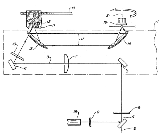
Referring to Figure 2, a rectangular plate 1 is rotated
at constant speed (by a motor, belt, and p~lleys, not
shown) in a horizontal plane about a vertical a~is 2.
The following components are mounted on the rotating plate
` and travel with it: two front surface mirrors 5 and 6
~Melles Griot 02MFG000); a 150 mm focal length lens 7
(Melles Griot OlLPX237); a quarter-wave retardation plate
10 (Melles Griot 02WRM013-632.8); a paraboloidal
reflector (axial cross-section 13~ with a clearance
notch to permit a laser beam to pass through; a second
paraboloidal reflector 14 without a notch; and an
aperture plate 15 (0.25 mm thick with a 2 mm hole
centrally located a~out the rotational a~is3. The
paraboloidal reflectors 13 and 14 have a focal length
o 10.2 mm (Aero Research Associates 484-001). These are
off a~is segments of parabolic reflectors mounted with the
optical a~is of their parent parabolas nearly coincident.
Beneath the rotating assembly is a polarized He-Ne laser
18 ~5 mW, Melles Griot 05LHP151). The laser beam goes
through a first quarter-waYe retardation plate 8 (same
as 10~, iE reflected off a front surface mirror 4
~same as 5 and 6), and passes through a second
guarter-wave retardation plate 9 tsame as 10~ Each
quarter-wave plate (8, 9 and 10) is mounted so that
it can be rotated in a plane perpendicular to the laser
beam for adjustment and can be locked in a position when
correctly adjusted. When the laser beam emerges from
plate 9, it is coincident with the a~is of rotation 2
of the ro~ating optical assembly.
~RD-82
2~22~,~
- 38 -
; The optical ~ignal obtained from the scatter at cuvette
: surface 12 is sensitive to the polarization of the
i; incident light. This signal dependence upon the incident
light polarization has been observed in other forms of
optical measurement such as fluorescence, raman
spectroscopy, plasma clot detection, and reflectivety. If
a rotating mirror is used to scan a plane polarized light
beam across a series of targets or samples, the plane of
the polarized light rotates about the a~is of the light
beam as this beam of light is scanned. Thus each position
of the rotating scanning mirror illuminates each target
with a different polarization alignment. It was
surprizingly discovered that, a uniform polarization
condition can be maintained independent of the scanning
mirror position by converting the light incident on the
mirror to circularly polarized light before scanning and
then reconverting it to plane polarized light after
redirection.
The three quarter-wave plates were used in this embodiment
because the optical signal obtained from the scatter at
cuvette surface 12 is sensitive to the polarization of
the incident light. Thus in order to assure uniform
results about the circle of cuvettes, the beam must be in
the same polarization condition at all positions. To
achieve this, a laser 18 producing a plane polarized
beam was employed. This beam is given circular
polarization by passing it through a properly oriented
quarter-wave retardation plate 8. A second retardation
plate 9 i~ provided to permit fine tuning to compensate
for imperfections in the characteristics of the first
retardation plate. A third quarter-wa~e plate lO on the
rotating member is used to produce plane polarized light
with ths electric field parallel to the plane of cuvette
surface 12. While retardation plates were used in this
~RD-82
2~2~;~
- 39 -
e~periment, magneto-optic devices may also be used to
obtain the same effects on polarization.
,~
The lens 7 is used to converge the laser beam 3 from
0.8 mm diameter as it enters the lens to 0.2 mm diameter
at the total internal reflection surface 12. The small
diameter facilitates multiple readings, which are averaged
to improve instrument precision.
A plate 19 with receptacles for forty cuvettes is
mounted a~ove the rotating optics assembly. The
receptacles are disposed in a circle whose cen~er is the
axis of rotation of the rotating optics assembly. Ons of
the cuvettes is shown at 11.
The pads 107 and 109 enable precise location of the
cuvette in the receptacle. (The surfaces 111 and 113
protect surface 21 from dirt or damage.) As the optical
assembly rotates, it presents the laser beam and the
receiving optics to each cuvette in turn. At each cuvette
a plurality of readings is obtained as the assembly moves
past the optical face of the cuvette.
Referring to Figure 3, the laser beam 3 enters face
21, travels through the transparent plastic material to
surface 22, where it undergoes total internal
reflection, and e~its the cuvette at surface 24.
However, as the beam exits surface 24 a small percentage
of the laser energy is reflected due to the refractive
index mismatch of air and plastic. Since this reflected
energy ~s significantly large relative to the signal
energy, it is important that it be directed away from the
detector~ Surface 24 is therefore angled so that the
reflection goes toward surface 101 of the cuvette.
Similarly, surface 101 is angled so that the reflection is
~RD-82
- ~o -
directed away from the detector, specifically, to a second
point on surface 24. The angles of surface 24 and surface
101 are designed so that many reflections take place in
the protrusion defined by surfaces 24, 103, and 101:
specifically, surfaces 24 and 101 converge slightly as
they approach surface 103.
At each reflection, most of the energy escapes the
plastic, so that by the time the reflecting beam reaches
surface 103, virtually all the energy is gone, and only a
negligible amount ever returns to the detector. Thus the
protrusion d~fined by surfaces 24, 103, and 101
constitutes a light trap which protects the integrity of
the ~ignal.
A light trap of this configuration was surprisingly found
to almost completely remove the unwanted illumnination
from the cuvette after it had generated the signal. Many
forms of spectroscopy, e.g. nephalometry, fluorescence,
ramman spectroscopy and absorbance spectroscopy, employ an
illuminating beam of light. Errors in measurement in
these forms of spectroscopy result when the illuminating
beam is reflected off the air cuvet~e interface while
exiting the cuvette and further excite signal in the
sample or enter the receiving optics directly. A light
trap of this design can eliminate these errors. This
light trap design is based on multiple reflections and
thus functions over a wide range of wavelengths. While
anti-reflective coatings can perform a similar task this
light trap functions for a wide range of wavelengths, is
le~s expensive, as it requires no post molding processing
and does not involve coatings that can interfere with
subseguent protein coating of a disposeable.
ORD-82
..
....
~:
, - 41 - 2~2~2
A mask 105 blocks any light -~hich is e~iting sur~ace 101
from reaching the detector. The analyte solution is
~'; contained in the well 20. Surface 22 is maintained in a
r, vertical orientation so that particles do not settle on
~ 5 it. The signal-generating scatter occurs at 12.
,: The laser beam 3 is introduced into the entry face 21
slightly below the normal to the surface, so that any
surface reflection of the laser beam is directed away from
the receiving optics. The beam is refracted at the entry
surface so that it impinges on the surface 22 at an
angle from the normal to surface 22 greater than the
critical angle. Thus it is totally internally reflected.
The sector (of a complete paraboloid) which is used at
13 (Figures 2 and 4~ to collect the optical signal
generated at the total internal reflection surface 12 is
determined by the requirement to be at an angle greater
than the critical angle and whereby only light scattexed
bac~ward toward thP light source is detected. Areas of
the paraboloidal reflector 13 at lesser angles are
masked with matte black pressure-sensitive paper tape.
Thus only light originating from the total internal
reflectance critical angle 25 to rays originating
parallel to surface 12, shown as ~3 in Figure 2, are
accepted.
The paraboloid is located so that the scatter source 12
is at its focus. Rays originating at the ~OCU5 of a
39 paraboloid ar~ reflected parallel to its asis, so the
signal light 23 to 25 (Figures 3 & 4) is transmitted
as a beam 17 toward the second paraboloid 14. tSee
Figure 4 for detail.
ORD-82
~222~
- 42 -
Rays parallel to the axis of a paraboloid converge at the
focus after reflection. The second paraboloid 14
concentrates the signal energy at its focus, where the
aperture plate 15 is mounted. The aperture plate
prevents stray light (not originating from the scatter
source 12), which is not focused by the receiving
optics, from reaching the photodetector 16. (Hamamatsu
Corporation S1723-04).
Although the use of single high F number parabola to image
point sources is well known in telescopes, it is also well
known that a low F number parabola, while efficient at
collecting li~ht, greatly distorts the image of light
sources larger than single points. It was surprisingly
discovered that two parabola of the same focal length,
when placed with their verticies away from each other,
open ends facing each other, create canceling
distortions. In this confiyuration, even realistically
sized sources of light are accurately imaged. The
efficient light gathering ability of this configuration is
applicable to light scatter, fluorescence, ramman
spectroscopy, plasma clot detection and other optical
measurements and imaging. As this is a reflective optical
system, it works well with a large range of light
wavelengths. When Yiewing a light source through the
plane surface of a cuvette, distortion of the image occurs
due to refraction at the cuvette air interface. Part of
this distortion can be canceled by tilting the parabolas
but it was surprisingly discovered that a segment of an
elipse chosen to have one of its verte~ to focus lengths
equal to the parabola it replaces cancels substantially
more of the distortion caused by the cuvette, rendering an
image spot 1/3 as large and 9 times as intense, as
determined by computer analysis. An eliptical mirror of
this type is easily fabricated by machining on a lathe
)RD-82
` ~ . ' ~ ~ ! . . .`. .
_ 43 _ ~ ~222~
with equipment similar to that used to fabricate the
parabola it replaced. Thus, using a properly shaped and
positioned ellipsoidal reflector near the cuvette corrects
i~ distortion caused by the refraction of light at the flat
surface of the cuvette.
An optical encoder (Sumtak Model LHF-050-2000, not shown)
was attached to the rotating optical assembly. The output
of this encoder was used to provide rotational information
to an IBM PCAT and digital data acquisition syst~m. As
the laser/detector optics assembly 1 passes under each
cuvette the digital data acquisition system/computer
digitized and stored the average of approximately 100
signal readings of the amplified output of the detector
16, taken as the internal reflection surface 12 is
scanned. Thus the average of approximately 100 readings
obtained from each cuvette was stored with each rotation
(one revolution-appro~imately every 2 seconds). The
readings were taken only from the center third of the
cuvette to avoid errors arising from the inside radius of
the cuvette well or the cuvette side walls. The computer
also stored a reading proportional to the laser output,
from another detector, and readings of a low scattering
region (laser impinging on black anodized aluminum cuvette
holder ring 19) and high scattering region (teflon block
mounted in place of cuvette). These readings were used to
compensate for variations in laser intensity and for
detector drift. When the computer monitored the cuvettes
for 10 minutes it ganerated a 5th order least squares
appro~imation equation to the scatter signal vs. time data
for each cuvette, subtracted the signal at time - 0
seconds from the curve and integrated this equation vs.
time for each cuvette. This integral was then correlated
with analyte concentration.
~5
ORD-82
2~2~
- 44 -
EXAMPLE 1
Hepatitis Virus S~rface Antiqen Test
Polycarbonate cuvettes, as depicted in Figure 3, were
coated with mouse monoclonal anti-hepatitis surface
antigen antibodies by incubating 200 microliters of a lO0
microgram per ml solution of antibody in 0.01 M phosphate
buffered saline, pH 7.4, overnight at room temperature
followed by three aspirate fill steps with 300 microliters
of 0.05 M Hepes/Tris, pH 8.3 buffer. The cuvettes were
then overcoated with 300 microliters of 0.05 M Hepes/Tris,
pH 8.3, buffer containing 1~ bovine serum albumin for 60
minutes at room temperature, washed twice with 300
microliters of overcoating solution, incubated 15 minutes
at room temperature with 300 microliters of 3% trehalose
in 0.05 M Hepes/Tris, pH 8.3 buffer, aspirated, dried in
room air and stored at room temperature in a desicator
below 20% relative humidity. The cuvettes were then
mounted in an instrument similar to that depicted in
Figure 2 with the modification that the cuvette was
rotated 180 about the illuminated surface 12 and the
laser entered the cuvette while propagating away from the
a~is of rotation, still following the path inside the
cuvette depicted in Figure 3.
Seventy-two microliters of standard, prepared by adding
the appropriate amount of hepatitis surface antigen from
(Merck) to a negative serum pool, was dispensed into
cuvettes by an automated pipetter and allowed to incubate
in the enclosed 37C instrument for five minutes. The
pipetter then dispensed 54 microliters of buffer
(containing 2.0 M potassium chloride, 2% bovine serum
albumin, 50 micrograms per ml of normal mouse IgG and
~ 35 0.05% sodium azide dissolved in 0.05 M sodium barbital
:
)RD-82
:, ~ ; .
2~22~2
- 45 -
buffer at pH 8.5) and 180 microliters of 105 nm diameter
0.1% monoclonal anti-hepatitis surface antigen coated gold
colloid suspension (in ten mM Hepes buffer, pH 7.0,
containing 0.05% sodium azide, 300 mM mannitol, and 0.5%
bovine serum albumin) at a rate sufficient to mix the
fluids in the cuvette. The light scattered by each
cuvette was then recorded for the ne~t ten minutes. The
time integral of the fifth order linear regression curve
fit of the light scatter vs. time data was reported for
each cuvette. The average of th~ signal from five of the
sis z~ro standards (one was 14 standard deviations from
the mean of the other five) and the average of the
duplicate standards correlated proportionately with the
hepatitis surface antigen present, as can be seen from the
following data:
HEPATITIS SURFACE
ANTIGEN CONCENTRATION MEAN SIGNAL
20 0 1.7358
0.1 ng/ml 2.2376
0.2 ng/ml 2.9421
0.4 ng/ml 3.99235
0.6 ng/ml 5.0442
250.8 ng/ml 6.72185
1.0 ng/ml 7.0187
1.5 ng/ml 9.31175
2.0 ng/ml 10.7365
2.5 ng~ml 14.04~4
305.0 ng/ml 24.9279
10.0 ng/ml 47.4585
ORD-82
'' , ,:
2022?,~2
- 46 -
EXAMPLE 2
Anti-Hepati~is Çore Antiqen Human An~ikQdy_Test
Polycarbonate cuvettes, as depicted in Figure 3 were
coated as in Example 1 ~Second Set of Experiments), with
recombinant hepatitis core antigen using a 5 microgram per
ml coating solution. The cuvettes were then dried, stored
and mounted in the instrument (Figure 2~, which was
enclosed and equipped with 37 air circulation.
Seventy-two microliters of the appropriate sample or
control were added to separate cuvettes by an automated
pipetter and allowed to incubate for 5 minutes, after
which time 54 microliters of assay buffer ~consisting of
1% bovine serum albumin and 1 M NaCl dissolved in pH 7.4
phosphate buffered saline~ and 180 microliters of a 0.1%
suspension of 105 nanometer diameter mouse monoclonal
anti-human IgG coated colloidal gold suspension was
dispensed with sufficient velocity to mis the contents of
the cuvette. The light scattered by the cuvette was then
recorded for the next 10 minutes. The time integral of
the fifth order linear regression curve fit of the
scattered light vs time data was reported for each cuvette
as signal. The mean signal of replicates of each serum
sample correlated with the presence of anti-hepatitis core
antigen employing a cuttoff of 3 standard deviations above
the mean of the negative control, as shown ~n the
following data:
, ~
ORD-82
' "'
.
'~
~222~
- 47 -
SAMPLE ~ OF TEST
TYPE REPI,ICATES MEAN OUTCO~E
Negative control 4 0.9524 S.D.=0.51
5Positive control 4 60.2099 S.D.=6.S
Negative sample 1 2 2.1671
Positive sample 1 2 10.483
Positive sample 2 2 41.058 +
Positive sample 3 2 33.494 +
10Positive sample 4 2 2.6043
Positive sample 5 2 74.2235 +
EXAMPLE 3
Plasma Clot Detection
An apparatus similar to that de~cribed in E~ample 1 and
Figure 2 was employed to determine the clotting time ~PT
or Prothrombin time) of plasma samples. (This apparatus
employed the three one-quarter wave retardation plates, as
described above, to maintain polarization of the scanned
laser beam and also employed two paraholoidal mirrors to
collect light scattered by the clotting samples for
quantitation.) Rectangular cuvettes (single cuvettes
broken from a Roche Cobas cuvette ring) were mounted in
the apparatus in place of the molded cuvette shown in
Figures 2 and 3. Laser light impinging on the vertical
surface of this cuvette was refracted at the surface of
the cuvette below the critical angle and thus propagated
through ~he sample in the cuvette, exiting on the opposite
side from the illumination. Light scattered, by the
proteins in solution in the sample, in a direction
substantially toward the illuminations was gathered by the
paraboloidal mirror nearest the cuvette (13, Figure 2) and
concentratPd by the second parabola ~14, Fiqure 2) through
~RD-82
- 2~22~
- 48 -
the apperture (15, Fiqure 2) on the detector (16, Figure
2). Average readings of each scan of the cuvette were
reported as in the previous Examples 1 and 2 as the optics
rotated once every 1.1 second over the 120 second reading
period. As the plasma clotted, the fibrin strands formed
scattered light much more efficiently than fibrinogen
monomer and thus the scattered light tracked the
progression o~ clot formation with the point of inflection
of the plot considered the clotting time. All reagents,
cuvettes and the apparatus were maintained at 37 degrees
C. The test was performed by pipetting 200 microliters of
Ortho Brain Thromboplastin 50BT), lot # OBT 996 into the
cuvette followed by 100 microliters of sample (either
Ortho Plasma Coagulation control level I lot # lPC 284 or
level III lot # 3PC 748). The cuvette was mixed by
rapidly aspirating and dispensing with the sample
pipette. The instrument readings were then started.
Figure 10 shows the data obtained from the clotting of a
level I control with a point of inflection clotting time
of 10 seconds. Figure 11 shows the data obtained from the
clotting of a level III control with a point of inflection
clotting time of 23 seconds. These times are in good
agreement with data collected on an Ortho Diagnostics
Koagulab 32-S coagulation instrument with the same
reagents.
ORD-82
. ` , ' ` .
~22~J~2
- 49 -
Third Set of ExPeriments
EXAMPLE 1
Test for Digo~in in Whole ~lood Sample
Using Scatter by Colloidal Gold of Light
Incident at Non-Total Inte~nal ~eflectance Anales
The apparatus used in this E~ample is shown in Figures 5
and 6. Tests were performed on an acrylic cartridge 26
(Arden Medical Systems) with a channel for the sample to
flow through. The surface of the channel was coated with
polyclonal goat-anti-digo~in antibody (A~lantic Antibody)
by standard passive adsorption methods. Light 27 from a
He-Ne laser 28 illuminated a portion of the coated
surface 29 at an angle almost normal to the surface. At
this angle of illumination, th4 light prop~gated into
aqueous samples rather than producing an evanescent wave
in the sample. Light scattered ~rom the coated surface
29 was collected by a lens 30 and focussed onto an
optical fiber 31, which guided the light to a
photomultiplier tube 32 (PMT). The output voltage of
the PMT was digitized and recorded by a computer 33.
The detection system (lens and fiber) was carefully
positioned and oriented so a~ to collect only light
scattered from the coated surface 29 which propagated
through the acrylic at angles betwe~n the plane of the
surface and the critical angle.
Colloidal gold was prepared as described above (diameter
appro~imately 40 nm.) and was coated with digoxin~bovine
IgG. Whole blood was spiked with various amounts of
digo~in. Samples containing equal mi~tures of spiked whole
blood and digoxin-colloidal gold were pipetted into the
ORD-82
~ f?~
- 50 -
cartridge and the resulting light scatter was monitored
over time.
Figure 7 shows typical results. In curve A, the sample
contained no free digo~in, and the light scatter signal
increased continuously during the seven minutes shown, as
the digo~in-colloidal gold bound to the surface. In curve
B, the whole blood sample contained appro~imately 4.5
~g/ml of digo~in which competes with the
digoxin-colloidal gold for sites on the coated surface,
and as a result the scatter signal leveled of~ after a few
minutes. (The curves have been offset so that they start
at the same level for comparison.).
In this example, the sample was below the coated surface.
When the sample is above the coated surface, the settling
of red blood cells causes large spurious scatter signals.
For whole blood assays, applicants have found that it is
preferable to orient the coated surface such that gravity
pulls the red blood cells away from the surface, or at
least does not pull them towards the surface.
EXAMPLE 2
Observation of Immunological Binding of
Colloidal Gold with LED_a~_Liqht Source
The apparatus used in this Example is shown in Figure 8.
The tests were performed on the same acrylic cartridge
34 as in ~xample 1 (Third Set of E~periments). The
light source 35 was a red light-emitting diode (LED,
~tanley ER-300, 300 millicandela, 660 nm. peak wavelength,
30 nm. half-width). Light from the LED was focused on the
coated ~urface 36 by a 10X microscope objective.
Scatter from the surface was focused by a lens 37 on an
~D-82
2~2~2~2
- 51 ~
aperture 38 and then to a PMT 39 with the same optical
alignment conditions described in Example 1 (Third Set of
E~periments). The LED output was electronically modulated
at 1 KHz and the PMT output was demodulated at the same
frequency by a lock-in amplifier 40 (PAR 128A) to
improve signal-to-noise performance of the apparatus.
The reagent used in this Example was 40 nm. colloidal gold
coated with goat-anti-mouse antibodies (Janssen
"AuroProbe~). This reagent was pipetted into cartridgss
coated with Mouse IgG or Bovine Serum Albumin (BSA).
Figure 9 shows typical results. Curve C shows a
substantial increase in scatter signal as the GAM-gold
binds to a cartridge coated with MIgG. Curve D shows
little response when the cartridge was coated with BSA.
ORD-82