Note: Descriptions are shown in the official language in which they were submitted.
-` 2 02 4 9 ~
PATENT i:
IMPROVED METHOD AND APPARATUS FOR PROVIDING
INTRAPERICARDIAL ACCESS AND INSERTING
INTRAPERICARDIAL ELECTRODES
.': ' .
'' '''`'.''~, ` ''
,"'"' ~
, : '." ~;
` Backround o~' the Invention ;
The present invention relates to a method a~d ; `~
apparatu- ~or provlding intraporicardial accQss with ;~
a ~inimal a~ount Or surg-ry and, ~or- particularly,
cono-rn-d with an improved t-chnique ror extending
a guid- wir- through th- p-ricardial wall In its
more 8p-ci~ic aspects, the inv-ntion is concerned
; with an i~proved ~othod ~or implanting deribrillation
l-otrod-- within th- p-rl¢ardiu~
~~r;~ Num-rou- ~ort- hav- b--n ~ad- to introduc-
plantabl- l-ctrod-- wlth a ~ini~al a~ount o~
lS ~ ~urg-ry Th~ ort- hav ar~-ct-d plac-m-nt both
intrap-rlcardi~lly and -xtrap rlcardlally Wh-r-
intrapcrlcardl~l plac-m-nt wa- provided, howev-r, the
,~J~ ; prlor ~rt ~ ort- havo ri~ked phy-ical trauma to the
h-art durlng placement The pre~ent invention is, ;~
de-lgnQd to minimizo this risk
Summarv O~r th- Inv-ntion
~`` ; Th- ~-thod of th- pr--ent lnv-ntlon provld~
acc-~- to th- lntorior o~ th- p-rlcardium through an ~'~J,"~
lnci-ion in the in~-rior bordor o~ tbe p-ricardlum
- ,i .", ".~
c " .;
~ ".. , .', ,,-~
~ 202~g~
p~NT
-2-
and a tunnel dissected between the pericardium and
the diaphragm. In the method, one jaw of a clamp-
like placement device is extended interiorly of the
pericardium through the incision and the other jaw is
extended exteriorly of the pericardium through the
tunnel. The ~aws include tubular guide elements with
aligned open distal ends curved toward one another.
once placed relative to the pericardium, the jaw
elements are moved to clamp the tissue of the
pericardium therebetween. A guide wire is then
extended through the tubular elements and the
pericardial tissue therebetween. Once the wire is so
placed, the clamp-like device is removed, leaving the
wire in place to facilitate access to the interior of
the pericardium.
In the method of placinq an electrode within the
pericardium, a guide cannula i8 extended over the
wire and into the pericardium. A primary electrode
i8 than passed through the cannula and into the
interior o~ the pericardium. A ~econdary electrode
may be inserted through the incision in the in~erior
border of the pericardium.
The apparatus of the invention comprises the
clamp-like placement device, including first and
second elongate ~aw elements, each of which has an -
open-ended tubular guide extending over the length -
thereof and terminating in an open distal end
extending laterally of the element. It further
aompri~e~ means for securing the ~aw elements
~0 together in a condition wherein the open distal ends
of the tubular guides are in aligned closely disposed
relationship to one another. In the preferred
" :
2~2~9Q~ -
-3-
embodiment, the apparatus further comprises means for
holding the ~aw elements in clamping engagement with
opposite sides of pericardial tissue and a secondary
open-ended tubular guide secured to and extending
along at lea~t one of the ~aw elements.
A prin¢ipal ob~ect of the invention is to ~
provide a method and apparatus for accessing the ---
interior of the pericardium with a minimum of surgery -~
and risk of physical trauma to the heart.
Another and more specific ob~ect of the
invention i~ to provide such a method and apparatus ;~
for extending a guide wire through the pericardial ;`
wall, without risk that the wire will effect physical ;~
trauma on the heart.
Still another ob~ect Or the invention iB to ;, ~,
provide an improved method for guiding an implantable ,
d-fibrillator electrode into the pericardium.
~; Yet another ob~ect of the invention is to
provide an app~ratus havlng a slmple mode of
operatlon whlch may be u~ed to pas~ a guide wire into
tho pericardium with a minimum of surgery.
A more specific ob~ect of the invention is to
provide such an apparatus having means to create a .~ ~
puncture through the wall of the pericardium. ^`~ '
A further ob~ect of the invention io to provide ; ~i
such an apparatuo having a secondary lumen adapted to
be used ror the introduction of other element~ into ~;
the pericardium.
~ ,,i i
',.":,'' '' ".."
2~2~90:~
PAT~NT
--4--
These and other ob~ects will become more
apparent when viewed in light of the following
detailed description and accompanying drawings. ~ -
Brief Description of the Drawinas
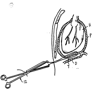
Fig. 1 is a front prospective view of the upper
chest region of a human body, with parts thereof
shown in section, lllustrating the electrodes of a
defibrillator which have been implanted in the
pericardium through use of the method and apparatus
of the invention;
Fig. 2 is a cross-sectional side view of the
body of Fig. l;
Fig. 3 is a side elevational view of a curved
prob- which may be used to dissect a tunnel between ;;
lS the pericardium and di~phragm in the method of the
present invention;
Fig. 4 i8 a perspective view illustrating a ~ ~
praferred embodiment of the intrapericardial access --
apparatu~
Figs. 5 to 15a are cross-sectional side views of
the upper region of a human body, sequentially
illustrating the st~aps of practicing the present
invention to first access the interior of the
perlcardium and then place defibrillator electrodes
therein;
`` 2~2~
P~TENT
-5-
Fig. 15b is a cross-sectional elevational view,
with parts thereof broken away, illustrating a
modification of the arrangement shown in Fig. 15a,
wherein the electrode within the anterior pericardial
space is secured to the pericardium through means of
a crimpable button;
Fig. 16 is an exploded perspective view of a
first alternative embodiment of the intrapericardial
access apparatus;
Fig. 17 is a perspective view of a second
alternative embodiment of the intrapericardial access
apparatus;
Fig. 18 is a cross-sectional view of the chest
region of a human body, illustrating the first
alternative embodiment access apparatus in the
process of being positioned to extend to either side
of the lower wall of the pericardium;
Fig. 19 is a cross-sectional elevational view
similar to Fig. 18, illustrating the first
alternative embodiment access apparatus in the
proces~ of being used to extend a guide wire through -~
the lower wall of the pericardium;
Fig. 20 is a side elevational view Gf a coiled
electrode which may be placed through means of the
method and apparatu~l of the present invention; and,
-~. :. i
Fig. 21 is a front elevational view of the Fig.
20 electrode, taken on the plane designated by line
21-21 of Fig. 20.
,~''''",''"''',''
~ 20249;~
PATENT
-6-
~etailed Descri~tion of the Illustrated Embodiments
The chest region of the human body shown in the
drawings i8 designated in its entirety by the letter
"C" and i8 illustrated to show the pericardium "P",
the heart l'H", the diaphragm "D" and the forward rib
cage ~a~. As shown in Figs. 1 and 2, an electrode
"El" is shown posteriorly positioned within the
pericardium and electrode ~E2~ is shown anteriorly
positioned within the pericardium. The electrodes
"E~" and ~Ea~ shown in Figs. 1 and 2 have been placed
through means of the method and apparatus of the
present invention. The proce~s for this placement is
described in detail in the following discussion.
The curved probe shown in Fig. 3 is designated
in its entirety by the numeral 10. This probe is
fabri¢ated of a riqid material, such a stainless
~teel, and finished 8e as to have a smooth exterior
surface. Ito purpose, as will become more apparent
from the following discussion is to enable a surgeon
to dissect a tunnel between the pericardium and
dlaphragm through a subxiphoid incision formed in the
che~t wall.
The preferred embodiment intrapericardial access
apparatus of Fig. 4 is designated in its entirely by
the numeral 12. This apparatus i9 of a forceps
construction embody.Lng upper and lower elongate ~aw
elements 1~ and 1~, re pectively, ~imilar to those
used for tenaculum forceps. The ~aw elements are
hlngedly sscured together for movement toward and
away from ea¢h other by a hinge pin 18. The distal
,
" '' ''
:
2 ~ 2 ~ 9 ~
PATENT
- 7 -
ends of the jaw elements 1~ and 16 are formed with
rigid aligned lateral extensions 20 and 22,
respectively. These extensions, as will become more
apparent from the following discussion, are pointed
and provided to clampingly engage the tissue of the
pericardium therebetween. Handles 2~ and 26 are
rigidly affixed to the ~aw elements 1~ and 16,
respectively, and terminate in thumb and finger
rings. Interengageable ratchet elements 28 and 30
are formed on the handles 2~ and 26 to selectively
lock the handles in a condition wherein the
extensions 20 and 22 are clampingly engaged with
pericardial tissue.
:.:
A first primary open-ended tubular guide 32 is
fixed to and extends over the outer side of the ~aw
element 1~ and terminates in a open distal end 3~ ~! ` ' ~''~''
extending laterally of the element. A second open-
ended tubular guide 36 is fixed to and extends over
the ~aw element 16 and terminates in an open distal
end 38 extending laterally of that element. The ends
3~ and 38 are axially aligned when the ~aw elements
are clampingly engaged with the pericardial tissue
and, in the preferred embodiment, are of such -
relative diameters that the end 38 may fit within the
end 3~ . The edges of the ends may be sharpened to
cut through the pericardial tissue upon being alamped
into engagement therewith.
A secondary open-ended tubular guide ~0 is fixed
to and extends along the guide 32. The guide ~0 ;
terminates in a bias cut open end short of the dis~al
end 3~. The purpose of the secondary lumen is to
provide additional access into the interior of the
pericardium for the insertion of instruments such as:
: 2~2~9~
PATENT
-8-
a secondary guide wire; an irrigation catheter; or a
fi~eroptic scope.
Figs. 5 to 15a seguentially illustrate the steps
o~ the inventive method in the process of accessing
the interior o~ the pericardium and implanting
defibrillator electrodes within the pericardium to
the posterior and anterior of the heart. At the
outset of the procedure, a subxiphoid incision ~2 is
formed in the chest wall. The method of the
invention i~ then carried out through the following
steps:
1. A pair of clamps or forceps 44 are used to
pick up the inferior border of the pericardium "P"
through the subxiphoid incision ~Fig. 5). This is
carried out under direct vision.
2. A curved probe, such as the probe 10 is
used to dissect a tunnel I~TI~ between the pericardium
"~" and the diaphragm "D", towards the posterior
aspect of the pericardium (Fig. 6). This is also
carried out under direct vision, while the inferior
border o~ the pericardium i8 held by the forceps ~.
3. A small nick 46 is cut through the inferior
border of the pericardium (Fig. 7). This is carried
out under direct vision, using a scalpel 48 while the
border of the pericardium is held with the clamp ~4. ~-
4. The intraE~ericardial access apparatus 12 is
inserted through the incision ~4 to extend the lower
~aw element 16 into the tunnel "T" and the upper ~aw
element ~ into the pericardium through the nick ~6
~Fig. 8). This step is carried out while the
202~0~
PATENT
_g_
inferior border of the pericardium is held by the
forceps ~, with the jaw elements of the apparatus 12
sufficiently spread to pass to either side of the
lower wall of the pericardium.
5. The ~aw elements of the access apparatus
are snapped together, trapping the tissue of the
pericardium between them ~Fig. 9). During this
process, the pointed lateral extensions 20 and 22 on
the elements pierce and grip surface of the tissue,
thus gripping and stabilizing the apparatus relative
to the periaardium. The ratchet elements 28 and 30
interengage upon snapping of the ~aws together to
hold the ~aws in the closed condition.
....
6. The next step is to pass a guide wire into
the pericardium and through the lower pericardial ~ , -
wall (Fig. 10). Depending upon the choice of the ;
surgeon, this may be achieved by slightly different
techniques. In one, a guide wire with a sharpened
tip is extended through the upper tubular guide 32 to
exit therefrom and pierce through the pericardial ~ ;
wall and pass into the lower tubular guide 36, from
whence it is extended out through the subxiphoid
incision. In another, after the apparatus i8 locked
in place, it is moved back and forth to form a
somewhat enlarged opening in the pericardial wall
where it is pierced by the extensions 20 and 22 and
then the apparatus is pulled backwards to position :~
the distal ends of the tubular guides 36 and 38 in
alignment with this hole. The guide wire, designated
S0, is then extended through the inner tubular guide
32, passed through the enlarged opening, and exited
out through the outer tubular guide 32. In yet
202~sn.~ ::
PATENT
--10--
another, sharpened edges 3~. and 38. on the distal
ends o~ the tubular guides are used to cut a hole
through the pericardial wall and then the guide wire
i8 passed from the upper tabular guide 32, through
S the hole and into the lower tabular guide 36. The
latter technique does not require a sharpened tip on
the guide wire. Regardless of which technique is
used, the guide wire 50 is passed from the
intrapericardial to the extrapericardial portion of
the apparatus to prevent any chance of myocardial
in~ury should the wire pass outside of the placement
apparatus.
. . .
7. The intrapericardial access apparatus 12 is
unsnapped and removed, leaving the guide wire 50 in
place IFig. 11). As 80 placed, the guide wire may be
u~ed for accessing the interior of the pericardium
~or any desired purpose. The steps hereinafter set
~orth are for the purpose of introducing
de~lbrillatlon electrodes into the intrapericardial
space.
8. A dilator ~2 and cannula S~ are threaded
over the guide wire, through the extrapericardial
tunnel "~" into the intrapericardial space (Fig. 12).
During this step, the dilator is first extended
through the hole pierced in the lower wall of the
pericardium to enlarge this hole and the cannula is
then extended thereover and through the hole.
Thereafter the dilat:or is withdrawn, leaving the
cannula in place.
9. With the cannula 5~ in place, the dilator
S2 is removed and the electrode 'IEl" is advanced
~ 2~9~
PATENT
through the cannula into position in the posterior
intrapericardial space (Fig. 13). The electrode "B~"
may be of the resilient helical coil type disclosed
in copending application S.N. 129,124, filed November
12, 1987. Such electrodes are capable of being
straightened into a generally rectilinear
configuration to facilitate advancing them through a
cannula and, when released, assume a three-
dimensional helical coil configuration as may be seen
in Figs. 20 and 21 herein.
10. ~fter the posterior electrode "El" is in ~ -
place, the cannula S~ is removed and a second cannula ~
56 having the electrode "E2" threaded therethrough is ;
advanced into the anterior pericardial space through
the inferior pericardial nick or incision ~6 (Fig. ;~
14).
11. The electrode "E" is passed through the
cannula 56 to unfurl into the anterior pericardial
space and the cannula 56 i5 then removed ~Fig. 15A). ~ ~ ;
The nick ~6 is then closed with a suture, thus
securing the electrode 11~21~ in place. The electrode
IIE~I' is held in po~ition by virtue of its passage
through the tunnel "T" between the pericardium and
the diaphragm.
As an alternative to suturing the anterior
electrode "F~" in place as described above, the
electrode may be held in place with a crimpable
annular button 58 (Fig. 15B) of the type disclosed in
copending application S.N. 120,590. With this
arrangement, a section of the second cannula,
designated 56a, is left in place around the electrode
~` 202~90,~
PATENT
-12-
"E~" and the button is crimped into engagement with
this section and sutured to the wall of the
pericardium.
.
The first alternative embodiment of the access
apparatus shown in Fig. 16 is designated in its
entirety by the numeral "60". This apparatus
comprises: an upper jaw element 62 of an open-ended
tubular configuration having an open distal end 6~
extending laterally therefrom; a lower jaw element 66
of an open-landed tubular configuration having an open
distal end 68 extending laterally therefrom; a T-
shaped block 70 fixQd to the element 62, said block
lncluding a tongue-like extension 72; and, a block 7
fixed to the ~aw element 66 and having a socket 76
therein for complimental receipt of the tongue-like
extension 72. The blocks 70 and 7~ are 80 positioned
relative to the elements 62 and 66 that the distal
endo 6~ and 68 as~ume an axially aligned condition
when the extension 72 i~ received within the ~ocket
76. The element 62 i8 50 proportioned relative to
the element 66 that the di~tal end 6~ may be received
within the distal end 66 when the extension 72 and
groove 76 are complimentally engaged. -~ ;
Tbe apparatus C0 is used in a manner ~ -
corresponding to that of the apparatus ~2, with the
exception that the iaw elements of the apparatus 60
may be inserted inte place individually and that the
apparatus includes no pointed lateral extensions,
such as the exten~ion~ 20 and 22. Figs. 18 and 19
Qhow the manner in which the apparatus 60 would be
placed to extend a guide wire through the lower wall
of the pericardium. It should be appreciated that
",,
~2~g~ ~ :
-13~
. .
the guide wire 50 would be provided with a sharpened
tip and extended from the upper jaw element 62
through pericardium and into the lower jaw element
66.
, . . . .
The second alternative embodiment apparatus of
Fig. 17 is designated in its entirety by the numeral
"60,". The parts of the apparatus "60," are similar
to those of the apparatus "60" and designated by like
numerals, followed by the subscript "a" as follows: - -
upper jaw element 62~; open distal end 64,; lower jaw
element 66,r open distal end 68,; block 70,; tongue- ~;
like extension 72,; block 74,; and socket 76,. The
block 70, i~ pivotally secured to the block 74, by a
hinge pin 78 and is moveable about this pin between
the open condition illustrated in Fig. 17 and a
closed condition wherein the distal end 7~, is
received within the distal end 68,.
The operation of the apparatus 60, corresponds to
that of the apparatus 60, with the exception that the
surgeon has the option of inserting the apparatus
into place as shown in Fig. 18 with the blocks 70, and
74, hingedly interconnected. Alternatively, he may
insert them individually and hingedly secure them
together after they are in place. ~ ~
:'. '
CONCLUSION -
From the foregoing detailed description, it is
believed apparent that the present invention provides
a method and apparat:us whereby intrapericardial
ac¢ess may be provided with minimal surgery and risk
of physical trauma to the heart. It should be
understood, however, that the invention is not
,~, 2~2~g~
P~TENT
-14-
intended to be limited to the specifics of the
described embodiments, b~t rather is defined by the
accompanying claims.
'.''' '.;':.:.':.: '
: ,', ~ ;, , ,
,, .' ;'
"'.." -'';.