Note: Descriptions are shown in the official language in which they were submitted.
2~27632
EYE DROPPER GUIDE
This invention relates to an eye dropper guide having a
surrounding base contoured to rest on the orbital margin
~bony rim] of the orbit [eye socket], so that an individual
may drop medicine into their own eye(s) without difficulty.
PRIOR ART AND BACKGROUND OF INVENTION
, . _ , .. .... _. _, .. ,, ~ .. . ._ .. ...... _ _.. _ .. ~ . .. _ .
Eye dropper funnels are known.
U.S. Patent 2,237,862 issued April 8, 1941, to Burhans,
teaches a eye cup with eye bowl 5 connected by bore 8, to
open ended hollow base 7, cover by diaphragm 9.
U.S. Patent 2,352,610 issued July 4, 1944, to Bonilla,
teaches a eye cup with eye bowl 11, connected to squeeze
bulb 10 by flap valve 23, bulb 10 has exterior port 17. The
device can be used to wash or treat the eye with fluid.
U.5. Patent 2,767,711 issued Oct. 23, 1956 also to
Ernst, teaches a eye cup for eye medication with bowl 5, and
removable or piercable closure cap (lid or diaphragm) 6,
bowl 5 has circumferential bead 13 to fit the eye socket.
U.S. Patent 2,920,624 issued Jan. 12, 1960, to Lerner
et al., teaches eye drop dispenser ~or medication having a
frame 10 with a margin 14 to fit the eye, pivotally mounted
spigot 24, communicates through conduit 32 to flange 16
adapted to engage a medication container. Spigot 24 allows
drop dispensing.
U.S. Patent 3,106,898 issued Jan. 16, 1962, to Erwin,
teaches a series of eye medicators, the broadest has bowl 10
with flange 11 to fit the eye, connected by a passage to
exterior Punnel 14. The claimed invention includes a valve
24 or 40 exteriorly operated by stem 23 or 37. Another form
has cup 44, with threaded passage 45, to receive threaded
2~27632
neck 47 of dispensing container 48, these may be moulded
together.
U.s. Patent 4,111,200 issued Sep. 5, 1978, to Sbarra,
t:eaches an eye medicator, with bowl 13 to fit the eye,
c:onnected by passage 13e to exterior snap portion 13b to fit
cap 12 of dispensing container 11. 13b and 12 are mutually
rotatable to allow or prevent communication through
alignable orifices.
U.S. Patent g,733,802, issued Mar. 29, 198B, to
Sheldon, teaches an eye medicator, conical transparent bowl
45 has opening 5g and top opening 52 adapted to receive
squeeze eye dropper.
As may be seen several such eye dropper funnels are
known.
It is an object of the invention to provide an improved
eye dropper guide. Other objects are hereinafter apparent
from the description, claims, and drawings.
DESCRIPTION OF_THE_INVENTION
In one aspect the invention is directed to an eye drop
guide comprising, a contoured base adapted to fit closely
the orbital margin [bony rim] of a human orbit [eye socket]
a wall extending upward therefrom to an aperture, which is
of a size to receive a nozzle of an eye drop container. The
aperture is at least about 30 mm above the nearest section
of the contoured base. Preferably the base has opposed
first and second ends. The first end is contoured to rest
ad~acent the nasal portion of the orbital margin. The
second end is contoured to rest a~ainst the temple portion
of the orbital margin. The base may have opposed lateral
portions intermediate of the ends, contoured to rest on the
brow and cheekbone portions of the orbital margin. The
aperture preferably has a midpoint from 25 to 33 mm outward
2027~32
of the furthest section of the first end. The aperture may
comprise a funnel member extending downward from the
aperture to receive a noz21e of an eye drop container. This
funnel has a tip at least 20 mm above the nearest section of
the contoured base. The aperture is preferably circular and
about 8 mm in diameter. The base may extend about 75 mm
from the first end to the second end. Preferably the
aperture is equidistant from the intermediate portions of
the base.
As the human orbital margin and its associated surround
is more or less invariant in the normal adult human,
regardless of sex, race and size ~except in abnormal
(pathological) development], a contoured base wauld be a
one-size-fits-all. The aperture should be appraximately
located between the rest position of the cornea and the
medial or inner canthus [inner corner of the eye], this is a
desirable location for eye drop introduction. The aperture
should be located at least 30 mm above the base to prevent
eye drop nozzle-eye lash interference and blinking. The eye
drop nozzle typically extends about 10 mm downward of the
aperture to within 20 mm of the base. The specific shape of
the contoured base allows it to be used for left and right
eyes interchangeably. The contoured base is advantageously
and conveniently bilaterally symmetrical about its long
axis. The advantage over the prior art is that a guide,
construoted in this way, automatically fits into position
with the dropper aperture over the best eye drop position.
~dvantageously a dropper funnel is associated with the
aperture, this may extend to within 20 mm of the base.
In another aspect the invention is an eye drop guide
comprising a contoured base adapted to fit closely the
orbital margin of a human orbit. The base comprises opposed
first and second ends. The first end is contoured to rest
ad~acent the nasal portion of the orbital margin, and the
second end is contoured to rest against the temple portion
of the orbital margin. The base has opposed lateral
2~27632
portions intermediate of the ends, contoured to rest on the
brow and cheekbone portions of the orbital margin. A wall
extends upward from this base to an aperture, which is
circular and about 8 mm in diameter. The aperture is at
least about 30 mm above the nearest section of the contoured
base. The aperture has a mid point about 29 mm outward of
the furthest section of the first end, and about 46 mm
inward of the furthest section of the second end. The
aperture comprises a funnel member extending downward from
the aperture to receive a nozzle of an eye drop container,
the funnel having a tip at least ~0 mm above the nearest
section of the contoured base. The aperture i9 equidistant
from the intermediate portions of the base. Optionally the
guide has eye drop container holder means attached to the
wall and circumambient the aperture. The holder means may
be flexible and include paired opposed arcuate flexible
flaps concentric to the aperture.
In a third aspect the invention is an improvement in an
eye drop guide having a base, an aperture and an
interconnecting wall, where the improvement comprises a
contoured base adapted to closely fit the orbital margin of
a human orbit. Preferably the base comprises opposed first
and second ends, the first end being contoured to rest
ad~acent the nasal portion of the orbital rim, and the
second end being contoured to rest against the temple
portion of the orbital rim, the base having opposed lateral
portions intermediate of the ends, contoured tc rest on the
brow and cheekbone portions of the orbital rim. The base
preferably extends about 75 mm externally from the first end
to the second end, internally the measurement approximates
65 mm.
In a fourth aspect the invention is an improvement in
an eye drop guide having a base, an aperture and an
interconnecting wall where the improvement comprises the
aperture being of a size to receive a nozzle of an eye drop
container, and being at least about 30 mm above the nearest
2~2~632
section of the base. Optionally the aperture comprising a
funnel member extending downward from the aperture to
receive a nozzle of an eye drop container, the funnel having
a tip at least 20 mm above the nearest section of the
contoured base. Conveniently the aperture is circular and
about 8 mm in diameter.
In a fifth aspect the invention is an improvement in an
eye drop guide having a base, an aperture and an
interconnecting wall, comprising eye drop container holder
means attached to the wall and circumambient the aperture.
Preferably the holder means is flexible and may include a
plurality of flexible flaps. More preferably the flexible
flaps are arcuate. These flexible arcuate flaps may
concentric to the aperture, conveniently there are two
flaps.
The precise form of the wall between base and aperture
is not critical. For manufacturing purposes it is
practically, conveniently and advantageously of the smoothly
sloping form of the type called domed. As would be
appreciated by those skilled in the art a variety of wall
forms may be utilized. In practice the wall rises to the
aperture and thus approximates to pyramidal with the apex at
the apertur~.
DESCRIPTION_OF THE PREFERRED EMBODIMENTS
Preferred embodiments are indicated in the drawings
where:
Fig. 1 shows an anterior (front elevational) view of
the bones forming the orbital margin ~bony rim] of the human
orbit [eye socket];
Fig. 2 shows a lateral (side elevational) view of the
bones forming the orbital margin of the human orbit;
Fig. 3 shows a longitudinal sectional view of an
embodiment of the invention;
Fig. 4 shows a transverse sectional view of the
2027~32
embodiment of Fig. 3;
Fig. 5 shows a plan view of the embodiment of Figs. 3
and 4;
Fig. 6 shows a view of a human eye;
Fig. 7 shows a view of a further embodiment of the
invention in use;
Fig. 8 shows a view of the embodiment of Fig. 7 when
not in use.
The general description of the invention is now
expanded by reference to the drawings, which illustrate
preferred embodiments of the invention.
The numeral 10 indicates the human eye socket in Figs.
1 and 2, the orbital margin [bony rim] of human orbit [eye
socket] 10, is formed by frontal bone 12, which forms the
brow portion of the margin, maxilla or super maxillary bone
14, which forms part of the cheekbone portion of the rim,
and zygomatic or malar bone 16, which forms most of the
cheekbone portion of the margin, and also the temple portion
of the margin. Nasal bone 18, which forms the bridge of the
nose, is ~ust outside the orbital margin, and is often
loosely con~idered by laymen to constitute part of the
orbital margin or to demarcate it.
Anatomically and medically the orbit is considered to
be quadrilateral pyramidal in form, which is a very inexact
geometrical description. Precise and accurate geometric
description is neither warranted, nor would it be
intelligible. It is easier to consider the salient features
of the orbital margin.
When the elevational frontal outline is considered one
finds the superior margin starting at the nasal end has a
shorter concave rise joined at midbrow to a longer concave
decline to the temple end and a closely similar but not
identical form is observed at the inferior margin, with a
more flattened shorter concave rise ~oined roughly at mid
2~2~632
cheekbone to a longer concave decline to the temple. The
mid cheekbone is slightly further from the nasal end than
the midbrow. The similarity is more pronounced if the
immediate surround of the orbital margin is considered
rather than the inner edge.
When the top or bottom plan outline i8 considered one
finds a convex outline. The superior margin starting at the
nasal end has a short convex portion joined at midbrow to a
longer less convex portion to the temple end. The inferior
margin again has a shorter convex portion ~oined at mid
cheekbone to a longer less convex portion to the temple end.
Again the mid cheekbone junctlon is further from the nasal
end than the midbrow junctions. The midbrow ~unctions
coincide, whereas the mid cheekbone junctions are adjacent
but not coincident. The similarity is more pronounced if
the immediate surround of the orbital margin is considered
rather than the inner edge.
In life these bones are fleshed, and the eye ball lies
withln these bones. Despite considerable human variation in
size, as well a~ ~exual and racial differentiation, adult
fleshed human eye sockets differ remarkably little in
dimension, extending when viewed in front elevation some 52
mm horizontally and some 35 mm vertically, these dimensions
may differ marginally perhaps by up to +2 mm. Also the eye
soc~et extends backwards some 30 mm, again with perhaps +2
mm variation, from its forward inner rim, adjacent the nose.
In practice the orbital margin [bony rim] of the adult human
orbit [eye socket] may be reasonably be viewed as of
standard size and dimensions, sufficiently 80 as to be more
or less interchangeable.
Although the orbital superior and inferior margins are
different in outline by constructing a contour, which has
approximates to the vertical convexities and horizontal
concavities of the human orbital margin, it is possible to
construct a contour which fits substantially closely about
2~27~32
the orbital margin and its immediate surround. A whole
family of related contours of these properties can be
constructed by those skilled in the art.
Typically this contour is of distorted ellipsoidal
form, when the contour is a smooth continuous curved
perimeter. Viewed from above the ellipsoid has a shorter
more convex nasal ellipsoidal portion merging smoothly into
a longer less convex temple ellipsoidal portion. Viewed
from the side it has shorter more concave nasal portion
merging smoothly into a longer less concave temple portion.
Family of suitable ellipsoidal contours are hard to define
precisely, but are easily recognizable by those skilled in
the art and extremely easy to test for fit on human
subjects. As would be appreciated by those skilled in the
art, the contour need not be a smooth continuous curved
perimeter, although this is more convenient for moulding and
the like.
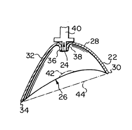
In Figs. 3 to 5, numeral 20 indicates the eye dropper
guide with domed wall 22, having median aperture 24 and base
26 designed to fit against the human orbital margin. Wall
22 has shorter inner dome wall 28 extendiny to inner edge
30, which fits against the nose, and longer outer dome wall
32 extending to outer edge 34, which fits against the
temple. Aperture 24, may have internal wall 36, forming a
funnel adapted to receive nozzle 38 of eye drop bottle 40,
to allow drops to fall onto the eye, the rest position of
the cornea is indicated at 42, which extends slightly
(typically about 2 to 3 mm) within the perimeter of base 26.
Measured externally from inner edge 30 to outer edge 34,
base 26 is preferably about ~5 mm long which i8 of suitable
size to fit the adult human orbital margin or limit,
internally inner edge 30 is about 65 mm from outer edge 34.
This length may be varied slightly as long it is sufficient
to cover the orbital margin. Inner dome wall 28 extends
about 25 mm along base 26, aperture 24 extends about 8 mm
along base 26, and outer dome wall 32 extends about 42 mm
2~27632
along base 26. Base 26 is concave in profile extending
inward a maximum of about 10 mm from (imaglnary) line 44.
The size of aperture 24 and optional funnel wall 36 is
adapted to receive standard eye drop bottles, as those
skilled in the art would realize the dimension of aperture
24 may be varied to fit different standards of eye drop
bottles, about 8 mm is preferred for standard North American
eye drop bottles. The top of dome wall 22, adjacent
aperture 24 i5 about 45 to 50 mm above or outward of
limaginary) line 44 extending from inner edge 30 to outer
edge 34. The perimeter of base 26 is concave to fit around
the orbital margin and extends about 10 mm upward or outward
of line 44. The top of dome wall 22 adjacent aperture 24 is
about 30 mm above base 26, and thus cornea 42, nozzle 38
typically extend~ inward some 10 mm, and is thus 20 mm above
base 26, slightly less above cornea 42, when present funnel
wall 36 similarly extends downward to 20 mm above base 26.
This minimum distance while not absolutely critical, is very
much preferred both to avoid the drop(s) missing the eye
through cau~ing a blink reaction through nozzle-eyelash
contact, and to avoid hitting the eye with the nozzle.
Fig. 4 shows dome wall 22 in transverse cross section
above cornea 42, wall 22 has lateral walls 44 terminating in
edge 46, and opposed lateral wall 48 terminating in edge S0.
In this instance edge 46 rests on cheekbone 52, and edge 50
on brow 54, lower eyelid 56 and upper eyelid 58 partially
cover eye 60. Edges 46 and 50 are about 45 mm apart, with
aperture 24 symmetrically between them, this is both
convenient and practical, as eye drop guide 20 can then be
used with right and left eyes indifferently. The distance
between edges 46 and 50 must be such that they rest outside
the orbital margin, and that guide 20 is not easlly
displaced by movement during the insertion of eye drop
bottle 40. In theory aperture 24 could displaced up to
about 4 mm toward the upper eyelid, and up to about 2 mm
toward the lower eyelid without ill effect, as the lower
eyelid moves less.
~2~632
The general position of aperture 2g and optional funnel
wall 36 surrounded by dome wall 22 is shown in Fig. 5, the
perimeter of base 26 is generally of oblong or ovoid
outline, the precise geometric shape of this perimeter is
not of itself critical, provided that it snugly fits around
the rim of the eye socket and is not easily dislodged by
using an eye drop bottle. In Fig. 6 is shown open eye 60 in
the rest position with centred cornea 42 and inner ~oin 62
of eyelids, preferred drop deposition area 64 is between
cornea 42 and inner join 62. This is the area above which
aperture 24 and nozzle 38 is most preferably located in use.
The centre of aperture 24 is most preferably located about
25 mm from the inner edge 30 of dome wall 22, less
preferably this may be varied up to about 4 mm inward or
outward.
Optionally a flexible eye drop bottle holder 66 may be
provided adjacent aperture 24, at the top of dome wall 22,
typically bottle holder 66 consists of paired arcuate
flexible flaps 68 and 70, concentric with aperture 24,
connected to dome wall 22 by pro~ections 72 and ~, in use
bottle 40 is thrust down into aperture 24 and is gripped by
flaps 68 and 70.
In use the patient lies down or rests the head
horizontally. The patient then places the guide 20 over one
eye with the inner edge 30 against the nose and rest of the
perimeter of base 26 against the eye socket rim, reducing
light incidence on the eye. This makes the eye relax, the
drop bottle is then introduced into aperture 24 and squeezed
as many times as necessary, alternatively a dropper proper
can be used. When it is necessary to drop into the other
eye guide 20 is then placed over the other eye in the same
fashion, and the drop bottle introduced into aperture 24 and
squeezed. This guide allows patients to medicate themselves
without difficulty, it would also allow other persons to
medicate patients easily. This is a considerable advantage
as it is difficult for either the patient or others to
~2~32
administer eye drops.
As would be appreciated by those skilled in the art the
guide can be constructed from a variety of conventional
materials by conventional methods. Wood, plastic, glass and
metal may be utilized. The guide itself may be transparent,
opaque, or translucent. It i5 preferred that the guide i8
opaque to allow the eye to relax and open as fully as
possible. It is less preferred that the guide i5
translucent, and even less preferred that the guide is
transparent.
As those skilled in the art would realise these
preferred illustrated dimensions, details and components can
be subjected to substantial variation, modification, change,
alteration, and substitution without affecting or modifying
the function of the illustrated embodiments.
Although embodiments of the invention have been
described above, it is not limited thereto, and it will be
apparent to persons skilled in the art that numerous
modifications and variations form part of the present
invention insofar as they do not depart from the spirit,
nature and scope of the claimed and described invention.
11