Note: Descriptions are shown in the official language in which they were submitted.
~ 2027~4~
8UBCUTANEOU8 DEFIBRILLATION ELECTRODE~
BACKGROUND OF THE INVENTION
The present invention relates to field of electrical
defibrillation, including cardioversion, and more particularly to
the structure for an electrode used in implantable defibrillation
systems. The term "defibrillation", as used herein, includes
cardioversion which is another technique involving relatively
high energy delivery, as compared to pacing, as well as other
aspects of defibrillation therapy such as the monitoring of
cardiac electrical activity (sensing) when not delivering high
energy impulses.
Defibrillation is a technique employed to counter
arrhythmic heart conditions including some tachycardias, flutter
and fibrillation in the atria and/or ventricles. Typically,
electrodes are employed to stimulate the heart with electrical
impulses or shocks, of a magnitude substantially greater than
pulses used in cardiac pacing. One defibrillation approach
involves placing electrically conductive paddle electrodes
against the chest of the patient. During cardiac surgery, such
paddles can be placed directly against the heart to apply the
necessary electrical energy.
More recent defibrillation systems include body
implantable electrodes. Such electrodes can be in the form of
patches applied directly to epicardial tissue, or at the distal
end regions of intravascular catheters, inserted into a selected
cardiac chamber. U.S. Patent No. 4,603,705 (Speicher et al), for
example, discloses an intravascular catheter with multiple
electrodes, employed either alone or in combination with an
epicardial patch electrode. Compliant epicardial defibrillator
electrodes are disclosed in U.S. Patent No. 4,567,900 (Moore).
~ 202~744
Epicardial electrodes are considered the most efficient,
in the sense that less energy is required for defibrillation as
compared to either chest contact paddles or intravascular
catheter electrodes. However epicardial electrode implantation
is highly invasive, major surgery, since it is necessary to enter
the chest cavity, which typically involves spreading of adjacent
ribs or splitting of the sternum. This procedure presents a risk
of infection. Further, implantation and attachment place
physical constraints upon the nature of electrode. These
electrodes must be either quite small, or extremely compliant and
resistant to fatigue, as they maintain conformal fit to the
contracting heart.
Generally, larger defibrillation electrodes are
considered more desirable, since they reduce the impedance at or
near the electrode. Sensing artifacts also are reduced for
larger electrodes. However, larger electrodes are difficult to
attach to the epicardium, as they must conform to the heart
during the contractions associated with normal cardiac activity.
Subcutaneous electrodes are more easily implanted, at less risk
to the patient. In a defibrillation electrode or any other
implanted device, however, increasing the size generally
increases discomfort and surgical risk to the patient.
Increasing the size of a defibrillation electrode affects
its electrical performance. Conventional electrodes are subject
to "edge effects" arising from the non-uniform distribution of
electrical energy when the electrode receives the pulse. In
particular, current densities are greater at the edges of the
electrode than at interior regions of the electrode. An attempt
to counter the edge effect is disclosed in U.S. Patent No.
4,291,707 (Heilman et al). A series of circular openings,
2Q27~44
. ~
through an insulative layer framing a conductive screen, are said
to substantially eliminate the edge effect by the additional
exposure of the screen. Another problem encountered in larger
electrodes is the resistance across the length (largest linear
dimension) of the electrode, leading to unwanted voltage
gradients across the electrode which can degrade electrode
performance.
Therefore, it is an object of the present invention to
provide an implantable defibrillation electrode with a large
effective surface area to lower the impedance at or near the
electrode, without causing undue patient discomfort.
Another object is to provide a defibrillation electrode
that has a large effective area, yet is easier to implant and
readily conforms to the contours of its implant location.
A further object is to provide a defibrillation electrode
structure enabling a relatively large size while reducing the
non-uniform field distribution associated with conventional
electrodes.
Yet another object is to provide defibrillation
electrodes of sufficient size and effectiveness to enable
transthoracic delivery of defibrillation pulses, with an
implanted system.
SUMMARY OF THE INVENTION
To achieve these and other objects, there is provided a
body implantable tissue stimulating electrode. The electrode
includes a plurality of flexible, electrically conductive
electrode segments having a nominal width and a length at least
five times the nominal width. A means is provided for
mechanically coupling the electrode segments with respect to one
another whereby each of the segments, over the majority of its
2~27~44
length, is spaced apart from each one of the other segments by a
distance of at least 1.5 cm. A means is provided for
electrically coupling the electrode segments for substantially
simultaneous reception of the tissue stimulating electrical
pulses from a pulse generating means. Consequently the electrode
segments, when receiving the tissue stimulating pulses, cooperate
to define an effective electrode area incorporating the electrode
segments and having a width of at least 1.5 cm.
In one preferred configuration, the electrode segments
are linear and in parallel spaced apart relation, all extending
in a longitudinal direction. The mechanical and electrical
coupling means can be a transversely extended distal portion of
an elongate, electrically conductive lead. The lead is connected
to each of the respective first end portions of the electrode
segments along its distal region, and connected at its proximal
end to a pulse generating means. Preferably an electrically
insulative layer covers the lead, leaving the electrode segments
exposed, to define a substantially rectangular "phantom" area or
effective electrode area.
Alternatively, the electrode segments can radiate
outwardly from a common junction, typically at the distal end of
the lead or conductive coupling wire from the pulse generating
means. While the coupling wire is covered with an insulative
material over the majority of its length, a distal end portion of
the co~pling wire can be left exposed, to provide one of the
electrode segments.
Yet another approach involves a single electrically
conductive wire or path, with portions of the path providing the
spaced apart segments. As an example, the path can be arranged
'- 2~2~4
~ _ .
in a serpentine configuration in which segments are parallel to
and aligned with one another, side by side. Alternatively, the
conductive path is formed as a spiral. In either event, adjacent
segments are spaced apart from one another a distance
substantially greater than their width, preferably by an order of
magnitude or more.
In a preferred example, elongate electrode segments about
30 cm long and with a nominal width of 0.5 mm extend
longitudinally, aligned with one another and spaced apart from
one another by about 3 cm. One end of each electrode segment is
mounted to the distal end portion of a conductive lead to a pulse
generator. At the opposite, free end of each segment is an
enlargement such as a loop or flared end, formed to minimize
local high current densities due to the previously described edge
effects. The combination of a large phantom area with multiple
conductive segments reduces non-uniform current distributions.
The best results are achieved with highly conductive
electrode segments. Accordingly, the segments are preferably
formed of low resistance composite conductors including drawn
braised strands (DBS), drawn filled tubes (DFT) and the like,
coated with platinum or another metal from the platinum group,
e.g. iridium, ruthenium or palladium, or alternatively with an
alloy of one of these metals. The strands can be formed of
titanium or platinum. A suitable filled tubular conductor is
composed of a silver core within a stainless steel tube. The
electrode segments can be formed of single wires, pluralities of
wires in a braided or twisted configuration, helically wound
coils, or a woven mesh or screen. In some embodiments,
particularly those employing the woven screen, it is further
~O~7~
desirable to include an insulated backing to more positively position the electrode segments
with respect to one another.
It has been found that highly conductive electrode segments reduce any voltage
gradient across the electrode, with the separate segments simultaneously receiving a
5 defibrillation or other stim~ tion pulse. The separate segments thus cooperate to act as a
single "patch" electrode, having an effective surface area equal to that of a rectangle or other
polygon cont~ining all of the segments. As an example, an electrode formed as a row of five
parallel electrode segments spaced apart from one another by 3 cm, each segment being 10
cm long, would have a rectangular phantom or effective area slightly larger than 120 (twelve
10 times ten) square cm. Yet, as compared to a continuous rectangular patch electrode
measuring ten by twelve cm, the branched segment electrode in accordance with the present
invention is easier to implant, reduces the high current density regions, and more easily
confirms to the thorax or other surface to which it is attached. In fact, branched
arrangements of segments can provide effective defibrillation electrode areas in the range of
from 100 to 200 square cm, while enabling easy implantation.
Thus, in accordance with the present invention there is disclosed a use of a
body implantable tissue stim~ ting electrode assembly in a process for applying defibrillation
pulses to a human heart, the process including the following steps:
(a) implanting a first compliant electrode in a patient, proximate the
20 pleural cavity and the rib cage, and on a first side of the thoracic region of the body;
(b) implanting a second compliant electrode in the body, proximate the
pleural cavity, and the rib cage, and on a second side of the thoracic region opposite the first
side, with
-6-
'~ 202774~
,_
at least a portion of the heart between the first and second
electrodes;
(c) implanting a defibrillation pulse generator; and
(d) electrically coupling the first and second
electrodes to a defibrillation pulse generator and providing
defibrillation pulses from the pulse generator across the first
and second electrodes.
If desired, one or more electrodes implanted proximate
the pleural cavity and rib cage can be used in combination with
lo one or more coil electrodes mounted on an intravascular catheter,
preferably positioned in the right atrium and the right ventricle
of the heart, with the distal end of the catheter near the apex
of the right ventricle.
As compared to the entry into the chest cavity normally
associated with implanting epicardial electrodes, transthoracic
placement of subcutaneous electrodes as outlined above is
substantially less invasive, preserves the integrity of the rib
cage and the pleural cavity, and reduces risk of infection.
Nonetheless, other implant locations, including direct
attachment to epicardial tissue, can be employed in accordance
with the present invention, to achieve relatively large effective
electrode areas while maintaining patient comfort with
substantially more uniform distribution current density.
IN T~E DRAWINGS
i For a further understanding of the above and other
features and advantages, reference is made to the detailed
description and to the drawings, in which:
Figure 1 is a top plan view of a defibrillation electrode
constructed in accordance with the present invention;
2 0 2 7 ~ 4 ~/1
"_
Figure 2 is a sectional view taken along the line 2-2 in
Figure l;
Figure 3 is a sectional view taken along the line 3-3 in
Figure 1;
Figure 4 is a top plan view of an alternative embodiment
electrode constructed in accordance with the present invention;
Figures 5-9 illustrate alternative constructions for
electrode segments of the electrodes;
Figure 10 is plan view of another alternative embodiment
electrode constructed in accordance with the present invention;
Figures 11-13 illustrate further alternative
configurations of the electrode of Figure 9;
Figure 14 is a top plan view of another alternative
embodiment electrode;
Figures 15, 16 and 17 illustrate a further embodiment
electrode;
Figure 18 is a top plan view of yet another embodiment
electrode;
Figure 19 is a schematic representation of the electrical
field between a continuous patch electrode and an electrode
having segments, but in which the segments are too close to one
another;
Figure 20 is a schematic representation of the electrical
field between two electrodes constructed according to the present
invention;
Figure 21 is a plot of intraelectrode impedance as a
function of the spacing between adjacent segments of each of the
electrodes, for electrodes with from two to four segments; and
- 8 -
2n~77~
,~,,,
Figures 22, 23 and 24 diagrammatically illustrate
alternative implantation approaches for defibrillation systems
incorporating electrodes embodying the present invention.
DETAILED DESCRIPTION OF THE PREFERRED EMBODIMENTS
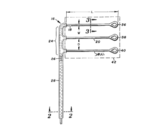
Turning now to the drawings, there is shown in Figure 1 a
defibrillation electrode 16 including three parallel and spaced
apart electrode segments 18, 20 and 22. Each of the segments has
a length (L in the figure) substantially longer than its width
(W), e.g. 30 cm long with a nominal width preferably about 0.5
mm. Generally, the width should be within the range of from
0.25-5 mm. Adjacent segments are spaced apart a distance (D)
substantially greater than the nominal width, e.g. 3 cm. This
center-to-center spacing should be at least 1.5 cm, and
preferably does not exceed 30 cm.
Electrode segments 18, 20 and 22 are fixed at respective
first ends to a distal end portion 24 of an electrically
conductive lead 26. The lead conducts electrical pulses to the
electrode segments from a pulse generator (not shown) coupled to
the proximal end of the lead. Lead 26 at the distal end
structurally supports the longitudinally extended electrode
segments in the transversely spaced apart configuration shown.
The electrically conductive portion of lead 26 is
surrounded by an electrically insulative cover or sheath 28,
preferably constructed of a body compatible polymer, e.g. a
medical grade silicone rubber or polyurethane. As seen in Figure
2, the lead includes a composite conductor formed of a core 30 of
silver surrounded by a tube 32 of stainless steel. This type of
composite conductor is known as drawned field tube (D~T) of MP35N
(brandname) alloy available from FWM Research Products of Fort
Wayne, Indiana. Further, a coating 34 of platinum is applied
7 ~ ~
over the stainless steel, preferably by ~uLLeril1g or other deposition process. While
preferably pl~timlm, coating 34 also can consist of another metal from the pl~timlm group
(e.g. iridium, ruthenium and p~ lillm) or an alloy of these metals. Insulative sheath 28 is
contiguous with and surrounds the pl~tinllm layer.
As seen in Figure 3, the construction of electrode segment 22 (and likewise
segments 18 and 20) over substantially all of its length is substantially similar to the
construction of the conductive portion of lead 26. Thus the segments also are highly
electrically conductive. Pl~timlm coating 34 provides a further advantage for the segments,
which are not covered by the insulative sheath. In particular, the pl~timlm coating when
applied by vapor deposition provides a microtexture which subst~nti~lly increases the reactive
surface area of the electrode segments, to reduce near field impedance of the electrode (the
term "near field" impedance refers to the voltage losses associated with the electrode due to
chemical and field effects). The reduced interface impedance increases the ratio of bulk
impedance to the total system impedance as measured between the stimlll~ting electrode and
the indifferent or signal return electrode. Thus, more of the voltage drop occurs across
tissue, where it is useful for causing the desired stim--l~tion, with proportionately less of the
voltage drop occurring at the electrodes where it is non-productive. This enables a reduction
in overall potential or pulse duration, in either event reducing the required energy for
defibrillation .
-10-
.~,
~ ~ ~ 7 F~ ~ ~
Given adequate separation between segments 18, 20 and 22, the current
distribution is made more unirollll. To further counter any current density differentials due
to edge effects at the ends of segments 18, 20 and 22, loops 36, 38 and 40 are formed at
these ends, respectively. Alternatively, the ends can be flared or otherwise enlarged, and
5 remain subst~nti~lly free of undesirable concentrations of high current. Such enlargements
also facilitate implant, as they tend to positionally fix the electrode segments.
Because the electrode segments are electrically common, the electrodes receive
and transmit defibrillation pulses simult~neously. The electrode segments are sufficiently
near one another to function in concert, providing an effective area or phantom area
10 incorporating the segments, as in~ic~ted in broken lines at 42. In other words, electrode
segments 18, 20 and 22 define a generally rectangular effective area, with substantially
greater compliance to contours and movements of body tissue, as compared to a continuous
patch electrode. In addition, the spacing between electrodes performs an important electrical
function by producing a subst~nti~lly more unirollll current distribution than that of a
15 continuous patch electrode. Patch electrodes are known to have regions of very high current
density around their outside edges, and regions of low current density at their centers. By
using a segmented electrode, with segments propelly spaced apart from one another, much
higher ~;ulre~ can be delivered to the central region of the effective or phantom area
because current is able to flow between adjacent segments. This results in a more uniro
20 electrical field across the heart.
Figure 4 illustrates an alternative embodiment defibrillation electrode 44
including five elongate electrode
-11-
202 7 ~ ~
.
......
segments 46, 48, 50, 52 and 54, each with a preferred width and
substantially greater preferred length as described in connection
with electrode 16. Each of electrode segments 46-54 is part of a
wire mesh pattern 55 and extends longitudinally. Transversely
extended end portions 56 and 57 of the pattern couple the
segments to a lead 58. An insulative sheath 62 surrounds lead 58
from electrode 44 to the proximal end of the lead. An
electrically insulative backing 64 supports mesh pattern 55. The
mesh pattern is covered by an insulative layer 66. Slots 68, 70,
lo 72 and 74 are formed in backing 64 and layer 66 between adjacent
electrode segments.
Figure 5 illustrates an alternative form of composite
conductor known as DBS (drawned braised strands), available from
FWM Research Products, Fort Wayne, Indiana. As shown, a silver
core 73 is surrounded by six stainless steel wires 75. The
structure is heated and drawn to braise all wires together. The
results is a solid, continuous composite conductor composed of a
silver core and a stainless steel outer shell or tube.
Figure 6 illustrates an alternative construction for the
electrode segments of either electrode 16 or electrode 44,
involving a plurality of composite conductors 76 in a twisted
configuration. Each of the conductors can include a silver core
within a stainless steel tube coated with platinum as previously
described. Alternative composite conductors for single and
multi~le wire arrangements include platinum or titanium ribbon or
wire, clad with platinum. The twisted construction enhances
flexibility and resistance to fatigue in the electrode segments.
Other alternatives include braided or knitted wires.
Figure 7 shows another alternative construction for the
electrode segments, in the form of a woven mesh or screen 78 on
- 12 -
.
2~7~i~
an electrically insulative backing 80. This type of electrode
segment construction is particularly well suited for epicardial
positioning, e.g. with electrode 44 in Figure 4.
Another alternative segment construction, shown in
Figures 8 and 9, involves a flexible, electrically insulative
cylindrical core 82 of polyurethane, medical grade silicone
rubber, or other suitable body compatible material. Core 82 is
surrounded by an electrically conductive coil winding 84,
preferably a wire or composite cable such as illustrated in
Figure 2. The helically wound coil conductor provides the
greatest flexibility and fatigue resistance of any of the
arrangements discussed, and for this reason is preferred in the
case of direct epicardial attachment, or any other implant
location in which the lead segments are subject to continued or
repeated muscular contraction or other abrupt tissue movements.
A disadvantage, relative to other embodiments, is that a helical
coil electrode segment, as compared to other segments of equal
length, involves a substantially longer conductive path with less
tensile strength.
All of the alternative constructions provide electrode
segments which are highly compliant, first in the sense that they
readily adjust to the contours of body tissue at the implant site
when they are implanted, and secondly over the long term, in
continually conforming to the tissue during muscular contractions
and other tissue movement.
Figure 10 illustrates a further embodiment defibrillation
electrode 86 including electrode segments 88, 90 and 92 formed as
branches, radiating or extended outwardly from a common junction
and stress relief area 94. Junction 94 is positioned at the
distal tip region of a lead 96 to a pulse generator (not shown),
- 13 -
2~2~7~4
."~ .
and includes a conductive portion surrounded by an insulative
sheath 98. The conductive region of the lead and the electrode
segments can be constructed as previously described.
The stress relief portion of the electrode is
electrically insulative and covers portions of the segments,
leaving exposed portions of the segments spaced apart from one
another and defining an effective or phantom area 100 shown by
the broken line. As before, segments 88-92 have a nominal width
preferably about 0.5 mm, and are longer than they are wide, for
example by at least a factor of five. At the free ends of the
segments are respective masses or bodies 102, 104 and 106. The
bodies are constructed of an electrically conductive, plastically
deformable material such as platinum or gold and, as seen in
Figure lo, include slots 108 slightly wider than the thickness of
segments 88-92. Each body is applied to the free end of its
respective electrode segment by inserting the free end within the
respective slot and pinching the body to frictionally secure the
body to the electrode segment. Bodies 102-106 thus provide
enlargements at the free ends of the segments to reduce the
chance for high current densities at the free ends, and provide a
means of fixation of the free ends.
Figures 11-13 schematically illustrate alternative
configurations for electrode 86. More particularly, Figure 11
illustrates a clamp 110 for electrically and mechanically
couplilng two intersecting cables 112 and 114. Cable 112 is part
of lead 96, with a distal portion of the lead providing center
segment 90. Electrode segments 88 and 92 are opposite portions
of cable 114. An extension 116 of electrically insulative sheath
98 covers clamp 110 and portions of cables 112 and 114, leaving
the segments exposed.
- 14 -
~ 2~2~794
,, ,
In Figure 12, segments 88, 90 and 92 extend radially from
a crimping member 118 at the distal end of lead 96.
Alternatively, segment 90 is the distal end of the lead, in which
case the remainder of the lead, crimping member 118 and portions
of the electrode segments are provided with an insulative
covering 119.
In Figure 13, crimping member 118 secures electrode
segments 88, so and 92 to the distal section 120 of lead 96.
Insulative sheath 98 leaves distal section 120 exposed, so that
it functions as a fourth electrode segment.
Figure 14 shows a further embodiment defibrillation
electrode 122 including a lead 124 having a distal end 126 formed
in a curved, serpentine configuration. An insulative sheath 128
covers the lead and leaves the distal region exposed. Further
insulation covers curved portions of the electrode at 130, 132
and 134, thus to define four parallel segments or length-portions
136, 138, 140 and 142 aligned with one another and side by side.
Figures 15, 16 and 17 disclose alternative serpentine
electrode configurations including an electrode 144 with a wire
mesh or screen 146 on an electrically insulative backing 148.
Figures 15 and 16 illustrate a conductive path 150 including
parallel electrode segments 152, 154, 156 and 158. The distal
end of segment 158 is enlarged at 160 to counteract edge effect
current densities.
' In Figure 17, an electrode 162 includes a serpentine
conductive path 164 formed between a pair of generally
rectangular electrically insulative layers 166 and 167. A
serpentine opening in layer 166 exposes part of a wire mesh layer
168. Slits in the patches at 170, 172 and 174 allow the patch to
conform to the site of implant. Selected parts of the conductive
- 15 -
~ 202~4~
'_
path can be covered with insulation if desired, to leave just
parallel segments exposed.
Figure 18 discloses yet another embodiment defibrillation
electrode 176 in which a single conductive path 178 at the distal
end of a lead 180 is formed into a spiral. The path can be a
coated composite cable or a wire mesh or screen as previously
described, with a similar nominal width in the radial direction.
The pitch of the spiral, i.e. radial spacing (D) between adjacent
arcs in the spiral, is preferably about 3 cm. Thus the effective
electrode area encompasses the outermost arc of the spiral, as
indicated by the broken line at 182. The spiral includes at
least two complete turns or length-portions as shown, with each
turn forming an arcuate electrode segment to provide respective
radially inward and outward segments 184 and 186.
Regardless of the particular embodiment, electrodes
constructed in accordance with the present invention provide a
substantially larger effective or phantom area than previously
practical for implantable defibrillation electrodes. One reason
for this is the spacing between adjacent electrode segments,
resulting in more compliant electrodes, both in the sense of
matching contours in body tissue, and "dynamically" in responding
to muscular contractions and other sudden or rapid tissue
movement, with virtually no risk of fatigue. Another feature
permitting the large size is the highly conductive electrode
segme~ts and lead distal end or other feature electrically
coupling the electrode segments. This ensures an acceptably low
voltage gradient across even relatively large electrodes.
As previously noted, a large but segmented electrode
structure results in a substantially more uniform current
distribution, as compared to conventional continuous patch
- 16 -
2~277~
..",
electrodes. Figure 19 schematically illustrates an electrical
field, in broken lines, between a continuous patch electrode 187
and an electrode composed of parallel, spaced apart wires or
segments 189. Adjacent segments 189 are quite close to one
another, e.g. spaced apart from one anther a distance of about 5
mm. Because of the low impedance between adjacent segments 189,
there is virtually no potential difference between these segments
and intervening tissue. Most of the current flow is along the
end segments 189, and very little occurs near the intermediate
segments or between segments. Consequently, the electrode formed
of segments 189, much like electrode 187, exhibits a non-uniform
current distribution, with very high current density at the
outside edges and low current density along the medial region.
In Figure 20, the electrical field between two electrodes
with respective segments 191 and 193 exhibits a substantially
uniform current density across each electrode. Again the field
is shown in broken lines, and illustrates the importance of
sufficient spacing between adjacent electrode segments. More
particularly, the segments of electrodes 191 and 193 are spaced
apart from one another a sufficient distance for intervening
tissue to provide substantial electrical impedance between
adjacent electrode segments. Thus, each of segments 191 and 193,
including the intermediate segments, responds to the opposite one
of the electrode pair, permitting current densities, over the
central regions of these electrodes, substantially equal to the
current densities at their edges.
Figure 21 shows the relationship between the spacing
between coils or adjacent and parallel electrode segments, and
impedance, for groups of two, three and four segments as shown at
195, 197 and 199, respectively. In all cases the impedance is
- 17 -
2 Q ~
highest when adjacent segments are closest together. In all
cases, increasing the spacing from 1 cm to the preferred 3 cm
reduces impedance, and the cases show some further improvement as
spacing is increased beyond 3 cm. For any selected spacing, the
four segment electrode exhibits the lowest impedance, which is
not surprising in view of the fact that larger electrodes
generally exhibit lower impedance.
Thus, it has been found that electrode performance is
substantially improved, in terms of reduced impedance as well as
uniformity of the electrical field, when the spacing between
adjacent segments is at least 1.5 cm. The upper limit of spacing
is less strict, and subject to physical (size and patient
comfort) constraints rather than electrical performance
constraints. Within these limits, the optimum spacing depends
upon the materials employed and the intended location of implant.
Generally, however, a spacing of 3 cm between adjacent electrode
segments has been found satisfactory.
Figure 22 schematically illustrates an implanted
defibrillation system including spaced apart electrodes 188 and
190, for example similar to electrode 16. The defibrillation
system further includes a pulse generator 192, and leads 194 and
196 connecting the pulse generator to electrodes 188 and 190,
respectively. Both of the electrodes are subcutaneous and
outside of the rib cage, in the thoracic region. The electrodes
are on opposite sides of the heart 198. More particularly,
electrode 188 is positioned to the left of, and anterior with
respect to, the heart. Electrode 190 is posterior with respect
to the heart, and to the right of the heart. Such transthoracic
application of defibrillation pulses requires electrodes having a
large surface area, achieved in accordance with the present
- 18 -
~0~774~
invention by the spaced apart electrode segments of each
electrode. Pulse generator 192 is also mounted anterior and to
the left of heart 198, below electrode 188. The pulse generator
can incorporate circuitry for sensing cardiac electrical
activity, in which case electrodes 188 and 190 are used in
sensing such activity as well as delivering defibrillation
pulses.
Figure 23 discloses a defibrillation system in which an
electrode 200 constructed in accordance with the present
invention is coupled to a defibrillation pulse generator 202 by a
lead 204. Another electrode 206, also constructed according to
the present invention, is applied directly to epicardial tissue.
Electrode 200 is positioned inside of rib cage 207, and can be
within the pleural cavity if desired. Stimulation occurs across
the heart, with electrode 200 to the left of the heart and
electrode 206 at the right ventricle.
Figure 24 shows a defibrillation electrode system
including an electrode 208 positioned anterior of and to the left
of the heart 210, as in Figure 22. A second electrode 212 is
provided as a coil, near the distal end of an intravascular
catheter 214 in the right atrium and terminating at the apex of
the right ventricle.
Regardless of the location of implant, electrodes,
constructed in accordance witll the present invention provide
relatively large (in the range of 100-300 square cm) effective
areas, yet readily conform to contours and contractions or other
movement of body tissue. The narrow electrode segments are
provided with end loops or other enlargements to counteract high
current densities due to edge effects and to provide fixation.
The present lead configurations further allow a subcutaneous
2027~4
implantation outside of the rib cage, with effective
defibrillation energy production due to large virtual sizes based
on the phantom areas incorporating the electrode segments.
What is claimed is:
- 20 -