Note: Descriptions are shown in the official language in which they were submitted.
2o29469
ELECTRODE ENDoTRAc~r~Ar~ TUBE
BACKGROUND OF THE INVENTION
The present invention relates generally to
electrodes for detecting electromyographic (EMG) signals of
the laryngeal muscles, and more particularly to electrodes
which are mounted on an endotracheal tube.
The recurrent laryngeal nerves, hereinafter referred
to as the laryngeal nerves, course through the neck to the
intrinsic laryngeal muscles, hereinafter referred to as the
laryngeal muscles. There are two laryngeal nerves, one on
the left side and one of the right of the neck. Each nerve
controls a set of laryngeal muscles, including a vocal cord.
Damage to laryngeal nerves is a common complication
of neck surgery. When the anatomy is relatively normal, the
course of a laryngeal nerve along the neck is usually
discernible. However, the presence of abnormal tissue, such
as tumor, inflammation or trauma may make anatomic dissection
of the nerve without damage nearly impossible.
If a laryngeal nerve is damaged during surgery,
paralysis of the related laryngeal muscles can occur.
Paralysis of the laryngeal muscles results in loss of speech
and may also disrupt breathing by preventing air passage
through the trachea. There is, therefore, a need for an
apparatus which will aid in the location of the laryngeal
nerves. It is also desirable to have an apparatus which
warns a surgeon when contact is made with a laryngeal nerve.
During surgery a tube is usually placed through the
patient's nose or mouth and into the trachea, passing between
the sets of laryngeal muscles. This endotracheal tube is
used to ventilate the lungs and may also be used to provide
anesthesia. Most endotracheal tubes include an inflatable
cuff surrounding the tube. Once the tube is inserted into
the trachea, the cuff is inflated to prevent air from
escaping by passing between the tube and the trachea wall.
.~
202946~
-- 2 --
One prior art method of locating a laryngeal nerve
uses an endotracheal tube having an additional cuff located
to be adjacent both sets of laryngeal muscles when the tube
is placed in the trachea. See, e.g. Engel P.M., et al. "A
Device For The Location and Protection of the Recurrent
Laryngeal Nerve During Operations Upon the Neck." Surgery,
Gynecology, and Obstetrics, 152:824-826, 1981. The addi-
tional cuff is inflated and connected to a device for
detecting pressure changes. A probe delivering an electric
charge is used to stimulate the laryngeal nerve. When the
probe contacts the nerve, the related set of laryngeal
muscles contract causing a pressure change within the cuff,
which can be detected by the pressure sensing device. Thus a
surgeon can locate a laryngeal nerve by using the probe to
stimulate various portions of the neck until a response is
noted on the pressure sensing device.
The pressure sensitive device, however, does not
operate satisfactorily. It can only detect relatively large
movements of the laryngeal muscles. Thus it may not be
sensitive enough to register all responses in the muscles
when the nerve is electrically stimulated.
The pressure sensitive device also may be unable to
detect the indigenous electric discharge which occurs when- a
laryngeal nerve is manipulated. Laryngeal nerves emit an
electrical impulse which travels to the related set of
laryngeal muscles when the nerve is manipulated or is
contacted by a surgical instrument. The impulse from contact
or manipulation is generally smaller than that which can
occur by use of outside electrical stimulation. Thus the
pressure sensitive device may not be able to warn a surgeon
when damage to a laryngeal nerve is imminent due to contact
with a surgical instrument.
Another method of locating a laryngeal nerve is the
use of electrodes emplaced directly into a related laryngeal
muscle. See, e.g., Lipton R.J. et al., "Intraoperative
Electrophysiologic Monitoring of Laryngeal Muscle During
Thyroid Surgery." Laryngoscope, 98:1292-1296, 1988. These
20294 6~
-- 3 --
electrodes are connected to an EMG machine which measures
changes in voltage in the muscle. A number of different
electrode types are known in the art, including needles,
needle pairs and hooked wires. These devices are capable of
detecting electrical changes in a laryngeal muscle caused by
external electrical stimulation of the related laryngeal
nerve. These devices are also sensitive enough to detect the
electrical changes which occur in a muscle when the related
laryngeal nerve is stimulated by manipulation, and, thus can
be used to inform a surgeon when contact is made with a
laryngeal nerve. These devices are undesirable, however,
because they are difficult to accurately emplace in the
muscle. A medical technique requiring a high degree of
expertise must be mastered by a surgeon before these
electrodes can be used.
Another device for measuring EMG activity in the
laryngeal muscles comprises a tube substantially thinner than
an endotracheal tube, and containing electrode wires which
extend from the tube's interior through the tube wall and
then circumferentially around the tube. The tube is placed
into the esophagus, which is located just behind the
trachea. This tube monitors the posterior laryngeal muscles,
the muscles at the back of the trachea, through the front or
anterior esophagus wall. See e.g., Fujita et al., "A New
Surface Electrode for Recording from the Posterior
Cricoarytenoid Muscle". Laryngoscope, 99:316-320, 1989. The
circumferential electrode device is designed for use while
the patient is awake, to measure EMG activity during normal
breathing and speaking, although it could also be used during
surgery.
The circumferential electrode device is inadequate
for use in locating and protecting the laryngeal nerve for a
number of reasons. First, because each set of laryngeal
muscles is located adjacent a respective right or left side
of the trachea, the circumferential electrode device allows
measurement of EMG signals from only one of the two sets of
laryngeal muscles. More particularly, the device is thin and
20294 69
-- 4 --
must be placed against either the right or left side of the
anterior esophagus wall. Since the device contacts only one
side of the anterior wall, it can only monitor the set of
laryngeal muscles in the trachea closer to that one side.
Second, placement of the thin tube is difficult because it
requires that the thin tube be positioned in the esophagus
without any direct visualization. Additionally, a cir-
cumferential electrode configuration could not be used on an
endotracheal tube employed for ventilation during surgery,
because that kind of tube is relatively thick; and a given
electrode surrounding the thick endotracheal tube would
unavoidably be in contact with both sets of laryngeal muscles
at the same time.
SUMMARY OF THE INVENTION
There is provided in accordance with the present
invention an endotracheal tube which performs the functions
of ventilating the lungs during surgery and monitoring the
EMG signals of the laryngeal muscles. The endotracheal tube
comprises a flexible tube having a distal end and a proximal
end. The tube contains a main lumen for ventilating the
lungs, an inflatable cuff and a thin lumen for inflating the
cuff. The thin lumen is located in the wall of the tube and
is attached to a fitting for connecting the thin lumen to an
air source for inflating the cuff.
The tube contains one or more electrode wires which
run in a direction parallel to the central axis of the
tube. Each electrode wire is insulated against electrical
contact at a first wire portion located between the ends of
the tube. Insulation may be achieved by embedding the first
wire portion within the wall of the endotracheal tube. An
uninsulated second wire portion, located between the tube's
distal end and the first wire portion, lies exposed on the
surface of the endotracheal tube permitting electrical
contact to be made by the second wire portion.
202q469
5 --
When an endotracheal tube is inserted in a human
patient, the distal end of the tube must be located above the
point where the trachea splits to communicate with both
lungs, thus insuring that both lungs are ventilated. The
tube must be inserted far enough into the trachea, however,
so that the cuff is located below the laryngeal muscles. The
cuff must also be located low enough so that when it is
inflated it does not push against the laryngeal nerves which
run up the neck, near the upper portion of the trachea,
toward the laryngeal muscles. Pressure by the cuff could
damage the laryngeal nerves. When the distal end of the
endotracheal tube is above the split in the trachea and the
cuff is below the location of the laryngeal muscles, the tube
is properly positioned.
The second or uninsulated portion of each electrode
wire is positioned on the tube so that the uninsulated
portion contacts a set of laryngeal muscles, particularly a
vocal cord of that set, when the endotracheal tube is
properly positioned. The uninsulated wire portion must be
long enough so that contact with the laryngeal muscles can be
easily accomplished. The uninsulated portion must not,
however, be so long that it contacts parts of the patient's
anatomy other than the laryngeal muscles. Contact with other
parts of the patient's anatomy, such as the tongue or the
pharyngeal muscles, could cause the electrode wire to receive
EMG signals from sources other than the laryngeal muscles. A
given length and location on the tube of the second wire
portion will suffice for virtually all adults. However, the
length and location of the second wire portion may differ for
an electrode endotracheal tube intended for use in children
or individuals who have other than the normal sized anatomy
usually found in adults.
The electrodes are capable of detecting EMG signals
of two distinct types. The first type of signal is one
produced when a laryngeal nerve is stimulated by an electric
probe. When a voltage is applied on or near a laryngeal
nerve, the electrical pulse is carried to the related set of
- 2029469
-- 6
laryngeal muscles through the nerve. The electrode endo-
tracheal tube can detect that electric pulse when it is
transferred into the related laryngeal muscles. Thus a
surgeon can locate a nerve by electrically stimulating
various portions of the neck during surgery and noting
whether a response is detected by the electrode wire
contacting the related laryngeal muscles.
The second type of EMG signal which the electrode
endotracheal tube will be able to detect is one caused by
physical manipulation of a laryngeal nerve. When laryngeal
nerve tissue is manipulated or contacted by a surgical
instrument, the nerve emits an electrical pulse. That pulse
is carried to the related laryngeal muscles via the nerve.
Thus, when a surgeon manipulates or contacts a laryngeal
nerve, an EMG signal can be detected in the related laryngeal
muscles. In this way the invention can be used to alert a
surgeon when potentially damaging contact with a laryngeal
nerve is imminent.
Located near the proximal end of the tube are
conventional mechanisms for connecting the electrode wires to
a device for processing EMG signals.
The electrode endotracheal tube also has the
advantage of being able to alert doctors when the endo-
tracheal tube is either too proximal or too distal in the
trachea to allow proper ventilation. When the patient is
under a light level of anesthesia, EMG signals associated
with breathing can be detected in the laryngeal muscles. If
those signals are not detected when the patient is initially
intubated, i.e. when the endotracheal tube is inserted, it is
an indication that the tube is not properly placed.
The present invention also has the advantage of
being easy to accurately place. Endotracheal tubes have been
used in surgery for many years, and many doctors, particu-
larly anesthesiologists, are already skilled in the insertion
of those tubes. Thus there is already a large group of
medical professionals who can properly place the electrode
endotracheal tube.
2029469
.
7 --
Other features and advantages are inherent in the
electrode endotracheal tube claimed and disclosed or will
become apparent to those skilled in the art from the
following detailed description in conjunction with the
accompanying diagrammatic drawings.
BRIEF DESCRIPTION OF THE DRAWINGS
Fig. 1 is a perspective of an embodiment of an
electrode endotracheal tube constructed in accordance with
the present invention;
Fig. 2 is a sectional view taken along line 2--2 in
Fig. l;
Fig. 3 is a sectional view taken along line 3--3 in
Fig. l;
Fig. 4 is a sectional view taken along line 4--4 in
Fig. l;
Fig. 5 is a fragmentary, vertical sectional view of
the neck and chest region of a human body showing the
placement of the electrode endotracheal tube;
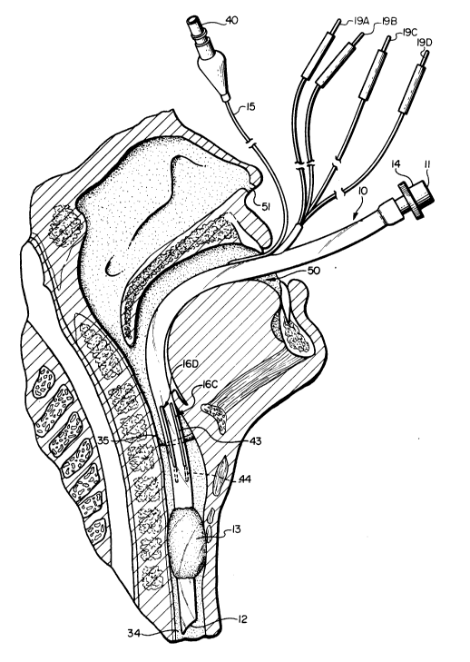
Fig. 6 is a fragmentary, sectional view of the head
and neck region showing the placement of the electrode endo-
tracheal tube; and
Fig. 7 is a schematic diagram showing the steps
employed in processing an EMG signal from an electrode.
DETAILED DESCRIPTION OF THE INVENTION
Referring initially to Figs. 1-4, indicated
generally at 10, is an electrode endotracheal tube
constructed in accordance with an embodiment of the present
invention and comprising a flexible, non-electrically
conducting tube having a proximal end 11 and a distal end
12. Tube 10 has a main lumen 20 for transporting gases to
and from the lungs.
2029469
8 --
At proximal end 11 is a fitting 14 for connecting
tube 10 to a respirating machine (not shown) which injects
and withdraws air from the lungs. A cuff 13 is located near
distal end 12. Cuff 13 is shown in an uninflated condition
in Fig. 1 and can be inflated by use of a cuff inflating
conduit 15, which is attached to a source of compressed air
(not shown) by a fitting 40. Cuff inflating conduit 15
communicates with a lumen 25 located in the wall 24 of tube
10 (Figs. 2-4), and lumen 25, in turn, communicates with cuff
13. Wall 24 is defined by inner surface 22 and outer surface
23.
Associated with tube 10 are four electrode wires
indicated generally at 16A, 16B, 16C and 16D, each composed
of an electrically conducting material and each running from
a location between the two tube ends 11 and 12 toward distal
end 12. The term "wires" includes any type of electrically
conducting lead suitable for use as an electrode, including
metal paint, metallic tape or metal strips. Wires 16A-D run
in a direction parallel to the tube's central axis 21. Each
electrode wire has a first portion 42, located between
proximal end 11 and distal end 12, and insulated against
electrical contact. In the embodiment of the invention
depicted in Figs. 1-4, each first wire portion 42 is embedded
within tube wall 24 to insulate wire portion 42 from
electrical contact.
A second wire portion 43 is located between distal
end 12 and first wire portion 42, on outer surface 23 of tube
10. Each second wire portion 43 is uninsulated and capable
of forming an electrical contact. Each wire may have an
optional third portion 44 embedded within wall 24 between
distal end 12 and second wire portion 43. Each wire is
embedded at third portion 44 to keep second portion 43 in
place on outer tube surface 23. Other expedients can be used
to insure that second portion 43 remains in place, so long as
those other expedients permit electrical contact between
second portion 43 and adjacent laryngeal muscles.
- 2029469
g
In a preferred embodiment of electrode endotracheal
tube 10, second wire portion 43 begins 10 cm from distal end
12 and ends 3 cm closer to proximal end 11. In other
embodiments of electrode endotracheal tube 10, second wire
portion 43 may begin 8 to 12 cm from distal end 12 and end 2
to 4 cm closer to proximal end 11.
As best shown in Figs. 2-4, wires 16A-D run in
pairs, two along the tube's left side, e.g. 16A and 16B, and
two along the tube's right side, e.g. 16C and 16D. When an
electrode pair is used to monitor the EMG activity in any
muscle, those electrodes are called a bipolar electrode
pair. It is possible to use single or monopolar electrodes
to monitor muscles, however, the use of a bipolar electrode
provides greater accuracy. The workings of electrodes and
the advantages of bipolar over monopolar electrodes are well
known to one skilled in the art.
Electrical connecting plugs l9A, l9B, l9C and l9D
are used to connect wires 16A-D to an EMG processing machine
(not shown). Any means capable of forming electrical contact
such as ports, alligator clips or insulated wires with bared
ends could be used with the present invention instead of the
depicted plugs.
Fig. 5 shows electrode endotracheal tube 10 properly
inserted into the trachea of a patient. Tube 10 is inserted
between a set of left laryngeal muscles 35 and a set of right
laryngeal muscles 36 and into the trachea 34. Distal end 12
is located within trachea 34 above the junction 37 where the
trachea splits into two passages, each going to a separate
lung. Cuff 13 is located below laryngeal muscles 35 and
36. Second wire portions 43 are in contact with laryngeal
muscles 35 and 36. Wires 16A and 16B are in contact with
left laryngeal muscles 35 and wires 16C and 16D are in
contact with right laryngeal muscles 36. The right recurrent
laryngeal nerve 31 is shown located beneath the thyroid 32.
The left recurrent laryngeal nerve 30 is exposed, as
depicted. Cuff 13 is shown inflated to prevent air from
escaping between the wall 33 of trachea 34 and tube 10.
--- 2029469
-- 10 --
Fig. 6 shows tube 10 inserted orally through mouth
50 and passing into trachea 34. Another embodiment of the
present invention would use a slightly thinner endotracheal
tube which could be inserted nasally through a nostril 51 and
into trachea 34.
Fig. 7 is a schematic diagram showing the steps that
could be used in processing EMG signals from an embodiment of
electrode endotracheal tube having a bipolar electrode
pair. The electrode endotracheal tube 10 could be used to
provide a variety of information. The processing steps
described below constitute only one way of using the informa-
tion detected by the electrode endotracheal tube.
Each electrode is connected to a conventional,
commercially available EMG processing machine such as the
Nicolet Nerve Integrity Monitor-2 as described in the Nicolet
Nerve Integrity Monitor-2 User's Guide, March, 1988, Nicolet
Biomedical Instruments, Madison, Wisconsin, and the
disclosure therein is incorporated herein by reference. A
device of this type can process the EMG signals and provide
information to the surgeon in the form of a record or an
alarm.
A voltage detected by electrode 16A at left
laryngeal muscles 35 is amplified by the processing circuit
at 57. The voltage reading at a tissue site is dependent on
a number of factors. Changes in voltage can be caused by
electrical stimulation to the nerve controlling that tissue
site or by contact or manipulation of that nerve. A
reference electrode 55 (not depicted in Figs. 1-6) is
attached to a reference site 56 on the patient's body (e.g.
the chest, arms or legs). It is desirable to have a
reference electrode 55 to screen out electrical impulses,
from outside sources, which can be detected throughout a
patient's body. The voltage detected at the reference site
is amplified by the processing device at an amplification
device 58. The two amplified signals from 57 and 58 are then
compared at a comparison device 60 and any difference in
those signals is stored at 62. The second electrode in the
-- 202946~
11 --
electrode pair, electrode 16B, also receives a voltage from
left laryngeal muscles 35, which is transmitted to the
processing machine, which amplifies the voltage at an
amplification device 59 and compares it with the signal from
reference electrode 55 at a comparison device 61. Any
difference in those voltages is stored at 63. Signals from
storage 62 and storage 63 are then compared at a comparison
device 64.
The signals from comparison device 60 and comparison
device 61 each represent the difference in voltage between
laryngeal muscles 35 and reference site 56. The signals from
comparison device 60 and comparison device 61 will be
slightly different since the voltage reading at each point in
left laryngeal muscles 35 will be slightly different. Any
similarities between the signal from comparison device 60 and
the signal from comparison device 61 will have been caused by
voltage at reference site 56. Any difference in voltage will
be registered at comparison device 64, and this will be the
result of an EMG signal in the laryngeal nerve. The signal
from comparison device 64 can then be displayed at a display
device 65 or recorded at a recording device 66 in a
conventional manner in order to provide a surgeon with
information on EMG activity in the laryngeal muscle.
Detection of EMG signals in right laryngeal muscles
36 would be similarly carried out using electrode wires 16C
and 16D.
A monopolar electrode works in a manner similar to a
bipolar pair. Only a comparison at 60 would be made,
however. Thus a monopolar electrode may result in EMG
signals whose source is the reference site rather than a
laryngeal muscle.
In order to monitor the EMG activity in both the
right and left sets of laryngeal muscles, the electrode or
electrode pair on each side of the endotracheal tube must be
connected to a separate channel of an EMG processing
machine. Thus, a device built according to the teachings of
- 2029469
- 12 -
the present invention can be used to monitor both laryngeal
nerves without having to adjust the endotracheal tube.
Electrode endotracheal tube 10 can be used to
provide information on whether the tube has been properly
placed within trachea 34. If the second wire portion 43 is
not in contact with left laryngeal muscles 35, the electrode
wires 16A and B will not be able to detect EMG signals within
these muscles. One way of determining whether electrode
endotracheal tube 10 is properly placed is by making use of
the normal EMG signals from the laryngeal muscles associated
with breathing. When a patient is under a light level of
anesthesia, which conventionally occurs just before and
shortly after the surgical procedure, these normal signals
can still be detected in the laryngeal muscles. If the
normal signals are not detected, it is an indication that the
tube is not properly placed. Once the tube has been properly
placed, the patient can be put under a deeper level of
anesthesia. Under this deeper level, the normal EMG signals
will cease, so that any EMG signal detected by the electrode
endotrachea tube will have been caused by stimulation of a
laryngeal nerve.
The electrode endotracheal tube can also be used to
determine whether the laryngeal muscles are functioning
properly prior to extubation or removal of the endotracheal
tube. This is important because, if the laryngeal muscles
are not functioning after a patient is extubated, breathing
can be disrupted.
As previously discussed, damage to a laryngeal
nerves can cause paralysis of the related laryngeal muscles,
but paralysis can also be caused by damage inflicted to the
laryngeal muscles upon intubation or by damage to that part
of the brain which controls the laryngeal muscles. When
patients are extubated, they are usually under a light level
of anesthesia, thus the normal EMG signals associated with
breathing can be detected in the laryngeal muscles. If these
normal signals cannot be detected, the doctors can be
prepared to take remedial measures when the tube is removed.
`- 202946~
- 13 -
The foregoing detailed description has been given
for clearness of understanding only and no unnecessary limi-
tations should be understood therefrom, as modifications will
be obvious to those skilled in the art.