Note: Descriptions are shown in the official language in which they were submitted.
'~ ~
W O YO/10417 ~ ~ f~ .J~ 2 PCT/~S90/01334
DEFLECTAB~E-END ENDOSCOPE ~ITH DEtACHABLE SHAFT ASSEM8LY
;,
- gackground of the Inve~lon:
This invention relates generally to endoscopes
and more particularly to such instruments which have a
flexible shaft and a deflectable end portion controlled
5 via a mechAnism at the proxlmal end.
Endoscopes are used in various medical and
industrial applications for viewing unaccessible
interior features of cav~ties, tubes or conduits, such
~s body organ~. The present invention i8 particularly
concerned with endoscopes o~ flexible and small outside
;~diameter shafts of less than 0.15 inch, useful, for
example, a~ ureteroscopes, hysteroscopes, angioscopes,
choledochoscope~, and cystoscope~.
~`The prior ~rt i5 replete with endoscopes which
incorporate an end portion which can be de~lected, by a
user, via a control mechanism at the proximal end o~ the
;idevice. Such endoscopes are characterized by various
structural configurations which enable a user to control
;;the de~lection o~ the distal tip o~ the end portion
through an ~ngle ~rom approximately 0 degrees to 180
degrees. Ihe following patents are examples of
endoscope~ having a deflect~ble tip:
4,653,476 ~onnet
4,580,551 Siegmund
4,577,621 Patel
4,353,358 Emerson
3,788,304 Takahashi
Other structures are shown in the following
additional patents:
30 3,426,663 3,948,251 4,483,326 4,616,630
3,470,876 4,063,796 ~,503,842 4,617,915
3,572,325 4,066,070 4,503,843 4,630,598
3,610,231 4,175,545 4,543,090 4,633,882
. . , ..
,
: , . ~ ,.
,:
WO 90/1041'~ , PC~IIJS~0/01334
2-
3,726,2724,176,662 4,557,253 4,646,722
3,7~8,3044,178,920 4,557,254 4,650,467
; 3,799,1504,203,430 4,561,427 4,6~1,202
; 3,7g9,1514,245,624 4,566,437 4,651,718
3,856,0004,271,845 4,567,882 4,653,476
3,880,1484,277,168 4,557,621 4,676,228
~ 3,892,2284,294,233 4, ~86, g23 4,685,449
3,897,7754,446,444 4,593,680 4,686,963
: 3,91S,1574,447,227 4,601,705
Summarv of the ~nvention
The present invention is directed to an improvedendoscope having a deflectable tip configured so as to
have a small outer diameter of approximately 0.15 inch
or le~s.
lS More specifically, the present invention is
directed to ~ flexible sha~t subassembly comprised of a
conduit having a deflectable end segment at lts distal
end which can be controlled by a manually operable
mechanism on a handle subassembly. The shaft subassembly
further includes a cone subassembly at its proxim~l end
including means for structurally connecting to the
handle suba~sembly. Illumination fibers, nn imaging
fiber, a pull wire and a working channel extend through
the shaft subaæsembly fro~ its distal end to terminals
in the cone subassembly ~or interfacing to the handle
sub~ssembly. A preferred handle subassembly comprises an
optical component which allows for the viewing of an
image emanating from the shaft subassembly imaging
fiber, ~ deflection mechanism which incorporates a means
for physical attach~ent and axial displacement o~ the
shaft subassembly pull wire, And ~ locking mechanism for
attaching the shaft subassembly to the handle
subassembly.
In accordance with one aspect of the present
invention, a series of discrete substantially aligned
cutouts ~re formed ~n the body o~ the shaft subassembly
end segment. The geometry of the cutouts vary
, : . : .
: ' !
W090/1~17 ~ PCT/US90/013
~3~
progressively along the length o~ this deflectable end
segment to produce a gradual dlstal tip deflection
profile as the end segment is forced to bend by an axial
force on the pull wire. The cutouts are preferably of
5 substantially triangular shape in a plane containing the
pull wire and imaging fiber.
In accordance with a preferred embodiment of the
invention, the body of the deflectable end segment is
`~ comprised of an elongated flexible multi-lumen ~ody
covered tightly with a smooth, thin elastomeric sheath.
The individual lumens in the body provide passageways
for the illumination and lmaging fibers, pull wire and a
relatively large working channel useful for movement of
fluids and/or for passing diagnostic or therapeutic
instrument~.
In accordance with a preferred embodiment, the
;~ conduit connecting the deflectable end port~on to the
cone subas6embly comprises an elongated flexible conduit
formed o~ two elongated counter wrapped ~lat ribbon
coils covered with an elastomeric 6heath over their
entire axial length. This conduit configuration provides
protection ~or the encased illumination and i~aging
fibers and working channel against stresse~ resulting
from the flexure, tension or compre~sion imposed upon
the shaft subassembly during operation o~ the endoscope.
In accordance with still a further aspect of the
preferred embodiment, the handle subassembly provide~ a
means to mate with the shaft subassembly and form the
complete endoscope unit. Contained wlthin the handle
subassembly are: the means to align and optically mate
the shaft subassembly imaging fiber bundle with a
viewing optics and a focusing mechanism which can be
ad~usted through the rotation of a screw mechanism: the
means for securing the pull wire contained in the shaft
subasæembly to a component whose axial movement i8
controlled by the rotation of a screw mechanism: and a
means for securely locking the cone subassembly to the
, , . ~ . - ~
.. : . .. . . . .
- ~ .: ,., : . . .. . :
PCl`~US90/01 334
~ ~ {~ $ ,.
-4-
handle subassembly in fixed orientation, e.g. via a
bayonet mount, whereby the flexure of the deflectable
distal end segment is in a plane oriented perpendicular
; to a plane defined by a working c~annel port and light
! :; s post on the cone ~ubassembly.
In accordance with still a further aspect of the
preferred embodiment, the shaft subassembly and handle
subassembly are provided with watertight seals at all
externally exposed mating interfaces to prevent a
compromise of internal components resulting from usage
and sterilizations.
Brief Description of the Drawinqs
Figures lA and lB are respectively side
elevation views of the mating shaft ~ubassembly and
handle subassembly of the endoscope in accordance with
~ the present invention.
¦ Figures lC and lD are respectively end views of
the sha~t subassembly and handle subassembly of Figures
lA and lB.
Figure lE is a side elevation view o~ the mated
shaft and handle ~ubassemblie~ in accordance with the
present ~nvention.
Figure 2 is an isometric view of the shaft
subassembly deflect~ble end segment.
Figure 3A i~ a lateral cross sectional view of
the deflectable end 6egment taken substantially along
the plane 3A-3A of Flgure 2.
Figure 3B lo a longitudinal cross sectional view
of the deflectable end ~egment taken substAntlally along
the plane 38-3~ of Figure 2.
Figure 3C ls an exploded view of the end portion
ta~en substantially along the plane 3C-3C of Figure 2.
Figure 3D is an exploded view of the distal tip
of the deflectable end portion shown in Figure 3B.
Figure 4A is a longitudinal cross sectional view
of the conduit ln the shaft fiubassembly taken
substantially along the plane 4A-4A in Figure lE.
!. . : , ~ .. - : "" ' '
WO90/10417 ~ J~ PCT/US~/Q13~
_5_ .
Figure 4B is a lateral cross sectional view of
.
the conduit in the shaft subassembly taXen ~ubstantially
~ along the plane 4B-4B ln Figure lE.
; Figure 5 is a longitudinal cross sectional view
Sof the cone subassembly taken substantially along the
plane 5-5 of Figure lC.
Figure 6A is a lateral cross sectlonal view of
; the cone subassembly taken ~ubstantially along the plane
6A-6A of Figure lA.
10Figure 6B is a lateral cross sectional view of
the cone subassembly taken substantially along the plane
6B-6B in Figure lE.
Fiqure 6C i8 a lateral cross sectional view of
the cone ~ubassembly taken substantially along the plane
156C-6C in Figure lA.
Figure 7 is a longitudinal cross sectional view
of the handle subassembly taken gubstantlally along the
plane 7-7 in Figure lB.
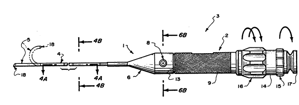
20Attentlon ls initlally dlrected to Figures lA
and lB which respectlvely illustrate a shaft subassembly
1 and a handle subassembly 2 which mate together to form
a complete endoscope 3 as deplcted in Figure lE.
Although endoscope 3 as illustrated in the drawing is
25particularly configured for use as a ureteroscope, it
should be understood that the feature~ Or the invention
are also applicable to endoscopes configured for other
applications. Briefly, the endoscope 3 is comprised of
two primary subassemblies; namely a shaft subassembly 1
30and a ~andle ~ubassembly 2 which can be readily
operatively attached to each other. The handle
subassembly 2, normally held in the grip o~ a user, has
a rigid ~tructure wlth moveable controls while the shaft
subassembly 1, whlch is partially inserted into a cavity
35~.g. a human organ, has a smooth flexlble structure.
The shaft æubassembly 1 in accordance with the
invention is basically comprised of ~n elongated
, . , ~, :; . . . . . :, .:.. , .... , . , . :
PCI`/US90/0~ 334
L r
-6-
flexible condult 4 having a deflectable end segment 5 at
lts distal end ~nd a rigid cone suba~embly 6 at its
- proximal end. The shaft subassembly 1 lnternally
.
i contains elongated lllumination fibers, an imaging
S fiber, a pull wire and a working channel ~not shown in
Figures lA-lE) to be discussed hereinafter. All of these
elongated elements terminate in the cone subassembly 6.
The cone su~assembly 6 includes a working channel port
7, configured with a female luer fitting, 6tandard in
the medical industry for allowing connection of syringes
and other tubing adapters to the internal working
channel. The fitting is preferably angled with respect
to the longitudinal axis of the shaft subassembly for
ease of insertion of the diagnostic and/or therapeutic
instruments. The subassembly 6 further includes a
lightpost 8 preferably comprising an industry standard
male terminal having a fine polished end face for
coupling a light source to the internal lllumination
fibers. An imaging fiber bundle extends through the
rigid post 9 and i8 terminated at its end by a small
ferrule 10. A pull wire i8 guided through the inner
partfi of the cone 6ubassemb1y and i~ securely attached
to a llfter 11 which can be axially translated to
deflect the end segment 5 as depicted in phantom ln
Figure lE. The cone subassembly 6 also includes a male
bayonet connector 12 configured around the lifter 11,
for interconnection with a female bayonet connector
mounted at the distal end 13 of the handla subassembly
2.
The handle subassembly 2 comprises a cylindrical
body which contains an eyepiece 14 configured in a
focusing ring 15 which rotates around the central axis
of the handle subassembly, and a deflection control ring
16. The deflection control ring 16 is an internally
threaded mechani~m which rotates from a null positlon
(corresponding to an axially aligne~, zero deflection
orientation of the distal end segment 5). A~ the rlng 16
,
r~
. . , " ~,,,, , ~ .
.
WO90/l0417 ~ J~ PCT/US90/013~
is rotated in one direction, it axially transl~tes the
lifter 11 to pull the internal pull wire to gradually
deflect the distal tip 18 of the end segment 5. The
distal tip 18 can operatively be placed at an angle
between 0 degree~ to 180 degrees with respect to the
axis of conduit 4. If the deflection control ring 16 i8
rotated to any position the deflected dlstal tlp 18 will
remain at that position until the deflection control
rinq 16 is rotated again. The distal end 13 of the
handle subassembly 2 is internally terminated with a
hollow female bayonet connector which tightly mates with
its male counterpart 12 on the cone subassembly 6 when
coupled together by a user. The central portlon of the
proximal end 17 of the eyepiece 14 is conflgured with a
optical window for viewing the image emanating from the
imaging fiber bundle at ferrule 10.
Coupling the sha~t subassemb1y 1 to the handle
subassembly 2, is initiated by holdlng ln one hand, the
handle subassembly around the knurled portion 19 and
resetting the deflection control ring 16 to its null
! deflection position. With the shaft subassembly 1 held
in the other hand with proper orientation, the rigid rod
9 is inserted through the opening at the distal end 13
of the handle subassembly 2. To engage the two
connector~, the male and female bayonet connectors of
the two subassemblie~ are mated together and the shaft
subassembly iB rotated with respect to the handle
subassembly or vice versa until it locks. The two units
will then be securely coupled and ready for use. To
disconnect the shaft subassembly, rotate the deflection
ring to the null deflection position and reverse the
direction of rotation of the shaft subassembly with
respect to the handle subassembly.
Attention is now directed to Figure 2 which
illustrates a side elevation view of the deflectable end
segment 5 of the shaft assembly 1 o~ Figure lA in lts
relaxed straight configuration. ~he distal tip 18 of
., .~ .:
: r
~, go/1 041 7 PCI`/US90~01 3~U
-8-
segment 5 constitutes the exit point for the light
pumped through the illumination fibers 20, 21 from
lightpost 8 and a distal port for the working channel
22. It also provides a viewing window 23A where the
S ; image of the scene viewed is formed, by the distal end
- ob~ective lens 23 (Figure 3D). To further describe the
deflectable end segment 5, cross 8ections taken ~t
different positions and planes will be discussed in more
'~' ' detail.
Figure 3A shows a lateral cross section view
through section 3A-3A of the deflectable end ~egment s
of Figure 2. $he structure essentially comprises a
flexible multi-lumen, preferably thermoplastic tubing 24
(e.g. polyurethane of a moderate shore hardness). The
lS relatively large central lumen 22 defines the worXing
channel extending along the longitudinal axis of tubing
24. The tubing wall 25 may include empty channels 26, 27
extending ~long lts length to facilitate flexibility of
the deflectable end segment 5. one or more lumens 28, 29
are provlded ln the wall 25 extending along its length
for respectlvely accommodatlng lllumlnatlon fibers 20,
21. An additional lumen 30 extends through the tubing
wall 25 along the length thereo~ for accommodatinq an
imaging fiber bundle 31. In an alternative embodiment
lumens 28, 29, 30 may be replaced by a slngular kidney
shaped lumen running parallel to the tublng longitudinal
axis to accommodate simultaneously the lmaging fiber 31
and illumination fibers 20, 21. A further lumen 32
extends through the tubing wall 25 along the length
thereof and accommodates a pull wire 33, preferably
formed of stainless steel,. A tubular sheath 34,
preferably of thermoplastic elastomeric material,
tightly encases the assembled deflectable end segment 5
and extends throughout its length. The sheath 34 is of
relatively lower shore hardness than the mult~-lumen
tubing 24. All the through lumens shown in Figure 3A may
deviate in s~ape from the circular geometry to another,
. .
WOgO/10411 ~ PCT/US901013~
_g _ .
e.g. elliptical, to accommodate the same overall
functions of the deflectable end segment 5 of Figure 2.
Attention i6 now directed to Figure 38 which
; -shows A longitudinal cross section of the deflectable
- s end segment S. A series o~ di~creet substantially
aligned cutout6 3s are formed in the wall 25 of the
multi-lumen tubing 24 and cut into the working channel
22. The dimension~ of the cutouts are varied
progressively along the length o~ the tubing 24 in a
unique distribution. These cutouts are substantially
triangular in a longitudinal plane containinq the pull
wire 33 and the imaging fiber bundle 31. The cutouts
start relatively wide at the peripheral wall 25 of the
tubing 24 and narrows down towards the tubing axis.
15The distribution of the cutouts is structured to
provide lncreased bendability of the tubing 24
progressively toward the dist~l -end. In addition it
causes a predetermlned gradual deflection profile of the
distal tip 17 in response to the pull wire 33 being
pulled proximally. The cutouts geometry and dlstribution
w$11 also assure a contlnuous deflection of the distal
tip 11 from 0 degrees to 180 degrees, within a plane
defined by the straight longitudinal axi~ of the tubing
24, and the pull wire 33. As the deflection is
lnitiated, the cutouts will progresslvely start to
close. The lmaging fiber 31, and the shore hardness o~
the multi-lumen tubing 24, possess enough stiffness to
stralghten out the de~lected end segment ~ro~ lts
deflected configuration when the force on the pull wlre
33 ls released at the handle. If deflectlon is required
in any other plane, then the shaft subassembly 1 and
handle subassembly 2 are rotated as a unit to rotate the
deflectable end segment 5. The cutouts may alternatively
have geometries with cross sections other than
triangular e.g. rectangular, rounded, key-hole shape or
a comblnation of some or all of the mentioned
configurations. The cutouts separation and distribution
. .
. -;... ~: ,
-, . . . . . .
0/1041t ~ rcr/lJssotol334
, , --10--
with respect to either end of the tubing 24 may also
vary to obtain the same function of the deflectable end
segment S. The working channel 22 in the deflectable end
segment S and the conduit 4 are connected together to
form a continuous path for the fluids and instruments,
by means Or ~ thin wall metal sleeve 18A which is
securely ~onded into the proximal end of the working
channel 22 in the deflectable end segment S.
Attention is now directed to Figure 3C which
shows a lateral cross section view of the distal end 17
of the deflectable end segment. The structure of the
distal tip 18 consists essentially of a relatively hard
cured adhesive casinq 18A which securely bonds the
distal ends of the illumination fibers 20,21, imaging
lS fiber 31 and pull wire 33 to the wall 2S of the
multl-lumen tubing 24. The adhesive caslng 18A is
preferably shaped in a circular cross section, with
smooth outside surface finish, to match that of the
`i tubing 24 and to provide a smooth transition between the
two ~egments. The adhesive casing 18A also provides a
physical means to encase the illumination fibers 20, 21
and imaging fiber 31, and to secure the encasement to
the distal end of the tubing 24. The walls of the
adhesive casing 18A conf$gures a hollow channel
2S throuqhout its length to provide continuity of the
working channel 22.
The pull wire 33 is terminated and bonded at the
distal end of the tubing 24 (Figure 3D) while the
illumination fibers 20, 21, imaging fiber 31 and working
channel 22 extend beyond the tubing 24/casing 18A
~unction. $he magnified view (Figure 3D) of the distal
end shows that the surface of the distal tip is
moderately angled e.g. 45 degrees, in a plane
perpendicular to the plane containing the pull wire 33
and imaging fiber 31. The corners 38, 39 and the outer
edge 40 of the distal tip are preferably rounded and
polished to provide an atraumatic tip con~iguration to
- . . .
,, , , . . , -,~
- . .. ~ - . : .,;, :. '.. :. :
,.,
....
90/lWl7 ~ s~ PCT/US~/013~
facilitate movement inside delicate relatively soft
tissue surfaces, e.g. human organs. The atraumatic tip
also provides a means to reduce the difficulty faced
when inserting and passing the distal tip through tight
orifices or constricted space e.g. seals o~ lntroducers
and catheters which are normally used in clinical
setups. As al~o illustrated in Figure 3D, the imaging
fiber 31 is terminated with a metal sleeYe 41 which
encapsulates at its distal end an objective lens 23,
e.g. GRIN rod lens. The metal sleeve 41 provides a means
to align the longitudinal optical axis o~ the imaging
fiber bundle (to minimize coupling loss of the image
light rays). The distal end of the imaging ~iber bundle
is preferably cut and polished in a p~ane of 90 degrees
to its central axis prior to its assembly. The distal
end of the imaging fiber 31 and the lens 23 are securely
bonded with an optical adhesive which also bonds to the
inner surface of the metal sleeve 41. ~he imaging fiber
bundle, metal sleeve 41 and lens 23 form a lens
subassembly 57, which is securely bonded to the dlstal
tip 18. The front surface of lens subassembly 57 i~
placed flush against the distal end of the tip 18.
Attention is now directed to ~igure 4A which
shows a longitudinal cross section of the conduit 4 of
the shaft subassembly 1. The conduit 4 i8 comprised of
coil assembly including two tight wound ~lat metal
ribbon coils 42, 43 counter wrapped tight against each
other and around a common central longitudlnal axis. ~he
material of the co~ 18 i8 preferably spring temper~d
stainless steel. The distal end of the coil subassembly
i~ terminated with a thin metal bushing 44 that
partially encapsulates, and is bonded to the extended
windings of the inner coil 42. The insidc diameter of
the distal end o~ the bushing 44 is large enough to 81ip,
over the proximal end Or the deflectable end portion ~7.
Bushing 44 is securely bonded to the distal ~nd of the
conduit 4 and the proximal end o~ the flexible end
segment 5.
... . . .-. . . .. .
: ~ ;: ., ',., ~': . .-
' ' ':` ' ., . '~; ' `
:
W~90/1~17 ~ C~ I'J PCT/US90/013
-12-
The coll subassembly is covered by a dual layer
of sturdy flexible tubular material preferably comprised
of an elastomeric thermoplastic. The initial layer 46
~ covers the coil subassembly from the cone subassembly 6
5 extending distally along approximately half the length
of the conduit 4. A second layer 47 covers the first
layer 46 and extends from the cone subassembly 6
distally over the entire length of the coil subassembly
ending at the distal end of the bushing 44. Both
coverings 46, 47 have smooth inner and outer surfaces
and exhibit high elastomeric properties to sustain and
support the stresses induced while flexing the coil
subassembly. The coverings 46, and 47 enhance the
transfer of torque from the proximal end of the
endoscope to it~ distal end. In addition, they reinforce
the strength of the conduit to withstand the stresses of
tension, compresslon, pulling and torsion. A key feature
of the coil subassembly ls that it securely houses the
imaging ~iber, illuminatlon flber, worXlng channel and
pull wire throughout their length from the proximal end
of the deflectable end segment S to the distal end of
the cone subassembly 6.
Attention is directed now to Figure 4B which
illustrates a lateral cross 6ectlon o~ the proxlmal
portion o~ condult 4, showing the lateral di6tribution
of the imaging ~iber bundle 31, imaging fiber conduit
48, lllumination fibers 20, 21, pull wire 33 and working
channel conduit 49. All these elements are loose in the
inner space of the coil subassembly throughout its
length except at the proximal end where they are
securely bonded to the coil subassembly with an
exception o~ the pull wlre 33 which runs completely
loose.
Attention is now directed to Figure 5 which
6hows a longitudinal cross section of the cone
6ubassembly 6. The cone subassembly includes a rigid
structured housing 51 having a distal end 50 which
.: , .. ... . ..-. .. .. . . ..
:,. - : : :: .: :
- ~. ., ~ - ..
W090/l~l7 ~ $ ~ 2 PCT/US90~013
-13-
receives the proximal end of condult 4. ~he proximal end
of conduit 4 is ~ecurely bonded wlthin a through channel
S8. The cone subassembly 6 is essentially configured to
' accommodate the terminations of the imaging fiber
bundle, illumination fibers, working channel and pull
wire all of which emerge from the proxlmal end of the
conduit 4. The cone subassembly 6 al80 provides a
bayonet connector meàns by which the sha~t subassembly 1
is attached to the handle subassembly 2 in a quick and
reliable manner.
An extension of the working channel 59 ~oins the
working channel conduit 49 which is securely bonded to
the cone housing 51. The curvature in the working
channel extension 59 provides a smooth passage for
passing rigid and semi-rigid instruments. The imaging
fiber bundle 31 is allowed to extend straight out from
the proximal end Or the condult 4 and pass centrally
through the hollow rigid post 9 and ferrule 10. The
image fiber bundle is securely bonded to ferrule 10,
which provides mechanical protection for the fiber end
which is cut and polished 90 degrees to the proximal end
surface o~ the ferrule 10. The length Or the rigid post
9 and subsequently the length of the image flber bundle
31 contained therein is directly related to the proper
imaglng ~focus) distance between the ~errule 10 end
surface and the viewing lens located within the handle
subassembly 2. The concentricity of the proxlmal end o~
post 9 is maintalned to provide ~or proper and efflcient
centering o~ the polished end surface 62 of the image
fiber bundle 31 with respect to the viewing lens in the
handle subassembly 2 when the shaft subassembly 1 and
handle subassembly 2 are engaged.
Illumination fibers 20, 21 are routed from the
- proximal end of the conduit 4 through an opening 63 ln
the lateral side wall of the cone hou~ing 51 and
terminate ln the light post adapter 8. ~he light post
adapter 8 i~ preferably configured to be compatible with
: ' : ', ` . . ,
WO ~/10417
-14-
industry standard fittings. The illumination ~ibers 20,
21 are securely bonded wlth an hardcure adhesive 64 to
the light post adapter 8. The illumination fibers 20, 21
are then cut and polished at a 90 degree angle to the
lateral end surface 65 of the illumination post 8.
The pull wire 33 is passed through the central
area of the cone subassembly 6 and is securely attached
and bonded to the movable lifter 11. Figure 6A shows the
location of the pull wire AS it passes centrally through
the cone assembly 6. The pull wire 33 i8 offset to the
side of the central axis of the cone subassembly in a
plane perpendicular to the plane defined by the
illumination post 8., the working channel port 7 and the
central longitudinal axis of the cone subassembly.
Diametrically opposite to the pull wire, on the lateral
inside surface of the cone subassembly is a location key
54 which extends from the laterally centermost portion
of the cone subassembly proximaily to the eide of lifter
11. The key function i8 to maintain alignment of the
li~ter 11 wlth respect to the illuminatlon post 8 and
worklnq channel port 7. The ~ey 54 also prevents
rotatlonal movement of lifter 11 about the longitudinal
centerline of the cone subassembly. Located at the
proximal end o~ the lifter 11 are two radially
internally protruding pins 52, 53 (Figure 5) which
engage with mating slots in the deflection control
mechanism on the handle subassembly 2.
By rotating the deflection control ring 16 on
the handle subassembly 2, the lifter 11 will travel
smoothly and gradually in a longitudinal path pulling
directly behind it the pull wire 33 which in turn causes
the distal end segment 5 to bend. The cone subassembly 6
is configured with a bayonet connector mean~ 12 that can
be quickly coupled to its counterpart on the handle
subassembly 2. Set in a recess of the proximal end of
the rigid cone Sl is a elastomeric circular seal 67
which is ~queezed between the distal end o~ the handle
- :. : - : -: . : : . ::
- : :~ - : : . ~:
.. .... :: ;
W090/l~17 (~l` PCT/US90/013
-15-
subassembly 2 and proximal end of the shaft subassembly
1 when the two subassemblies are mated.
; Figure 6A shows the houslng Sl wall structure
which surrounds a key 54, the imaging fiber,
illumination flbers, wor~ing channel and pull wire, as
they emerge out of the proximal end of the cone housing
51 to their corresponding terminals. These elements are
bonded to the inside wall o~ the housing 51 within a
central base element 55. In addition, the rigid post 9
O i8 securely threaded and bonded to the central portlon
of the houslng Sl.
Attention 18 now directed to Figure 6B whlch
shows the extension of the key 54 into a cutout 66 in
the lateral wall o~ the lifter 11. The mating o~ key 54
with cutout 66 guarantees allgnment of the llfter 11
wlth respect to the previously mentioned components of
the cone subassembly and prevents rotation o~ the llfter
11 .
Figure 6C shows the location of the pull wire
wlthln the body ot the axlally movable lifer 11. The
pull wire is securely bonded to lifter 11 such that any
ax~al movement of the lifter 11 proxlmally or distally
causes a subsequent pull or release Or the pull wire 33.
Also shown ln Plgure 6C is a cutaway of the male bayonet
connector 12 which i8 to be mated wlth a female bayonet
connector located on the handle subassembly 2.
Attention 1~ now dlrecte~ to Figure 7 which
6how~ ~ cros~ sectlon o~ the ~andle subassembly 2. In
order to connect the shaft subassembly 1 to the handle
~ubassembly 2, the flber post 9 i8 lnserted into the
connector cavlty 71 o~ the handle subassembly 2 and slid
proximally unt~l the concentrlc portlon 6~ of the post 9
engages the optical module 72 of the handle subassembly
2. Concurrent with this engagement, the ~ale bayonet
pro~eotions 12 on the shaft subassembly are ~nserted
into rece~es between the female bayonet pro~ection~ 90
on the handle subassembly. The pins 52, 53 ln the llfter
~ ~90/~ 2 PCT/US90/013~
-16-
11 of the shaft subassembly are slid along longitudinal
slots in the shuttle 70 of the handle su~assembly. A
rotation of the shaft subassembly 1 with respect to the
handle subassembly 2 results in the locking of the male
and female bayonet connectors 12, 90 and the engagement
of the lifter pins 51, 52 within lateral slots 74 and 75
on the shuttle. By so structurally interconnecting, the
shaft and handle subassemblies are automatically
operationally connected enabling the user to manipulate
the control ring 16 on the handle subassembly for
purposes of deflecting the deflectable end segment 5.
The user may also manipulate the distance between the
lens in the optics module 73 and the proximal end of the
image fiber bundle 62 by turning the focusing ring 15.
The male bayonet connectors 12 on ths shaft subassembly
1 and the female bayonet connectors 90 on the handle
subassembly 2 are provided with slight angles such that
when engaged the handle subassembly and the shaft
6ubassembly are pulled together. The elastomeric seal 67
then becomes sandwiched between the proximal end of the
cone subassembly 6 and the distal end 69 o~ the handle
suba6sembly. This sandwiching results in the secure
loc~ing together of the two subassemblies.
The proximal end of the shuttle 71 is threaded
with external threads 76 which are mated with internal
threads 77 of the deflectlon control ring 16. The
deflection control ring 16 ~s axially constrained
between flanges 78 and 79. Therefore a rotation of the
deflectlon control ring causes a long~tudlnal movement
of the lifter 11 which results in displacement of the
pull wire 33 and subsequently a bendlng of the
deflectable end segment 5. The pitch angle of the
threads 76, 77 is chosen to allow for a 0-180 degree
bend of the segment 5 with less than a single rotation
of the deflection control ring 16. The pltch angle is
also chosen to minimize the amount of torque necessary
to rotate the deflection control ring thus providing
.. r.. . .. . . . . . . . . . . . . ....... .. . . . ..
t
~/10417 ~- ? o~i~ PCT/US90~013
.J iJ ~
-17-
enough axial tension on the pull wire ~ effect
deflection of distal end portion 5 in a discrete or
continuous fashion. Fiqure 7 illustrates an embodiment
ln which a pitch angle of 8 degrees is utilized, though
a lesser or greater angle could be tolerated. It is
appreciated that alternative embodiments could utilize
other structures, e.g. a cam as used ln zoom optical
lens, ln place of the threaded deflection control
mechanism 16 ~ust described.
An optics module 73 with ~ lens 82 and plane
wlndow 83 i8 provided ln the handle subassembly 2 for
focusing on the pollshed sur~ace 62 of the lmage fiber
bundle 31. The optical module 73 is provided with a
concentrlc lumen 72 which when mated wlth the concentric
proximal end 61 of the post 9 ensures alignment of the
optical fiber bundle 31 with the optical lens 82. The
optical module 73 is encased within focusing ring 15.
The focusing ring 15 is internally threaded 85. The
internal threads 85 are mated with external threads 84
on the union 80 between the ~ocusing mechanism and the
deflection mechanism. Rotation o~ the focusing ring with
respect to the handle 79 results in the longitudinal
displacement o~ the optics module with respect to the
polished proximal end o~ the image fiber bundle 62. It
is appreciated that alternative embodiments could use
other structures, e.g. a cam mechanism for the
longitudinal movement of the optics module.
Seals 86, 87, 88 and 89 are provided to protect
the interior of the handle subassembly ~rom water and
vapors which might otherwise compromise the handle
subassembly during use or sterilizations.
From the foregoing, it should now be appreciated
that an improved endoscope has been disclosed herein
comprised of ~ reusable handle subassembly and a sha~t
subassembly which can be readily attached and detached
~rom the handle subassembly. The connection between the
two subassemblies is characterized by a bayonet coupling
. -. ~
..
.. ; . . :. . ':
W~,0/10J17 ~ 2 rcT/us90/0l3~
-18-
whlch not only structurally connects the two
subassemblies but which automatically operationally
interconnects a shuttle mechanlsm in the handle
. suba~sembly with a lifter in the shaft subassembly for
S enabling a user to readily pull on a pull wire extending
through the shaft subassembly to a deflection end
segment. The shaft subassembly is further characterized
by the inclusion of inherent resiliency and tensioning
60 as to normally cause the deflectable end segment to
be allgned with the rest of the shaft subassembly when
line pull wire is in a relaxed state. The deflectable
end segment is characterized by a series of cutouts
whose dimensions progressively change from the distal to
the proximal end to facilitate gradual and smooth
bending of the deflectable end segment.
~, ~ ... :, - :
. . .. : :. .. :, .. - . . ...
.:
: ,
,