Language selection

Search

Patent 2031891 Summary

Third-party information liability

Some of the information on this Web page has been provided by external sources. The Government of Canada is not responsible for the accuracy, reliability or currency of the information supplied by external sources. Users wishing to rely upon this information should consult directly with the source of the information. Content provided by external sources is not subject to official languages, privacy and accessibility requirements.

Claims and Abstract availability

Any discrepancies in the text and image of the Claims and Abstract are due to differing posting times. Text of the Claims and Abstract are posted:

  • At the time the application is open to public inspection;
  • At the time of issue of the patent (grant).
(12) Patent: (11) CA 2031891
(54) English Title: INTRA-ARTERIAL STENT WITH THE CAPABILITY TO INHIBIT INTIMAL HYPERPLASIA
(54) French Title: DRAIN TUTEUR INTRA-ARTERIEL AYANT LA CAPACITE D'INHIBER L'HYPERPLASIE INTERNE
Status: Expired
Bibliographic Data
(52) Canadian Patent Classification (CPC):
  • 3/121
(51) International Patent Classification (IPC):
  • A61L 27/00 (2006.01)
  • A61B 17/00 (2006.01)
  • A61F 2/00 (2006.01)
  • A61F 2/82 (2013.01)
  • A61F 2/88 (2006.01)
  • A61K 51/12 (2006.01)
  • A61L 31/14 (2006.01)
  • A61N 5/10 (2006.01)
(72) Inventors :
  • FISCHELL, ROBERT E. (United States of America)
  • FISCHELL, TIM A. (United States of America)
(73) Owners :
  • CORDIS CORPORATION (United States of America)
(71) Applicants :
  • FISCHELL, ROBERT E. (United States of America)
  • FISCHELL, TIM A. (United States of America)
(74) Agent: GOWLING WLG (CANADA) LLP
(74) Associate agent:
(45) Issued: 2000-10-24
(22) Filed Date: 1990-12-10
(41) Open to Public Inspection: 1991-06-12
Examination requested: 1996-02-13
Availability of licence: N/A
(25) Language of filing: English

Patent Cooperation Treaty (PCT): No

(30) Application Priority Data:
Application No. Country/Territory Date
448,691 United States of America 1989-12-11

Abstracts

English Abstract


It is well known that radiation therapy can reduce the proliferation of
rapidly growing
cancer cells in a malignant tumor. The present invention utilizes a
radioisotope which is
integral to an arterial stent which can irradiate the tissue in close
proximity to the
implantation site of the stent in order to reduce the rapid tissue growth
caused by arterial
wall trauma resulting from balloon angloplasty or atherectomy.


Claims

Note: Claims are shown in the official language in which they were submitted.


What is claimed is:

1. An intra-arterial stent comprising a generally
tubular structure whose external surface engages the arterial
wall and which is patent throughout its entire interior
length, said stent being adapted to be inserted within
an artery of a human body at least part of said stent
being formed from a radioactive material which emits radiation
that can reduce the proliferation of cells in the arterial
call that are in close proximity to said stent.


2. The stent of Claim 1 in which the radioactive
material emitting the radiation is a radioisotope.
3. The stent of Claim 2 in which said radioisotope
is located at the core of said generally tubular structure
of the stem.
4. The stent of Claim 2 in which said radioisotope
is plated onto said generally tubular structure of the
stent.
5. The stent of Claim 2 including an outer coating
of anti-thrombogenic material.
6. The stent of Claim 2 in which said radioisotope
is a beta particle emitting radioisotope.


6


7. An intra-arterial stent comprising a generally tubular, thin-walled
structure adapted to be expanded radially outward against the wall of an
artery
in a human body at least part of said stent being formed from a radioisotope
material which emits radiation that can reduce the proliferation of cells in
close
proximity to said stent.
8. The stent of Claim 7 in which said radioisotope has a half-life of less
than 100 days.
9. An arterial restenosis prevention device comprising an elongated wire
having a radioactive tip inserted into an artery of a human body for temporary
placement adjacent a stenotic site within said artery for reducing the
proliferation of the growth of cells at said stenotic site.

7


10. The device of claim 9 wherein the radioisotope tip includes a
beta-particle emitter radioisotope.
11. A thin wire-like structure for use in the prevention of restenosis of an
artery following arterial trauma, the thin wire-like structure comprising a
reversibly slidable radioactive source in the form of a radioactive end
portion
tip insertable temporarily into the artery at the trauma site and serving when
so
inserted to prevent restenosis of the artery at the trauma site, wherein said
radioactive tip includes a beta-particle emitter radioisotope and emits
beta-particles within said artery.


8

Description

Note: Descriptions are shown in the official language in which they were submitted.


CA 02031891 1999-04-O1
INTRA-ARTERIAL STENT WITH THE CAPABILITY TO INHIBIT
INTIMAL HYPERPLASIA
This invention is in the field of intra-arterial stents that are used to
maintain patency of an arterial lumen typically subsequent to balloon
angioplasty or
atherectomy.
BACKGROUND OF THE INVENTION
Since the mid-to late-1980s, intra-arterial stents have found extensive use as
a
treatment to prevent restenosis subsequent to balloon angioplasty or
atherectomy. A
recurrent problem is that excessive tissue growth (intimal hyperplasia) at the
site of
the balloon dilation or atherectomy plaque excision results in restenosis of
the artery.
One possible solution to this problem is to coat the stent with an anti-
thrombogenic
surface so as to reduce platelet and fibrin deposition. This is described in
U.S. Patent
No. 4,768,507 issued September 1988, to Robert E. Fischell and Tim A..
Fischell
entitled "Intravascular Stent and Percutaneous Insertion Catheter System for
the
Dilation of an Arterial Stenosis and the Prevention of Restenosis". ich is
Although an anti-thrombogenic coating can
prevent acute thrombotic arterial closure and decrease the need for
anticoagulent
drug therapy, there is still an urgent need to decrease restenosis which is
caused by
intimal hyperplasia.
SUMMARY OF THE INVENTION
It is well known that radiation therapy can reduce the proliferation of
rapidly
growing cancer cells in a malignant tumor.
In accordance with one aspect of the present invention, a radioactive source
is utilized which is integral to an arterial stent which can irradiate the
tissue in close
proximity to the implantation site of the stmt in order to reduce the rapid
tissue
growth caused by arterial wall trauma resulting from balloon angioplasty or


2031891
atherectomy.
In accordance with another aspect of the present invention, a radioactive
source is utilized which is provided on a thin wire which temporarily places
the
radioactive source adjacent a stenotic site within an artery to reduce the
proliferation
of the growth of cells at the stenotic site.
BRIEF DESCRIPTION OF THE DRAWINGS
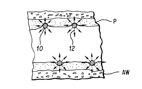
FIG. 1 is a cross section showing two turns of a radioisotope helical coil
spring
stent imbedded into a balloon dilated or atherectomized plaque within a human
artery.
FIG. 2 is a cross section through the spring wire of a helical coil spring
stmt
showing a radioisotope core material within a spring material.
FIG. 3 is a cross section through the spring wire of a helical coil spring
stent
showing a thin plating of radioisotope material on the exterior surface.
FIG. 4 is a cross section through a central core spring wire of a helical coil
sprang stent showing a radioisotope plating which is covered with an anti-
thrombogenic coating.
DETAILED DESCRIPTION OF THE DRAWINGS
As described in U.S. Patent No. 4,768,507, intra-arterial stents can be made
in the
form of a deployable helical coil spring. FIGS. S and 6 of the 4,768,507
patent
illustrate typical cross sections of such a spring wire, helical coil stent.
FIG. 1 of the present invention shows a cross section 10 of two turns of a
helical coil spring stent that has been fabricated from a pure metal or alloy
which has
been irradiated so that it has become radioactive; i.e., it is a radioisotope
. These two
turns are shown imbedded into plaque P within the arterial wall AW. The arrows
12
pointing outward from the cross section 10 indicate the omnidirectional
emission of
particles from the stmt wire. The purpose of this radiation is to decrease the
rate of
2


2031891
proliferative cell growth of the traumatized arterial wall AW (which growth is
termed
"intimal hyperplasia"). Thus it would be expected that restenosis, which
frequently
occurs after stent implantation, will be significantly reduced.
The radioisotope used for this purpose may be an alpha, beta or gamma
emitter. The half-life would ideally be between 10 hours and 100 days. An
optimum
emitter might be a beta emitting isotope such as vanadium 48 which has a half-
life of
lb days and only 8% of its emitted energy is from gamma radiation. The ideal
attribute of a beta emitter is that the radiation does not travel very far in
human
tissue. Thus only the tissue in close proximity to the radioisotope stent will
be
affected. Furthermore only moderate levels of radiation are desired since it
is known
that very high levels can cause injury to nonproliferating tissues.
Another method to make the material of the stent spring wire is from a metal
into which is alloyed an element that c:an be made into a radioisotope. For
example,
phosphorus 32, a 14.3 day half-life beta emitter, could be alloyed into steel
which
could be used for the stent wire.
FIG. 2 shows a stmt wire cross section in which a wire made from a
radioisotope core material 20 is formed within an outer covering 22 that has
the
attributes that are desirable for being a coil spring stent.
FIG. 3 shows a cross section of an alternative embodiment of the present
invention in which a radioisotope coating 30 is plated onto a spring material
core 32.
For example, the beta emitting isotope gold 198 (half life 2.7 days) could be
used to
coat any suitable spring metal material.
FIG. 4 shows a more complex stmt cross section in which a core 40 of some
material ideally suited for stents is plated with a radioisotope coating 42
which is, in
turn, coated with an anti-thrombogenic coating 42 such as carbon as described
in
U.S. Patent No. 4,768,507.
3

~
CA 02031891 1999-04-O1
Although helical coil spring stents have generally been described herein, the
concept of utilizing a radioactive material within the stent structure so as
to attenuate
intimaI hyperplasia is certainly applicable to any stent design. Furthermore,
the
temporary placement at the site of the vessel wall trauma of a radioactive
source
within the arterial lumen, for example a thin wire with a radioactive tip
which wire
can be withdrawn after a limited time is also envisioned:
~ Various other modifications, adaptations, and alternative designs are of
course possible in light of the above teachings. Therefore, it should be
understood at
this time that within the scope of the appended claims, the invention may be
practiced other<vise than as specifically described herein.
4
Representative Drawing
A single figure which represents the drawing illustrating the invention.
Administrative Status

For a clearer understanding of the status of the application/patent presented on this page, the site Disclaimer , as well as the definitions for Patent , Administrative Status , Maintenance Fee  and Payment History  should be consulted.

Administrative Status

Title Date
Forecasted Issue Date 2000-10-24
(22) Filed 1990-12-10
(41) Open to Public Inspection 1991-06-12
Examination Requested 1996-02-13
(45) Issued 2000-10-24
Expired 2010-12-10

Abandonment History

There is no abandonment history.

Payment History

Fee Type Anniversary Year Due Date Amount Paid Paid Date
Application Fee $0.00 1990-12-10
Maintenance Fee - Application - New Act 2 1992-12-10 $50.00 1992-09-28
Maintenance Fee - Application - New Act 3 1993-12-10 $50.00 1993-09-29
Maintenance Fee - Application - New Act 4 1994-12-12 $50.00 1994-09-27
Maintenance Fee - Application - New Act 5 1995-12-11 $75.00 1995-09-26
Request for Examination $200.00 1996-02-13
Maintenance Fee - Application - New Act 6 1996-12-10 $150.00 1996-10-04
Maintenance Fee - Application - New Act 7 1997-12-10 $150.00 1997-10-09
Maintenance Fee - Application - New Act 8 1998-12-10 $150.00 1998-10-02
Maintenance Fee - Application - New Act 9 1999-12-10 $150.00 1999-09-28
Final Fee $300.00 2000-07-19
Maintenance Fee - Application - New Act 10 2000-12-11 $200.00 2000-10-03
Maintenance Fee - Patent - New Act 11 2001-12-10 $200.00 2001-10-09
Registration of a document - section 124 $100.00 2002-02-12
Maintenance Fee - Patent - New Act 12 2002-12-10 $200.00 2002-10-08
Maintenance Fee - Patent - New Act 13 2003-12-10 $200.00 2003-10-16
Registration of a document - section 124 $100.00 2004-02-11
Maintenance Fee - Patent - New Act 14 2004-12-10 $250.00 2004-12-10
Maintenance Fee - Patent - New Act 15 2005-12-12 $450.00 2005-11-08
Expired 2019 - Corrective payment/Section 78.6 $650.00 2006-09-25
Maintenance Fee - Patent - New Act 16 2006-12-11 $450.00 2006-11-08
Maintenance Fee - Patent - New Act 17 2007-12-10 $450.00 2007-11-09
Maintenance Fee - Patent - New Act 18 2008-12-10 $450.00 2008-11-10
Maintenance Fee - Patent - New Act 19 2009-12-10 $450.00 2009-11-12
Owners on Record

Note: Records showing the ownership history in alphabetical order.

Current Owners on Record
CORDIS CORPORATION
Past Owners on Record
FISCHELL, ROBERT E.
FISCHELL, TIM A.
Past Owners that do not appear in the "Owners on Record" listing will appear in other documentation within the application.
Documents

To view selected files, please enter reCAPTCHA code :


To view images, click a link in the Document Description column. To download the documents, select one or more checkboxes in the first column and then click the "Download Selected in PDF format (Zip Archive)" or the "Download Selected as Single PDF" button.

List of published and non-published patent-specific documents on the CPD .

If you have any difficulty accessing content, you can call the Client Service Centre at 1-866-997-1936 or send them an e-mail at CIPO Client Service Centre.


Document
Description 
Date
(yyyy-mm-dd) 
Number of pages   Size of Image (KB) 
Description 1998-09-04 4 150
Description 1999-04-01 4 153
Description 1998-02-05 4 150
Abstract 1994-02-27 1 10
Claims 1994-02-27 4 53
Drawings 1994-02-27 1 16
Description 1994-02-27 4 133
Claims 1998-02-05 5 77
Drawings 1998-02-05 1 17
Claims 1999-04-01 5 97
Cover Page 1994-02-27 1 14
Representative Drawing 2000-09-26 1 8
Claims 1998-09-04 5 80
Drawings 1998-09-04 1 17
Claims 2000-04-17 4 71
Cover Page 2000-09-26 1 31
Prosecution-Amendment 1999-12-15 2 5
Prosecution-Amendment 2006-09-25 2 67
Correspondence 2000-07-19 1 29
Fees 2003-10-16 1 31
Prosecution-Amendment 2002-02-12 19 1,067
Prosecution-Amendment 1999-04-01 9 233
Fees 2000-10-03 1 29
Prosecution-Amendment 2000-04-17 4 83
Prosecution-Amendment 1999-05-03 3 96
Fees 1998-10-02 1 30
Fees 1999-09-28 1 30
Fees 2001-10-09 1 28
Assignment 1990-12-10 5 201
Prosecution-Amendment 1996-02-13 11 323
Correspondence 1991-04-24 10 273
Prosecution-Amendment 1999-01-28 2 5
Assignment 1990-12-10 5 202
Prosecution-Amendment 1996-02-13 17 520
Correspondence 1991-04-24 10 282
Fees 2002-10-08 1 37
Fees 1997-10-09 1 32
Assignment 2004-02-11 4 144
Fees 2004-12-10 1 29
Correspondence 2005-04-21 1 19
Correspondence 2005-09-19 1 15
Correspondence 2006-10-19 1 14
Fees 1996-10-04 1 35
Fees 1995-09-26 1 39
Fees 1994-09-27 1 43
Fees 1993-09-29 1 36
Fees 1992-09-28 1 32