Note: Descriptions are shown in the official language in which they were submitted.
~\q~s
DEVICE AND METHOD OF SEPARATING
AND ASSAYING WHOLE BLOOD
AB ST~CT OF THE DI SCLOSURE
A device and method of separating the
cellular components of whole blood from plasma
or serum and assaying the plasma or serum for a
soluble constituent. The d*vice includes a filter
pad, that separates the cellular components of
whole blood f rom the serum or plasma, in releas-
able contact with a test pad, that assays the
serum or plasma for a particular soluble consti-
tuent. The filter pad, contami~ated with the
cellular components, is detachable from the plasma
or serum-saturated te~t pad, thereby eliminating
assay interference by the cellular components of
whole blood. The method includes contacting the
whole blood with a ~est device including a filter
pad co~prising a suitable carri@r matrix optional-
ly incorporating a lectin, a thrombin, or a mix
ture thereof, such tha~ the c~llular csmponents
of the whole blood are separated from the plasma
or serum as the blood permeates through the filter
pad. The essen~ially cell-ree plasma or serum
then saturates a test pad that is iQ releasable
contact with the filter pad. A~ter the plasma
or serum saturates the test pad, the filter pad
is detached from the test pad, and the exposed
test pad is examined ~or a qualitative or quanti-
tative response to a particular soluble consti-
tuent of the whole blood.
MS-1588
- . . ,, :, , :, , :
, . . , ~ -
--1--
2 ~
DEVICE AND METHOD OF SEPARATING
AND ASSAYING WHOLE BLOOD
FIELD OF THE INVENTION
The present invention relates to a
device and method of separating the cellular
components of whole blood from the plasma or
serum, and assaying the plasma or serum for a
particular soluble constituen~. More particular-
ly, the present invention relates to an improved
method of removing cellular components from whole
blood by utilizing a filter pad comprising a
suitable carrier matrix optionally incorporating
a lectin, a thrombin or a combination theteof.
The essentially cell-free plasma or serum, in an
undiluted and unaltered form, then saturates a
reagent-impregnated test pad that is in releasable
contact with the filter pad. After the undiluted
plasma or serum saturates the test pad, the filter
pad, contaminatéd with the cellular components
of the whole blood, is separated from the test
pad and detached from the test device. The ex-
posed test pad then is examined for a response
to provide a prompt and accurate qualitative or
quantitative assay for one or more soluble consti-
2S tuents of the plasma or serum.
BACKGROUND OF THE INVENTION
.
Presently, numerous test devices areavailable to simply and rapidly analyze bvdyfluids for the presence or absence of a particular
MS-1588
: . , . ~ , :
, ~:
,~
-2 2~
soluble constituent. For example, tests are
available to detect glucose, uric acid or protein
in urine, or to detect glucose, triglycerides,
potassium ion or cholesterol in blood. Historic-
s ally, assays of a whole blood sample for a parti-
cular soluble constituent are the most difficult
tests to design.
The cellular components of whole blood,
and especially the red blood cells, are the pri-
mary interfering substances in assays for asoluble constituent of whole blood. Most simple
blood tests are chromogenic, whereby a soluble
constituent of the whole ~lood interacts with a
particular reagent either to form a uniquely-
colored complex or derivative as a qualitativeindication o~ the presence or absence of the
constituent, or to form a colored complex or
derivative of variable color intensity as a quan-
titive indication of the presence of the consti-
tuent. The deep red color of the whole bloodsample substantially interferes with these chromo-
genic tests, and therefore the highly-colored
red blood cells usually are separated from the
plasma or serum before the blood sample is assayed
for a particular soluble constituent.
The presence of red blood cells also
can interfere with various nonchromogenic blood
assays, whereby the assay results are either
inconsistent or, if consistent, are inaccurate.
~urthermore, other cellular components, including
the white blood cells, also can interfere in
standard chromogenic blood assays. Therefore,
to achieve a reliable assay for a soluble consti-
tuent of whole blood, it is essential ~o separate
the serum or plasma from the cellular components
MS-1588
,. . - :- ,:
.,
, : , ,
2 ~ 3
--3--
of whole blood prior to analyzing the whole blood
sample for a soluble component.
Conventionally, the plasma or serum is
separated from the cellular material of whole
blood by centrifugation. The cellular material
collects at the bottom of the centrifuge tube
and the supernatant plasma or serum is decanted.
Accordingly, the interfering cellular components
of whole blood are sufficiently removed such
that a substantial background interference is
avoided. However, the cen~rifuge method has the
major disadvantages of requiring a relatively
large blood sample, usually from about 0.1 ml to
about 5 ml, and a long centrifuge time of approxi-
15 mately S to 10 minutes. Furthermore, the centri- -
fuge method requires several manipulative steps.
Consequently, laboratory technicians possibly
can contact a potentially infectious blood sample,
or laboratory equipment contaminated by the rela-
tively large blood sample, and contract a disease.
Overall, the centrifuge method is most
suited for large, au~omated laboratories that
assay a multitude of blood samples, and for insti- -
tutions, such as hospitals, that do not require
assay results in a matter of minutes. However,
many small laboratories and private medical
o~fices do not have such a blood separator on
site. Therefore, simple chromogenic tests cannot `~
be performed quickly, safely and easily on site
and the whole blood sample must be sent to an
outside laboratory for efficient and safe separa-
tion and assay. As a result, the assay results
are not available in minutes but in hours or
days.
MS-1588
2 ~ 7 ~
_4_
Accordingly, investigators have continu-
ally sought a device and method of quickly, safely
and easily separating essentially all ~f the
interfering cellular components of whole blood
from the plasma or serum such that the identity
and concentration of soluble constituents in the
plasma or serum are not altered~ Conseguently,
an assay for a particular constituent of the
plasma or serum is trustworthy, accurate and
free from interference by the cellular components
of the whole blood. Investigators have provided
several methods and devices for separating the
interfering cellular components of whole blood
from the plasma or serum. However, each method
and device possessed at least one disadvantage
that made the method or device inaccurate, cumber-
some or impractical in assaying a whole blood
sample for a particular soluble component.
Methods other than centrifugation have
been used to separate the cellular components of
a small whole blood sample from the serum or
plasma. One of the simpler methods, as disclosed
by Adams et al in U.S. Patent No. 3,092,465,
uses a bibulous, or moisture absorbing, matrix
that is impregnated with a chromogenic testing
reagent and coated with a semipermeable barrier.
The semipermeable barrier retains the cellular
components of the whole blood sample and permits
passage of the smaller molecules and ions to
contact the chromogenic testing reagent impreg-
nated in the bibulous matrix. In the case of a
positive test, the essentially colorless plasma
or serum interacts with the chromogenic testing
reagent to produce a color in the bibulous matrix.
The color is observed by water rinsing or wiping
MS-1588
,. . : , ,: : ~
.
:,
,
-5-
away the cellular material retained on the se~i-
permeable barrier. However, the rinsing or wiping
technique is cumbersome and laborious, and assay
interference is possible if the red blood cells
are not completely wiped or rinsed from the semi-
permeable barrier. In addition, the possibility
of technician contact with the potentially infec-
tious blood sample is high.
Fetter in U.S. Patent Nos. 3,552,925
and 3,552,928 disclosed another method and device
to assay small whole blood samples for soluble
constituents. Fetter described a test devioe
having a bibulous matrix impregnated with a non-
volatile inorganic salt or an amino acid at a
first region on the matrix and impregnated with
a test reagent at an adjacent second region of
the matrix. A whole blood sample is introduced
onto the bibulous matrix such that the whole
blood first contacts the first region of the
bibulous matrix including the inorganic salt or
amino acid. The salt or amino acid precipitates
the cellular components from the blood, and the
plasma or serum then migrates to the test reagent-
impregnated second region of the bibulous matrix
for chromogenic interaction with the test reagent.
The salts or amino acids used in this process
effectively separate the red blood cells from
the whole blood sample, but also introduce con-
taminating ions or molecules into the plasma or
serum and precipitate a portion of the soluble
plasma or serum constituents. Therefore a quan-
titative assay for a soluble constituent of the
plasma or serum is unreliable. The Fetter method
does eliminate a distinct manipulative step in
the separation of the cellular ~omponents of
MS-1588
:
7 ~
whole blood from the plasma or serum. However,
the method suffers from the disadvantage that
the plasma or serum may no longer contain the
true concentration of the soluble constituents
of interest.
Another prior art method of separating
the cellular components of whole blood from the
plasma or serum is disclosed by Vogel et al, in
U.S. Patent No. 4,477,575, describing a process
and a composition for separating plasma or serum
from whole blood using a layer of glass fibers
having a defined average diameter and density.
In addition to the defined glass fiber parameters,
the amount of plasma or serum that can be separ-
ated is limited to at most 50%, and preferablyless than 30~, of the absorption volume of the
glass fibers. Otherwise, whole blood, containing
approximately 50~ filterable cellular material,
effectively clogs the glass fiber layer. There-
20 fore, the method requires a high ratio of hydro- ;
phobic glass fibers to whole blood volume.
Furthermore, in many prior art methods,
the whole blood is diluted before assaying for a
soluble plasma or serum constituent. The dilution
f whole blood is burdensome because an extra
manipulative step is required, and dilutisn intro-
duces the possibility of assay error because of
an incorrect dilution of the blood sample. The
possibility of technician contact with the poten~
tially infectious blood ample also is increased.
For example, German Patent No. 34 41 149 dis-
closed a method of separating plasma or serum
from whole blood by passing the whole blood
through a lectin-impregnated matrix that is re-
peatedly rinsed with a diluent to dilute the
MS-1588
, ' , ' ~ .: , ,
~ , , .~ ,
-7- ~ 7~
plasma or serum before the assay is performed.
However, the possibility of imprecise dilution
can result in an inaccurate assay for the plasma
or serum constituent of interest.
In developing a method and device for
separating and assaying small whole blood samples,
a primary consideration is the degree of sophisti-
cation of the technician performing the assay.
Often it is desirable to have relatively untrained
personnel perform routine assays and obtain accur-
ate quantitative results. Therefore, it is impor-
tant that the assay method include a minimum of
manipulative steps1 be free of possible interfer-
ences or contamination, minimize or eliminate
the possibility of the laboratory personnel from
physically contacting the blood sample, and pro-
vide for easy measureme~t. For instance, among
the several possible manipulative steps, the
dilution of the whole blood, or the plasma or
serum, prior to the actual assay introduces the
most probable step for assay error or personal
contact with the blood sample. Another commsn
manipulative error is the incomplete wiping or
rinsing of the ~ellular components of whole blood
from the sur~ace of a device that utilizes a
cell-impermeable membrane to separate the cellular
components from the plasma or serum of whole
blood.
Therefore, a need exists for a method
and device to efficiently separate and accurately
assay small volumes of whole blood. The method
preferably avoids a distinct manipulative step
to separate the cellular components from the
plasma or serum prior to the assay. Further-
more, in order ts avoid dilution errors, the
MS-1588
. .
,
2 ~
--8--
method preferably allows the assay of undiluted
plasma or serum. In addition, the device prefer-
ably avoids the use of complicated and expensive
multilayered test strips. It also is desirable
to have a blood separation and blood assay method
that shields the technician from contact with
the blood sample; that avoids the time delays of
the present methods; and that yields accurate
and reproducible results.
The ideal method includes withdrawing
a whole blood sample in "noninvasive" amounts,
such as a pin prick drop, and immediately deposit-
ing the undiluted whole blood sample on a single
separating-analyzing device, whereby the cellular ~ -
components are separated from the undiluted plasma
or serum and the presence or concentration of a
plasma or serum constituent is determined within
minutes. Alternatively, the separating-analyzing
device can contact a resh puncture wound and
thereby withdraw a blood sample from the wound
for analysis. Such a separation and assay method
and device would allow medical personnel to carry
out whole blood analyses on a more routine and
more confident basis.
Consequently, investigators have at-
tempted to develop test devices that incll~de an
element to separate, collect and retain the cellu-
lar components of whole blood. In addition,
some test devices were developed wherein the
cell-separating element can be physically discon-
nected from the test device and discarded before
assaying the plasma or serum for a particular
consti~uentl For example, the previously-mention-
ed Vogel et al U.S. Patent No. 4,477,575 dis-
closed a device wherein a glass fiber cell-separ-
MS-1588
,, , . :
. . ~ ~ ;, : :
~ 9 ~ 7 ;~
g
ating layer is disposed over a reaction layer,
such that the separating layer can be removed
from the reaction layer. The reaction layer
then can be examined for a response to a particu-
lar plasma or serum constituent. However, theglass fiber separating layer of the Vogel device
requires the disadvantageous high ratio of glass
fiber to blood described above. Consequently, a
specific volume of blood must be pipetted onto
the test device, thereby adding a time-consuming
manipulative step that can result in operator
error and erroneous assays. Such a manipulative
step also can lead to reduced operator safety
because of potential physical contact between
the operator and the blood sample.
Similarly, Rothe et al, in U.S. Patent
No. 4,223,089, described a dry phase test strip
to assay for ammonia wherein plasma or serum is
applied to a porous membrane over a reaction
layer. Upon contacting the plasma or serum, the
reagents in the reaction layer convert the urea
in the plasma or serum to ammonia. The gaseous
ammonia diffuses through a porous spacer below
the reaction layer to permeate an indicator layer
that changes color in response to the amount of
ammonia generated. This devi~e allows the top -
reaction layer to be physically disconnected
from the indicator layer to permit examination
of the indicator layer for a response to ammonia.
Rothe et al, in U.S. Patent No.
4,604,264, also disclosed a variation of the
features of Rothe e~ al U~S. Patent No. 4,223,089
and of vogel et al V.S. Patent No. 4,477,57S in
a hinged assay device. This particular device
of Rothe et al includes a separating layer con-
MS-1588
. . : .. ., , , , , . -, , .; . . .
,, ' ' ' . ''~ ~ `;'. , ':' ,
2 ~
sisting of a glass fiber fleece, wherein the
whole blood is applied near the end of the separ-
ating layer distant from the hinge area of the
device. As the blood permeates through the separ-
ating layer, the cellular components are separatedfrom the serum or plasma. The serum or plasma
then migrates towards the hinge of the device to
an area of the separating layer underneath a
reaction/indicator layer that is disposed above
the separating layer and secured to the separating
layer by a hinge. By pressing down on the reac-
tion/indicator layer, contact between the the
lower face of reaction/indicator layer and the
separating layer allows the reaction/indicator
layer to absorb the serum or plasma for reaction
with reagents in the reaction/indicator layer.
Assay detection, such as a color change, is
achieved by observation through a transparent
film disposed on the upper face of the reac-
tion/indicator layer.
Kennedy et al, in applicationPCT/US86/02192~ disclosed a disposable dry phase
test stick having a reactive area covered by a
semipermeable membrane. The semipermeable mem- -
brane separates cellular and particulate matter
from whole blood and allows the plasma or serum
to contact the reactive area. The semipermeable
membrane can be detached from the reactive area
to remove cellular components and particulate
matter from the device and to expose the reactive
area for examination of a response to a particular
analyte. The semipermeable membrane is a poly-
tetrafluoroethylene material made hydrophilic
with a surfactant or soap~like wetting asent.
However, the surfactant or soap like wetting
MS-1588
..... . .
- , ~ . , ~ , :. , - .
-11- 2~ 7~
agent can remove particular noncellular compon-
ents from the plasma or serum, such as potassium
ions, or can contaminate the plasma or serum.
Another prior art patent directed to
separating the cellular components of whole blood
from the plasma or serum is Rapkin et al U.S.
Patent No. 4,678,757, wherein a carbohydrate-
treated permeable carrier is used to separate
the cellular components of whole blood from the
1~ plasma or serum. The plasma or serum of the
whole blood then contacts a reagent-treated per-
meable carrier for assay of a particular blood
constituent. In this device, the cellular compon-
ents collect and are retained at the bottom edge
Of the carbohydrate-treated carrier, and the
plasma or serum per~eates through the carbohy-
drate-treated carrier to contact the reagents
necessary for the assay. Assay results are deter-
mined by observation through a transparent materi-
20 al covering the permeable layers. In the Rapkin ~-
et al. device, the layer collecting and retaining
the cellular components is not physically discon-
nected Erom the device before examining the device
for a response to a particular blo~d constituent.
In addition, Terminiello et al, in
U.S. Patent No. 4,774,192, disclosed a porous
membrane having a porosity gradient such that
the cellular components of whole blood are re-
tained in an area of the membrane having a low
porosity. The serum or plasma can flow through
the area o low porosity to contact assay reagent
components incorporated into an area of the mem-
brane having a high porosity. Furthermore, G.
Rayman and J.L. Day, in the publication "New
Device to Improve the Accuracy of Bedside Blood
MS-1588
' ' ' ' "~ ', , ' "' ' ':, ~ ' `'
,' ' ` . '
~3~
-12-
Glucose Tests", The Lancet, Nov. 12, l9g8, pp.
1107-1109, described a disposable test strip for
glucose wherein the surface of the strip is wiped
to clear the cellular components of the whole
blood from the strip. Neither device incorporates
an element to retain the cellular components of
the whole blood that then is physically discon-
nected from the assay element of the device. In
the Terminiello et al device, higher molecular
weight soluble plasma components, such as choles-
terol, may not completely permeate through the
low porosity area of the membrane; and in the
Rayman and Day device the cellular components `
may not be completely wiped from the surface of
the strip. Therefore, in each device, inaccurate
and unreliable assays may result.
Therefore, because of the disadvantages
present in the above-cited prior art methods and
test devices, it is apparent that a simple and
2~ effective method of separating the cellular com-
ponents of whole blood to provide essentially
cell-free, unaltered and undiluted plasma or
serum is needed. Accordingly, the method and
device of the present invention allows the safe,
accurate and economical assay of a whole blood;
or other biological fluid, sample for a particular
soluble component by utilizing a filter pad in
releasable contact with a reagent-impregnated
test pad to achieve essentially to~al separation
of the cellular components of whole blood from
the plasma or serum. The device allows the assay
of whole blood without resorting to lengthy and
expensive wet phase assays and without the need
of complicated and costly multilayered test
strips. In addition, the cell-free plasma or
.
~S-1588
;~
-13~
ser~m saturating the test pad is unaltered and
undiluted, thereby allowing a more accurate and
trustworthy assay for a soluble constituent.
The method and device of the present invention
also eliminate the disadvantages of hemocrit
sensitivity, technique sensitivity due to wiping
or rinsing the cellular components f rom the test
device and disposal of the cellular components.
Furthermore, in accordance with the
method and device of the present invention, after
the whole blood sample has saturated the filter
pad and the test pad, the filter pad retaining
the cellular components of the blood is physically
disconnected from the test device by peeling,
15 tearing or snapping the filter pad from the de- -
vice, and thereby exposing the test pad of the
device. The test pad, saturated with undiluted
and unaltered plasma or serum, then is examined
for a response to a particular plasma or serum
constituent by standard dry phase chemistry test
strip procedures.
In accordance with another important --
feature of the present invention, the device
essentially precludes contact between the techni-
cian and the blood sample. The blood sample is
absorbed into the filter pad in such a manner
that excess blood sample does not remain on an
outside surface of the device. In addition, the
cellular components do not have to be wiped or
rinsed from the device before examination of the
device for a response. Consequently, the device
essentially eliminates the possibility of contact
between the technician and a potentially infec-
tious blood sample.
MS-1588
: `: ~ , i: `
.
-14~ 3
As a result, the assay of plasma or
serum for a particular soluble constituent is
accurate and reliable because the interferences
attributed to the highly-colored cellular compon-
ents are essentially eliminated. The prior artmethods and devices rely either upon wiping the
cellular components from the surface of the ana-
lyte detection device or upon immobilizing the
cellular components, physically or chemically,
in an area of the analyte detection device distant
from the actual assay area, usually in a compli-
cated multilayered test strip. As will be demon-
- strated more fully hereinafter, the device of
the present invention provides an accurate and
economical method of first separating the cellular
components of whole blood from the plasma or
serum. Then, the element retaining the interfer-
ing cellular components is completely detached
from the test device to permit examination of
the exposed assay area of the device for a re-
sponse to a particular component of the plasma
or serum. The assay is detected by conventional
dete~tion techniques, either visual or instru-
mental, without the additional opacifying or
transparent layers often included in a multi-
layered device to help eliminate the interfering -~
effects of the cellular components of the whole
blood. Furthermore, the serum or plasma is dis-
tributed evenly throughout the entire tesL pad,
therefore the assay response is homogeneous
throughout the test area of the device. Conse-
quently, a simple and accurate assay detection
can be achieved at any position of the test area.
In addition, both the detached filtering element
and the test area of the device are discarded
MS-lS88
, ' ~
-15- ~ J
after use, thereby precluding contact between
the technician and the blood sample.
SUMMA~Y OF THE INVENT I ON
In brief, the present invention is
directed to a device and method of separating
the cellular components from whole blood and
assaying the undiluted and unaltered serum or
plasma for a particular solu~le constituent, or
constituents, without additional manipulative
steps. It has been found that separating the
cellular components of whole blood from the plasma
or serum by the method and device of the present
invention provides an essentially cell-free,
undiluted and unaltered serum or plasma sample
15 for immediate assay of a particular soluble con- :
stituent. The separation method and device do
not introduce contaminants into the serum or
plasma, and do not alter the compositional makeup
of the plasma or serum. Furthermore, the method
and test device of the present invention also
can be used to assay other biological fluids
having cellular or particulate matter that inter-
fere in an assay for a particular soluble consti-
tuent. In addition, the device includes an ele-
ment to separate the cellular components of thewhole blood from the plasma or serum that is
detachable from the device, thereby both prevent-
ing the inadvertent contamination of the serum
or plasma by the cellular components and facili-
tating the assay of the serum or plasma for aparticular constituent. The method and device
also essentially precludes technician contact
with a potentially infectious blood or other
biological sample.
MS-1588
.. . . ~ .
.. . .
'
-16- ~ ~?~ J r~ j~
In accordance with an important feature
of the present invention, the device includes a
filter pad, that separates the cellular components
of whole blood from the serum or plasma, in re-
leasable contact with a test pad that assays theserum or plasma for a particular soluble consti-
tuent. The filter pad comprises a suitable car-
rier matrix. In addition, the carrier matrix
can be impregnated with a separating reagent to
facilitate the separation of the cellular blood
components from the serum or plasma. In accord-
ance with the present invention, the filter pad,
contaminated with the cellular components of the
whole blood sample, is detachable from the plasma
or serum saturated test pad, thereby precluding
the cellular components from interfering with
the assay for a soluble constituent of the blood
or plasm~. The serum or plasma, in an undiluted
and unaltered foem, is assayed for a particular
soluble constituent by the specific indicator
reagent composition incorporated into the test
pad. Detachin~ the filter pad from the device
allows examination of the exposed tes~ pad for a
response, such as a color change, to a particular
soluble constituent of the whole blood. In addi-
tion, the present invention not only eliminates
technique dependence of the assay, such as elimi-
nating the manipulative step of wiping or rinsing
the cellular components from a multilayered test
dev~ce, but also assures that the proper volume
of plasma or serum contacts the test pad of the
device. Furthermore, technician contact with
the potentially infectious blood or other bio-
logical sample is essentially precluded.
MS-1588
~: .
~, . . : :
17 ~ ~ 3 ~ !~ 7 ~
The method includes first contacting a
whole blood sample with a test device comprising
a filter pad, including a suitable carrier matrix
and optionally impregnated with a separating re-
agent, and a test pad, including a suitable sub-
strate material incorporating an indicator reagent
composition. As used here, and herelnafter, the
expression ~separating reagent" is defined as a
chemical or mixture of chemicals that effects a
separation of the cellular components of whole
blood from the remaining soluble blood constitu-
ents. Likewise, as used here and hereinafter,
the expression ~indicator reagent composition~
is defined as a chemical or mixture of chemicals
causing an observable or detectable interaction
upon contact with the substance being detected.
The whole blood sample initially contacts only
the filter pad of the test device and permeates
through the filter pad wherein the red blood
cells and other cellular and particulate compon-
ents of the whole blood sample are separated
from the plasma or serum through the action of
the carrier matrix and, if present, the separating
reagent. The undiluted and unaltered plasma or
serum continues to permeate through the filter
pad to contact a test pad that is in releasable
contact with the filter pad~ After the plasma
or serum saturates the test pad, the filter pad
is physically disconnected, or detached, from
the test pad, such as by peeling, tearing or
snapping the filter pad f rom the device, and
discarded.
The assay of interest i5 performed by
the test pad of the device. The undiluted plasma
or serum interacts with the indicator reagent
MS-1588
~. . . . . . .
~ ~ .
-18- ~ ~ 3 ~ .J
cQmposition previously incorporated into the
test pad to produce a detectable change in the
test pad, such as a color change, to show the
presence or absence of a particular soluble con-
stituent or to permit a quantitative determina-
tion of the particular soluble constituent. After
detaching the filter pad from the device, the
exposed test pad is examined, either visually or
instrumentally, for a qualitative or quantita-
tive response to the particular soluble constitu-
ent of interest. After examination for a re-
sponse, the test pad of the device also is dis-
carded. Consequently, the assay for the particu-
lar soluble constituent of interest is achieved
by using a simple and inexpensive test device
that effectively separates the interfering cellu-
lar and particulate components from the test
sample without the need of multiple screening
and filtering layers and without manipulative
steps, such as wiping or rinsing, that can lead
to technician error or contact with the potential-
ly infectious blood sample.
Therefore, the present invention is
directed to a method and device for rapidly and
effectively separating the cellular components
from the plasma or serum of undiluted whole blood
and assaying the plasma or serum for a particular
soluble constituent. ~ore particularly, and in
accordance with an important feature of the pre-
sent invention, one or more, untreated or chemic-
ally-treated, filter pads are arranged to separ-
ate the cellular components from whole blood and
to allow the plasma or serum to pass onto one or
more test pads that are in releasable csntact
with the filter pad, or pads. After the test
~S-1588
.. ; , ,, . ~
. ., - ~ ' ' ~ '
"
~, :.
. -19~
pad is saturated with plasma or serum, the filter
pad is separated from the test pad, and the ex-
posed test pad is examined for a qualtitative or
quantitative response to soluble serum or plasma
constituents. Accordingly, assay interferences
attributed to the cellular components of whole
blood are eliminated, thereby achieving a more
accurate and more reliable serum or plasma assay.
In a preferred embodiment, the whole
blood contacts a test device comprising a filter
pad including a suitable carrier matrix impregnat-
ed with a thrombin. To achieve the full advantage
of the present invention, the whole blood contactc
a test device comprising a filter pad including
a suitable carrier matrix impregnated with a
lectin. The thrombin or lectin-impregnated filter
pad facilitates separation of the cellular compon-
ents of the whole blood from the plasma or serum
and does not add contaminating ions or molecules
to, or remove soluble constituents from, the
serum or plasma. The amount of lectin included
in the filter pad is in the range of from about
55 to about 40,000 units per cm3 of carrier matrix
material comprising the filter pad, wherein each
R unit~ is defined as the amount of lectin neces-
sary to agglutinate a two percent solution of
red blood cells within one hour of incubation at
25C. The amount of thrombin included in the
filter pad is in the range of from about 90 to
about 900 NIH (National Institute of Health)
units per cm3 of carrier matrix material compris-
ing the filter pad. The undilu~ed serum or plasma
passes through the filter pad of the device,
essentially unimpeded, to saturate the test pad
of the device and the assay of interest is per-
MS-1588
.
,,: :
.
:, . : . ,:, . ,: : -
, :. ~ :
~-~3
-20-
formed. The test pad of the device is in releas-
able contact with the filter pad and comprises
an indicator reagent composition homogeneously
incorporated into a suitable substrate material.
In accordance with an important feature of the
present invention, the particular plasma or serum
constituent of interest is detected or measured
without diluting the plasma or serum, and without
any wiping or rinsing of the test device or per-
10 forming any other manipulative steps. ~ -
Therefore, it is an object of the pre-
sen~ invention to provide a method and device to
quickly and effectively separate the cellular
and other particulate components from the plasma
or serum of small whole blood samples or other
biological samples.
It is also an object of the present
invention to provide a method and device for the
rapid, easy and effective separation of undiluted
and unaltered plasma or serum from the cellular
components of whole blood.
Another object of the present invention
is to provide a method and device to separate
the cellular csmponents of whole blood from the
plasma or serum, and then to assay the plasma or
serum for a particular soluble constituent without
additional manipulative steps.
Another object of the present invention
is to provide a method and device to eliminate
interferences attributed to the cellular compon-
ents of whole blood in assays of undiluted and
unaltered plasma or serum for soluble constitu-
ents. ;
MS-1588
, , . :
.
~ rrl3
-21-
Another object of the present invention
is to provide a method and device to eliminate
technician contact with a potentially infectious
whole blood or other biological sample.
Another object of the present invention
is to provide a method of separating the cellular
components from whole blood by utilizing an ag-
glutinizing compound, a coagulating compound, or
a combination thereof.
Another object of the present invention
is to provide a method of separating the cellular
components from whole blood by utilizing a lectin,
a thrombin, or a combination thereof.
Another object of the present invention
is to provide a new and improved test device for
the qualitative or quantitative analysis of a
small sample volume of whole blood.
Another object of the present invention
is to provide a new and improved test device to
qualitatively or quantitatively assay whole blood
that essentially eliminates assay interferences
attributed to the cellular components of the
whole blood sample.
Another object of the present invention
is to provide a test device for assaying an un-
diluted whole blood sample for a particular
soluble constituent comprising a filter pad to
remove the cellular components of the whole blood
in releasable contact with a test pad incorporat-
ing an indicator reagent composition, to assaythe plasma or serum for the particular soluble
constituent.
Another object of the present invention
is to provide a dry phase test strip device in-
cluding a test pad, comprising a substrate materi-
MS-1588
~,
7 ~
-22-
al capable of homogeneously incorporating anindicator reagent composition that interacts
with the constituent of interest in the serum or
plasma, in releasable contact with a filter pad,
comprising a suitable carrier matrix capable of
homogeneously incorporating a separating reagent
composition that interacts with the cellular
components of whole blood.
Another object of the present invention
is to provide a new and improved test device and
method of manufacturing the test device for sens-
ing the presence of a chemical compound in whole -- .
blood, wherein the test device comprises a filter :
pad in releasable contact with a test pad, such
that a whole blood sample contacting the filter
pad first is separated into cellular components
and into undiluted plasma or serum with the cel- ;
lular components remaining in the filter pad and
the undiluted plasma or serum permeating through
20 the filter pad; then, after the undiluted plasma :
or serum sufficiently contacts a test pad to
produce a detectable or measurable response, the
filter pad is detached from the test device to
allow either visual or instrumental examination
f the exposed test pad for a response to the
chemical compound of interest.
BRIEF DESCRIPTION OF THE DRAWINGS
The above and other objects, adva~- -
tages and novel features of the present invention
will become apparent from the followinq detailed
description of the present invention taken in
conjunction with the drawing wherein:
FIG. 1 is a partial side view of a
test device for separating the cellular components
of whole blood or other biological samples from
MS-1588
7 J
-23-
the plasma or serum and for assaying the plasma
or serum for a particular soluble constituent;
FIG. 2 is an end view of the test device
of FIG. 1 taken in the direction of arrows 2-2
of FIG. 1 showing the sample port for introducing
the whole blood or other biological sample to
the test device;
FIGS. 3 and 4 are a side view and a
top view, respectively, of the test device of
FIG. 1 before the filter pad is positioned in
releasable contact with the test pad:
FIGS. 5 and 6 are views similar to
FIG. 1 showing alternate embodiments of the test
device of the present invention;
FIGS. 7 and 8 are views similar to
FIG. 6 and FIG. 1 showing alternate embodiments
of the test device of the present invention useful
for assaying more than one soluble plasma or
serum constituent;
FIG. 9 is an expanded side view of the
assembly elements of a preferred embodiment of a
test device for separating the cellular components
of whole blood from the plasma or serum and for
assaying the plasma or serum for a particular
soluble constituent;
FIG. lOa-lOc illustrate the use of a
test device of the present invention in the assay
of a whole blood sample for a particular soluble
constituent; and
FIG. 11 is a plot of the Kubelka-Munk :~
function tK/S) vs. the cholesterol concentration
of standardized test samples assayed for choles-
terol with test devices of the present invention.
~S-1588
,; ~
2 ~ 7 ~
-24-
DETAILED DESCRIPTION OF THE INV~NTION
In accordance with the method of the
present invention, the cellular components of a
whole blood sample first are effectively separated
from the plasma or serum; then, the undiluted
and unaltered plasma or serum is assayed for a
particular soluble constituent, or constituents,
without any further manipulative steps. According
to the method and device of the present invention,
a small blood sample, usually a pin prick amount,
is sufficient to achieve separation of the cellu-
lar components and to assay the serum or plasma,
as opposed to the large milliliter size blood
samples required in the centrifuge method of
separation. Furthermore, the method and device
of the present invention eliminates the manipula-
tive steps that can cause technician contact
with the potentially infectious whole blood sam-
ple.
Surprisingly and unexpectedly, the
test device of the present invention essentially
completely separates the highly colored and inter-
fering red blood cells from the plasma or serum,
and then assays the plasma or serum for a parti-
cular soluble constituent, within minutes~ without
the additional ~ime-consuming and potentially
unhealthful manipulative steps of either centri-
fugation, decantation or plasma or serum dilution.
The method of the present invention provides
rapid and reliable whole blood assays on undi-
luted, unaltered, and noncontaminated plasma or
serum samples with a simple and inexpensive test
strip device. Overall, the method and device of
the present invention are ideally suited for
35 routine blood assays a~ home, in small labora- -
.:
MS-1588
:. ~ . , . :
- ~3~ ~77
-25-
tories or private medical offices, wherein the
number of assays usually is relatively low, but
accurate results are nevertheless required in a
short time period.
As will become apparent from the follow-
ing detailed description of the invention, the
method and device of the present invention are
especially suited for blood assays utilizing
chromogenic responses to determine the presence
or concentration of the various soluble con-
stituents of whole blood. Therefore, it is of
primary importance to remove the highly-colored
red blood cells from the whole blood sample in
order to achieve an accurate and reliable detec-
tion and measurement of the chromogenic response.Furthermore, any method or device for separating
the cellular components of whole blood from the
plasma or serum should quickly and efficiently
achieve cell separation; remove only the cellular
components and not the soluble plasma or serum
constituents; avoid contamination of the plasma
or serum with interferi~g, soluble ions or mole-
cules; ~nd minimize or eliminate hemolysis, where-
in the red blood cells rupture and release their
highly-colored components to the plasma or serum.
Due to the potentially infectious nature of the
blovd or other biological sample, the method and
device also should minimize, or eliminate, human
contact with the test sample.
In accordance with an important feature
of the present invention, it has been found that
the cellular components of whole blood are effec-
tively separated from the essentially colorless
plasma or serum by allowing the whole blood sample
to contact and permeate through a filter pad
~S-1588
-26-
comprising a suitable carrier matrix that option-
ally has been impregnated with, or otherwise :
includes, a suitable separating reagent. The
separating reagent assists the carrier matrix in
the removal of the highly-colored cellular com-
ponents of the whole blood sample to yield plasma
or serum that is amenable to a simple and accurate
determination of its soluble components.
In accordance with another important
feature of the present invention, the filter pad
of the test device is in releasable contact with
the test pad of the test device As the whole
blood permeates through the filter pad, the cellu-
lar components are separated from the whole blood
sample and collected and retained in the filter
pad. The unaltered serum or plasma then advances .
to, and saturates, a test pad that is in releas-
able contact with the filter pad. The test pad
comprises a suitable substrate material incor-
porating an indicator reagent composition that
interacts with the plasma or serum constituent
of interest to give a detectable or measurable
response. After the test pad is saturated by ~ -
the plasma or serum, the cell contaminated filter :
pad is separated from the test pad and detached
from the test device, thereby allowing either a ~ -
visual or an instrumental examination of the ~ :
exposed test pad for a response, such as a chromo-
genic response, to a particular plasma or serum -
30 constituent. :
Generally, the Eilter pad of the present
invention comprises a suitable carrier matrix ~:
capable of filtering the cellular components
from a whole blood sample. The carrier matrix
is normally a hydrophilic matrix capable of separ-
MS-1588
. .
-27-
ating the cellular components of whole blood
from the plasma or serum. Preferably, the carrier
matrix also is capable of incorporating a separat-
ing agent to facilitate removal of the cellular
components from the whole blood. In addition to
collecting and retaining the separated cellular
components, the carrier matrix, either untreated
or treated with a separating agent, should permit
the plasma or serum to permeate through the car-
rier matrix essentially unimpeded to contact thetest pad.
The carrier matrix also should permit
the whole blood sample to permeate through the
filter pad at a sufficient rate to allow adequate
time for efficient red blood cell separation,
yet rapidly enough to obtain blood assays rela-
tively quickly. In addition, the carrier matrix ;-
should not promote hemolysis, contaminate the
serum or plasma by serum or plasma-extraction of
components of the carrier matrix, remove serum
or plasma constituents by chemical or physical
interactions, or appreciably alter the undiluted
plasma or serum in a way to make the subsequent
blood assays inconclusive, inaccurate or doubtful.
Therefore, the filter pad of the~present
invention normally comprises a hydrophilic carrier ~
matrix, possessing the above-mentioned character- ;
istics, that allows the blood to move, in response
to capillary force~, through the carrier matrix.
.
The cellular components are separated from the
plasma or serum and retained by the carrier
matrix. The essentially unaltered serum or plasma
then continues advancing through the carrier
matrix to contact and saturate a test pad that
is in releasable contact with the filter pad.
MS-1588
2 8 2 ~ 3 ~ , 7 A 3
The carrier matrix can be any hydro-
philic material that allows only the essentially
cell-free plasma or serum to pass through the
filter pad to contact the test pad for analysis
of a particular soluble substituent. Suitable
hydrophilic carrier matrices include bibulous
and nonbibulous, fibrous and nonfibrous matrices,
like hydrophilic inorganic powders, such as silica
gel, alumina, diatomaceous earth and the like;
sponge materials; argillaceous substances; cloth;
hydrophilic natural polymeric materi~ls, particu-
larly cellulosic material, like cellulosic beads,
and especially fiber-containing papers such as
filter paper or chromatographic paper; and syn-
thetic or modified naturally occurring polymers,
such as cellulose acetate, polyvinyl chloride,
polyacrylamide, polyacrylates, polyurethanes, ~ -
crosslinked dextran, agarose, and other such
crosslinked and noncrosslinked water-insoluble ~ :
hydrophilic polymers. Similarly, other suitable
carrier matrices include fibrous and nonfibrous
matrices like a qlass fiber matrix; and syn- . :
thetic polymers, like polypropylene, polyethylene,
nylon, polyvinylidene fluoride and polysulfones.
The carrier matrix can have a pore
size of between about 0.1 u (microns) and about
50 u, and preferably between about 0~3 u and
about 10 u, to achieve efficient separation of
the cellular components and to permit the serum
or plasma to advance through the filter pad. To
achieve the full advantage of the present inven- .
tion, the carrier matrix has a pore size ranging
from about 0.5 u to about 8 u.
The filter pad of the test device can
include more than one carrier matrix, and the
MS-15~8
. ~ : , .
,: .. . ~ ,
.
~ ,7J` ~_3
-29-
carrier matrices can have different physical
characteristics and can be of different chemical
compositions or a mixture of chemical composi-
tions. The carrier matrix, or matrices, of the
filter pad also can vary in regards to smooth-
ness and roughness combined with hardness and
softness. However, in every instance, the carrier
matrix, or matrices, of the filter pad separates
and collects the cellular components of whole
blood, and allows the plasma or serum to pass
through the filter pad to the test pad unaltered
and essentially unimpeded. Therefore, regardless
of the exact composition of the carrier matrix
or matrices, the primary considerations are separ-
ation, collection and retention of the cellularcomponents of whole blood and transmittal of
substantially unaltered and undiluted plasma or
serum.
To achieve the full advantage of the
present invention, the carrier matrix comprises
a cellulosic material, such as paper, and prefer-
ably filter paper; or a glass fiber matrix. Both
filter paper and a glass fiber matrix possess
the properties required of a suitable carrier ~;
matrix of the pr~sent invention, plus the advan~
tages of abundant supply, favorable economics,
and a variety of suitable grades. Furthermore,
filter paper and glass fiber matrices are capable
of homogeneously incorporating a separating re-
agent in the filter pad, and of effectively separ-
ating the cellular components from whole blood.
As known to those skilled in the art,
filter paper and gla~s fiber matrices are avail-
able in a variety of thicknesses and porosities.
The thickness and porosity of the carrier matrix
MS-1588
, . : . :.
?J ~
_30_ --
in turn influence the effectiveness of the separa-
tion process. The thickness and porosity of the
carrier matrix are directly related to the ability
of the carrier matrix to separate the cellular
components from plasma or serum and to the time
required for the whole blood sampie to permeate
through the carrier matrix to separate the cellu-
lar components from the whole blood sample.
Therefore, if a carrier matrix of high porosity
is used, the thickness of the carrier matrix
should be sufficient to allow a minimum effective
contact time between the whole blood and the
filter pad in order to achieve an effective separ-
ation of the cellular components of the whole
blood. Conversely, if a carrier matrix of low
porosity is utilized, a relatively thin layer of --
carrier matrix can be employed. The proper bal- ~
ance between carrier matrix porosity and thick- ~-
ness, and a judicious selection of the type and
concentration of separating reagent impregnated
into the carrier matrix, is well within the ex- -
perimental ~echniques used by those skilled in
the art of pre~aring the test devices described
in the present specification.
A carrier matrix of sufficient thickness
and of sufficiently low porosity can separate
the cellular components from the plasma or serum
without incorporating a separating reagent into
the carrier matrix. For example, the effective
separation of the cellular components from a
sample of whole blood has been observed as the
blood permeates through an untreated carrier
matrix having sufficient thickness and sufficient-
ly low porosity. However 7 the separation of the
cellular components of whole blood from plasma
MS-1588
: , . .; , ~ :
"
.: ,
~31- ~ ~3 ~J~
or serum is not observed if the carrier matrix
is too thin or too porous. Therefore, if a thin
or porous carrier matrix is impregnated with a
suitable separating reagent, the cellular com?on-
ents are effectively removed and fixed in thecarrier matrix as the blood sample permeates
through the carrier matrix, and the plasma or
serum advances through the carrier matrix to
contact the test pad of the test device.
10It has been found that an untreated
carrier matrix having a thickness of at least
about 1 mm (millimeters), and preferably of at
least about 1.5 mm, effectively separates the
cellular components from a whole blood sample.
However, if a separating reagent is incorporated
into the carrier matrix to assist removal of the
cellular components from the serum or plasma,
the carrier matrix can have a thickness of at ,
least about 0.2 mm, and preferably at least about
0-3 mm. To achieve the full advantage of the
present invention, the filter pad comprises a
carrier matrix in the form of a pad, having dimen-
sions of, for example, approximately 0.25 cm.
(centimeters) by 1.0 cm to approximately 0.5 cm
by 2 cm, and a thickness of approximately 0.2 mm
to approximately 2 mm. A filter pad of such
dimensions has suff icient lenqth, width and thick-
ness for effective cell removal within a reason-
ably short time after the blood contacts one
30 end, or the topt of the filter pad. ~
If a test device of the present inven- ~;
tion has a filter pad of the above-defined dimen-
sions, a pin-prick amount of blood, such as about
0.02 ml (milliliters~, usually is a sufficient
amount of sample to provide a fast and accurate
MS-1588
,: . : : .
. ~ .:.
.. . .
2 ~ 3
-32-
assay for a particular soluble constituent. The
blood sample can be applied to the test device
dropwise or with a pipette, or, preferably, the
test device can contact a fresh puncture wound
and the blood sample is drawn into the test device
by capillary action. Appreciably increasing the
size of the filter pad substantially increases
the time of separation, and also requires a larger
blood sample. Furthermore, a relatively large
sample of blood can be used to inorease the speed
of serum transfer to saturate the test pad. In
accordance with another feature of the present
invention, the amount of whole blood sample con-
tacting the test device does not have to be pre-
cisely measured. However, care should be exer-
cised to avoid overloading the filter pad with
an excessively large blood sample such that a
portion of whole blood contacts the test area of
the test device.
As will be discussed more fully herein-
after, the filter pad preferably comprises a
carrier matrix in~orporating a separating reagent
to achieve essentially total removal of the cellu-
lar components of whole blood from the plasma or
s~rum. However, the prior art separating re-
agents, such as the inorganic salts and amino
acids disclosed by Fetter in tJ.S. Patent No.
3,552,925 and 3,552,928, have proven deficient
because contaminating ions or molecules are intro-
duced into the serum or plasma, and assayableplasma or serum constituents also are separated
from the plasma or serum.
Therefore, in accordance with an impor-
tant feature of the present invention, a separat-
ing reagent, comprising an agglutinizing agen t,
MS-1588
-33- ~ J~
a coagulating agent or a ~ixture thereof, is
incorporated into the carrier matrix such that
~he cellular components of the whole blood ag-
glutinate or are trapped as the blood sample
chromatographs through the carrier matrix. The
agglutinated or trapped cells become fixed, and
are collected within the carrier matrix, as the
plasma or serum continues advancing through the
filter pad to eventually contact and saturate
1~ the test pad of the test device.
As will become apparent from the follow-
ing detailed description of the invention, various
lectins or thrombin, used individually or in
combination, agglutinate or coagulate the cellular
components of whole blood to fix the cells within
the carrier matrix~ and to allow the undiluted
seru~ or plasma to advance, unaltered and essen-
tially unimpeded, to the test pad of the test
device. In addition, the lectins and thrombin
of the separating reagent do not promote excessive
hemolysis. Therefore, the red blood cells do ~ -
not rupture and their highly colored components
do not interfere with and mask the chromogenic
assays.
The lectins are proteins or glycopro-
teins that are known to aqglutinate, or clump,
cells and precipitate complex carbohydrates.
Lectins are isolated from a wide variety of na-
tural sources, including seeds, plant roots,
39 bark, fungi, bacteria, sea~eed, sponges, fish
eggs, invertebrate and lower vertebrate body
fluids, and mammalian cell membranes. The lectins
are often blood group specific, and have been
used in blood grouping, polyagglutination studies,
35 and various histochemical studies of normal and ;
~iS-1588 ~
-34- ~3~
pathological conditions. The lectins used in
the present invention preferably are not specific
to a particular blood group, or the general
utility of the process and device of the present
invention could be limited. Therefore, among
the lectins showing no specificity of blood group-
ing and that are suitable for use in the present
invention are Abrus precatorius (abrin, Jequirty
bean), Bauhinia purpurea (camels foot tree),
Caragana arborescens (Siberian pea tree), Codium
fragile ~Green marine algae), Canavalia ensiformis
~Con A, Concanavalin A, Jack bean), Glycine max
(Soybean), Lathyrus odoratus (Sweet Pea), Lens
culinaris (Lentil), Limulus polyphemus (Horseshoe
crab, Limulin), Lycopersicon esculentum (Tomato),
Maclura pomifera (Osage orange), Mycoplasma
gallisepticum, Perseau americana (Avocado),
Phaseolus coccineus (Scarlet runner bean),
Phaseolus vulgaris (Red ~idney bean), Phytolacca
americana (Pokeweed), Pisum sativum (garden pea),
Psophocarpus tetragonolobus (winged bean), Ricinus
communis (Castor bean), Solanum tuberosum (Potato),
Triticum vulgaris (Wheat germ), Vicia faba ~fava
beant broad bean), Vigna radiata ~Mung bean),
Viscum album (European mistletoe~, Wisteria flori~
bunda (Japanese wisteria), and other like, non-
blood specific lectins.
As will become more apparent herein-
after, the preferred lectins incorporated into
the carrier matrix of the filter pad are the
lectin from concanavalin A (jack bean), the lectin
from solanum tuberosum (potato), the lectin from
Triticum vulgaris (wheat germ), the lectin from
Bauhinia purpurea (camels foot tree) and the
lectin from Phytolacca americana (pokeweed). To
MS-1588
,
" .
,
:;
-~5~
achieve the full advantage of the present inven-
tion, the carrier matrix of the filter pad is
impregnated with the lectin from potato or the
lectin from pokeweed.
s In addition to, or in place of, the
above-described lectins, a coagulating agent
also can be used to effect separation of the
serum from the cellular components of whole blood.
Specifically, the enzyme thrombin, from bovine
plasma, has been incorporated into the carrier
matrix of the filter pad of the present invention
to successfully separate serum from whole blood
samples. The bovine thrombin effectively promotes
blood clotting such that the red blood cells are ~-
removed from the whole blood as the blood per-
meates through the impregnated carrier matrix.
Other thrombins useful according to the method
of the present invention include human thrombin,
goat thrombin, pig thrombin and sheep thrombin.
It also has been found that it is unnecessary to
immobilize the thrombin, or the lectins, onto
the carrier matrix and that an effective separa-
tion of the cellular components from a whole
blood sample can be achieved by using the lectins,
the thrombin, or a combination ~hereof.
As previously described, the device of
the present invention includes of one or more
filter pads, comprising a carrier matrix, or
matrices, either untreated or treated with a ~-
separating agent, to separate soluble serum or
plasma constituents from undiluted whole blood.
To achieve the full advantage of the present
invention, the filter pad comprises a carrier
matrix impregnated with a blood separating re-
agent, such as a thrombin or a lectin that is
MS-1588
:. ' ` : '`:
-36~
not blood group specific. The whole blood sample
contacts the filter pad of the test device, where-
by the cellular components of the whole blood
are separated from the serum or plasma as the
blood sample advances through the filter pad.
In accordance with an important ~eature of the
present invention, the serum or plasma then ad-
vances through the filter pad to contact a~d
saturate a test pad that is in releasable contact
with the filter pad The test pad of the test
device of the present invention comprises a suit-
able substrate material including a suitable
indicator reagent composition for the parti~ular
assay of interest. After the test pad is satur-
l ated with the plasma or serum, the filter pad is
separated from the test pad and is detached from
the test device, such as by peeling, snapping or `
tearing the filter pad from the test device.
The exposed test pad then is examined, either
visually or instrumentally, for a response to a
particular analyte of interest.
Specifically, the test device of the
present invention, and the positioning o~ the -
filter pad and the test pad on the test device,
2 is better understood by reference to FIGS. l
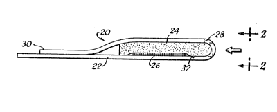
through 9. FIG. l shows a test device 20 includ- -
ing a filter pad 24 in releasable contact with a ~-;
test pad 26. Both the filter pad 24 and the
test pad 26 are securely adhered to a support
strip or handle 22. The test sample:is introduced
to the test device 20 in the direc~ion of the
arrow at the curved end of the test device 20
throu~h a sample port 28 to first contact and
permeate through the filter pad 24. The filter
pad 24 separates the cellular components from
MS-1588
.~;- . , :.:.: "
-37-
the whole blood sample, and the plasma or serum
advances through the filter pad 24 to contact
the test pad 26. ~fter the plasma or serum satur-
ates the test pad 26, an upper free edge 30 of
the support strip handle 22 is pulled to separate
the filter pad 24 from the test pad 26. By fur-
ther pulling the upper free edge 30, the support
strip or handle 12 is disjoined at a notch 32 to
detach the filter pad 24 and the upper free edge
30 from the test device 20 and to expose the
test pad 26 for visual sr instrumental examination
of a response to a particular anaylte of interest.
FIG. 2 is an end view of the test device 20 taken
in the direction of arrows 2-2 of FIG~ 1, and
15 more clearly shows the sample port 28 for intro- -~
ducing the blood sample to the test device 20.
FIG. 3 is a side view and FIG. 4 is a
top view of the test device 20 beore the support
strip or handle 22 is arranged to position the
filter pad 24 and the test pad 26 in releasable
contact. As will become more apparent herein-
after, in order to facilitate the quantitative -
determination of plasma or serum constituents~ -
it i5 preferred that the support strip or handle
22 be manufactured from a hydrophobic, nonabsorb~
tive material. In addition, the material used
in the manufacture of the support strip or handle
22 should be sufficiently pliable to allow the
test pad 26 to be disposed in releasable contact `-
with the filter pad 24. To achieve the full
advantage of the present invention, for the em-
bodiment illustrated in FIGS. 1 through 4, the
filter pad 24 and the test pad 26 preferentially
are impregnated with the separating reagent and
indicator reagent comPosition, respectively,
MS-1588
: .. .. : .
.
r
- ~ , .
-38- 2~
befo~e the filter pad 24 and the test pad 26 are
adhesively secured to the support strip or handle
22. Alternatively, the separating reagent and
the indicator reagent composition can be incor-
porated into the filter pad 24 and the test pad26 after the filter pad 24 and the test pad 26
are adhesively secured to the support handle 22,
but before the filter pad 24 and the test pad 26
are positioned in releasable contact.
FIGS. 5 and 6 are alternate embodiments
of test devices similar to the test device shown
in FIG. 1. In FIG. 5, a hydrophobic plastic
handle 42 of a test device 40 is adhesively se-
cured to a test pad 46. A filter pad 44 is ad-
hesively secured to a tab ~8. The tab 48 is
adhesively secured to the handle 42 such that
the test pad 46 is in releasable contact with
the filter pad 44, and such that the tab 48 has
a free, unsecured edge that can be pulled to
20 disconnect the tab 48 and the filter pad 4~ from .-
the handle 42 and the test pad 46. The filter '-
pad 44 is positioned to extend beyond the edge
of the hydrophobic plastic handle 42 and the
edge of the tab 48 to allow the test sample,
introduced in the direction of the arrow, to
contact the filter pad 44. After the test sample
permeates the filter pad 44, and the undiluted
and essentially cell-free plasma or serum contacts
and saturates the test pad 46, pulling the tab
48 detaches the tab 48 and the filter pad 44
from releasable contact with the test pad 46 and
allows examination of the test pad 46 for a re-
sponse to the analyte of interest.
A test device 60 shown in FIG. 6 is
similar to the test device shown in FIG. 1, how-
MS-1588
:' ' ' ''' ~'- ~ ~ '
. .
.. ~, . .
2 ~ 3
-39-
ever a sample port 68 of the test device 60 is
positioned above the filter pad 64, thereby allow-
ing ~he dropwise addition of the blood sample to
the test device 60 in the direction of the arrow.
The separation of the cellular components from
the plasma or serum is achieved as the whole
blood sample advances downward through the filter
pad 64. The essentially cell-free plasma or
serum then contacts and saturates the test pad
66 for an assay of the particular soluble consti-
tuent of interest. The filter pad 64 is separated
from the test pad 66 and detached from the test
device 60 in an identical manner to separating
and detaching the filter pad from the test device
illustrated in FIG. 1.
FIGS. 7 and 8 are alternate configura-
tions of a test device similar to the test devices ~-
shown in FIG. 6 and FIG. 1. In FIG. 7, a test
device 80 includes a plurality of sample ports
88, a plurality of filter pads 84, and a plur-
ality of test pads 86, thereby allowing the single
test device 80 to assay a whole blood s ampl e for
several soluble plasma or serum constituents.
In accordance with the test device shown in FIG.
7, each test pad 86 has incorporated therein a
different indicator reagent composition capable
of assaying for a particular soluble plasma or
serum constituent of interest. In the embodiment
illustrated by FIG~ 7, each test pad 86 must be
sufficiently spaced, or include a barrier 82
between each test pad 86, to avoid the serum or
plasma saturating a particular test pad 86 from
contacting a second test pad 86, otherwise the
different indicator reagent compositions in each
test pad 86 will commingle and yield a faulty
MS-1588
.. . ..
' ' ' . ' . ~. ' '"' ' ' " '. ",.' , ' . ','1' :
~J~
-40-
assay. A test device 100 shown in FIG. 8 is
similar to the test device of FIG. 1 and FIG. 7
except the test device 100 of FIG. 8 includes a
single filter pad 140 dispoqed over, and in re-
leasable contact with, all of the test pads 160,that are separated by barriers 120.
FIG. 9 illustrates the preferred embodi-
ment of the test device of the present invention
wherein a test pad 220 of a test device 200 is
securely affixed to a top face 240 of a hydro-
phobic support strip 210 by an adhesive layer
230. Similarly, a filter pad 270 is securely
affixed to a detachable envelope strip 250 by an
adhesive layer 280. The filter pad 270 is posi-
15 tioned on the detachable envelope strip 250 suf- .
ficiently close to a sample port 260 such that
contacting the sample port 260 of the test device
200 with a test sample allows the test sample
first to contact the filter pad 270. In addition,
in the test device 200~ the test pad 220 is posi-
tioned sufficiently distant from the sample port
260 such that the test sample contacts only the
filter pad 270 first and then contacts the test
pad 220 after the cellular componen~s of the
whole blood sample have been removed by the filter
pad 270.
The adhesive composition utilized in `~
the adhesive layer 230 and in the adhesive layer
280 can be any adhesive composition that possesses
sufficient tackiness to maintain secure contact
between the top face of the hydrophobic support
strip 240 and the test pad 220, and between the
filter pad 270 and the detachable envelope strip
250, such that the filter pad 270 is removed
with the detachable envelope strip 250, and such
MS-1588
,
;
. ~ -' . ' ~ , . , , '
:
2~3~7~j
--41--
that the test pad 220 remains attached to the
top face 240 of the support strip 210 when the
detachable envelope strip 250 is detached from
the test device 200. Consequently, the adhesive
layer 230 and the adhesive layer 280 should not
be adversely affected by contact with serum or
plasma or with a whole blood sample, and should
not include extractable components that could
contaminate the test sample and therefore lead
to inaccurate or untrustworthy assay results.
Examples of suitable adhesive compositions useful
in the adhesive layer 230 and the adhesive layer
280 include, but are not limited to, silicone-
based adhesives, rubber-based adhesives and
acrylic-based adhesives. Such adhesives are
commercially available and are well known to
those skilled in the art of designing dry phase
test strips.
The detachable envelope strip 250 then
is secured to a bottom face 290 of the support
strip 210 at a position on the bottom face 290
essentially beneath the test pad 220. A layer
of adhesive composition 300 used to secure the
detachable envelope strip 250 ~o the bottom face
290 of the support strip 210 is limited only in
that the adhesive composition should provide
sufficient adhesive strength to maintain the
detachable envelope strip 250 in contact with
the bottom face 290 of the support strip 210 and
also possess sufficiently low adhesive strength
to allow separation of the detachable envelope
strip 250 from the bottom face 290 of the support
strip 210 when an upper free edge 310 of the
detachable envelope strip 250 is pulled.
MS-1588
, : , .................... , , . :
, , ~ ~ . . j .,,
~q~S~
42 - -
~ he top face 240 of the support strip
210 also is adhesively secured to the detachable
envelope strip 250 and the filter pad 270 by
adhesive layer 320 that is applied to the filter
pad 270 and to the bottom face 325 of the upper
free edge 310 of the detachable envelope strip
250 such that the adhe~ive layer 320 does not
contact either the test pad 220, the whole blood
sample or the plasma or serum. It also should
1~ be noted that the adhesive layer 320 may prevent
any excess serum or plasma, or any excess whole
blood sample, from permeating through the filter
pad 270 to contact the top face 240 of the hydro-
phobic support strip 210. Accordingly, only the
test pad 220 is saturated with serum or plasma
and the top face 240 of the hydrophobic support
strip 210 is free of serum or plasma that could ;~
drip and contact the technician or could otherwise
interfere with the assay. Consequently, a more
accurate and reliable assay results, and the
technician is further protected from contacting
a potentially infectious blood or other biological
sample.
As previously stated, the particular
indicator reagent composition for the assay of
interest usually is incorporated into the test
pad 220 before the test pad 220 is adhesively
secured to the top face 240 of the support strip
210. The test pad 220 therefore comprises a
substrate material impregnated with an indicator
reagent composition suitable for the assay of
the soluble plasma or serum constituent of
interest. The substrate material of the test
pad can include any bibulous or nonbibulous
material known to those skilled in the art of
MS-1588
~- .... -
: -
,
-43~ J ~ ~
designing dry phase diagnostic cest strips. Simi-
larly, if the filter pad 270 includes a lectin,
a thrombin or a combination thereof, the ~arrier
matrix of the filter pad 270 usually is impreg-
nated with the lectin and/or thrombin before thefilter pad 270 is adhesively secured to the de-
tachable envelope strip 250. However, alterna-
tivelyt the indicator reagent composition can be
incorporated into the test pad 220 after the
test pad 220 is adhesively secured to th~ top
face of the support strip 210 or the lectin
and/or thrombin can be impregnated into the filter
pad 270 after the filter pad is adhesively secured
to the detachable envelope strip 250. In any
event, the indicator reagent composition is in-
corporated into the test pad 220 and the lectin
and/or the thrombin is impregnated into the filter
pad 270 before the detachable envelope strip 250
is adhesively secured to the bottom face 290 of
the support strip 210.
X~MPLE 1
IMPREGNATION OF THE CARRIER M~TRIX
OF THE FILTER PAD ~-
The agglutinizing agent, coagulating
agent or combination thereof, such as bovine
thrombin, the lectin from solanum tuberosum, the
lectin concanavalin A, or the lectin from
phytolacca americana, is solubilized in normal,
or isotonic, saline solution. A carrier matrix,
such as filter paper, like WHATMAN CCPS00 filter
paper, or a glass fiber matrix, like WHATMAN
PD107, available from Whatman Ltd., Maidenshead,
Kent, U.K., then i5 impregnated with the lectin-
and/or thrombin-containing saline solution either
by immersing the carrier matrix into the saline
MS-1588
. - ,: . ,
' ~ - . .~ . :
-4~
solution or by spraying the saline solution onto
sheets or precut strips of the carrier matrix.
The impregnated carrier matrix then is dried at
about 50C. for about 20 to about 40 minutes
prior to use.
It is not necessary to immobilize the
thrombin or lectin when impregnating the carrier
matrix as in Example 1. The simple drying tech-
nique is sufficient to maintain the lectin or
thrombin in place within the carrier matrix for
separation of the cellular components from whole
blood. The carrier matrix, after appropriate
sizing, e.g., 0.5 cm x 1.0 cm, then can be secured
to either a transparent or an opaque, hydro-
lS phobic plastic handle such as the support stripor handle 22 in FIG. 1, or the detachable envelope
strip 250 in FIG. 9.
Tests to determine the ability of the
coagulatiny agent bovine thrombin and the ag-
glutinizing agents concanavalin A, solanum tuber-
osum, triticum vulgaris and phytolacca americana
to agglutinate or clot blood were run at concen-
trations ranging from 90 to 900 NIH units/cm3 of
thrombin; 140 to 9200 units/cm3 concanavalin A;
4000 to 40,000 units/cm3 solanum tuberosum lectin;
115 to 1150 units/cm3 triticum vulgaris lectin;
a~d 57 to 570 units/cm3 phytolacca americana
lectin. In each case, a carrier matrix was im-
pregnated with a separating reagent in accordance
with the method of Example 1, and the carrier
matrix was securely affixed to a plastic handle
or substrate. An untreated pad of filter paper
was adhered adjacent to, and in contact with,
the impregnated carrier matrix. For each test
MS-1588
-
, " , ~. , ~ ,:
., ~ : ',' '' ' '' ':
~ ` ~
2~3~ ~J ~
-45-
the amount of plasma or serum separated from
whole blood was determined, and the time of separ-
ation noted. The whole blood was deposited on
the separating reagent-impregnated carrier matrix
and allowed to advance, chromatographically,
through the impregnated carrier matrix to the
untreated filter paper substrate. Table I in-
cludes the optimum concentration range found for
each separating reagent and the percent of sep-
arated plasma or serum tranferred to the untreatedpad of filter paper.
TABLE I
Percent of
Separating Optimum Concen- Plasma Ob-
15 Reagent tration Range served
Thrombin 300-450 NIH 40%
units/cm3
Con- 600 920 40
canavalin A units/cm3
Solanum 12000-16000 100%
tuberosum units/cm3
Phytolacca 570 units/cm3 60%
americana
Triticum 1150 units/cm3 ~ 80%
vulgaris
Identical tests were performed in the
presence of calcium chloride or manganese sulfate
because these salts have been proposed as blood
clottinq accelerators. However, when used in
conjunction with the lectin or thrombin separating
reagents, and at concentrations of 0.5%, 1.0%,
and 3.0% of calcium chloride or manganese sulfate,
no improvements in time of the separation or - -
efficlency of the separation was ob~erved. Simi-
larly, to improve the wetting of the carrier
MS-1588
-46-
matrix, the anionic surfactant, sodium alpha-
olefin sulfonate, was impregnated into the carrier
matrix, at about 0.1~ by weight, in addition to
the separating reagent. The wetting of the car-
rier matrix was improved, therefore reducing thetime for plasma or serum separation. However,
the amount of separated plasma or serum was not
increased. Identical results were observed when
a combination of calcium chloride and sodium
1~ alpha-olefin sulfonate was included with the
thrombin or lectin separating reagent. According-
ly, no appreciable benefits are observed if an
ino~ganic salt or surfactant is included in the
separating reagent, plus the inorganic salt or
the surfactant have the disadvantage of possibly
contaminating the plasma or serum and therefore
providing an inaccurate assay.
In accordance with another important
feature of the present invention, the filter pad
of the test device effectively separated the
ceilular components of whole blood from the plasma
or ~erum even in the presence o anticoagulants,
provided that the filter pad includes a separating
rea~ent comprising a lectin, a thrombin or a
combination thereof. Specifically, whole blood
containing anticoagulants, such as heparin or
ethylenediaminetetraacetic acid (EDTA), is amen-
able to the separation and testing method and
the device of the present invention. Therefore,
fresh blood samples can be treated with anticoagu-
lants and then tested by the process and device
of the present invention at a later date. In
accordance with another important feature of the
present invention, hemolysis is essentially elimi-
nated in separations utilizing lectins and was
MS-1588
' : .: , ' . ' ~ '
'; : '` ~`
. .
. .
-47- 2 `~
minimal in separations utilizing thrombin. The
degree of lysis in the thrombin examples was
such that the coloration from the highly colored
red blood cells did not materially interfere in
any subsequent assays for plasma- or serum-soluble
constituents.
In addition to an unexpectedly efficient
separation of the red blood cells from the plasma
or serum, the process and device of the present
invention allow a sueficiently large and assay-
able amount of plasma or serum to reach the test
pad of the test device. Furthermore, the plasma
or serum reaches the test pad of the test device
in an essentially unaltered form. Generally, ~ -
the proper amount of serum or plasma has reached
the test pad when the test pad is saturated with
plasma or serum. This is accomplished either by
using a sufficiently large whole blood sample to
assure plasma or serum saturation of the test
pad, or, preferably, by adjusting the relative
sizes of the filter pad and the test pad such ~ -
that the test pad is saturated with plasma or
serum. Generally, the size of the filter pad is
directly related to the predetermined blood sample
size and, if present, the separating reagent
utilized. A precisely measured whole blood sample
volume is not necessary. This process allows
for an essentially fixed amount of plasma or
serum to reach the test pad, and renders a more
accurate soluble constituent determination. The
variables of blood sample size, filter pad size,
test pad size and the amoun~ of indicator reagent
composition impregnated into the tes~ area easily
can be determined by those skilled in the art
after the particular agglu~inizer, coagulant or
MS-1588
, - ~ . .. , ,,, , . ~ .. , .. .~ , . . .
, ." . ~ . .
- . ~ 1 , , , ., :
~'~, , ' ,- ,,. ~ '
-48- - 2~
combination thereof is chosen as a separating
reagent for incorporation into the filter pad
and its separation efficiency determined.
In accordance with another important
feature of the present invention, essentially
none of the plasma- or serum-soluble constituents
of the whole blood sample are separated from the
plasma or serum along with the cellular compon-
ents, nor do intracellular components of the
cells contaminate the separated plasma or serum.
To demonstrate the utility of the present inven-
tion, assays were performed on the essentially
clear liquid that permeated the test pad after
the cellular components were separated by the
filter pad. Surprisingly, the method and device
of the present invention separated the plasma or
serum from the cellular components with no observ-
able increase in potassium ion or loss of choles-
terol, and with essentially no evidence of red
blood cells. In addition, the white blood cells
also were separated from the whole blood sample.
Table II summarizes the results of the assays
for potassium ion, cholesterol, red blood cells
(erythrocytes) and white blood cells (leuko-
cytes) on the serum or plasma obtained after redblood cell separation. Leukocytes and erythro-
cytes were assayed using test pads sensitive to
leukocyte esterase and the peroxidative activity
of hemoglobin, whereas cholesterol sensitive and
potassium sensitive test pads were used to assay
for those particular constituents. The assays
were performed on plasmas or serums obtained
from a fresh whole blood sample by incorporating
the separating reagents thrombin, the lectin
from solanum tuberosum, the lectin from phytolacca
MS-1588
~ , ,: ~ , . :
- ~ ~ 3 ~
-49- .
americana or the agent used in Fetter U.S. Patent
No. 3,552,925, namely sodium sulfate, into a
filter pad of the present invention.
TABLE II
PLASMA OR SERUM ASSAYS
Separating Potassium
Rea~entLeukocytes Blood Ion Cholesterol
Thrombin Negative Positive Positive Vnknown (lysi< :
blocked :
determination:
Solanum Very slight Slight Positive Positive
tuberosum positive positive
lectin
Phyto- Slightly Negative Positive Positive
lacca positive :
americana
lectin .
~0 :~
Sod~um Inconclu- Slight Positive Negative ~;
sulfate sive positive ~-
Positive - observable reaction
Negative - no observable reaction
From the data summarized in Table II,
it is seen that the salts u~ed in the Fetter
method precipitate the high molecular weight
constituents of plasma or serum, such as choles-
30 terol, therefore making such constituents unavail- -
able for assay at the test pad of the test device.
However, the lectin and thrombin separating re-
agents do not promote precipitation of high mole- -
cular weight plasma or serum constituents. Al-
though thrombin promoted sufficient lysis to
.
MS-1588
- . . : - - . . ......................... .
' ' : , ~ ~ ~ , :;
7~3
-50-
block the assay of cholesterol in serum, lysis
can be controlled by using normal saline, as
opposed to phosphate-buffer saline, when impreg-
nating a carrier matrix with the thrombin separ-
ating reagent solution. Hemolysis inhibitingagents also can be incorporated into the carrier
matrix used in the filter pad of the present
invention. Therefore, high molecular weight
components of plasma or serum can be assayed by
using any of the lectin or thrombin separating
reagents. Also, in certain instances, hemolysis,
or blood stainin~, may be desirable since assays
then can be performed on the constituents present
within the red blood cells.
In addition to the fast and efficient
separation of the serum or plasma from the cellu-
lar components of whole blood, and the essen- -
tially unimpeded migration of unaltered plasma
or serum to the test pad, the method and device
of the present invention allow a quantitative
assay of a plasma- or serum-soluble constituent
without dilution of the whole blood or plasma
and without interference from the highly-colored
red blood cells. Testing the undiluted serum or
plasma both omits a manipulative step and, more
importantly, eliminates the possibility of tech-
nician error and technician contact with the
potentially infectious test sample.
A suitable chromogenic indicator reagent
composition is impregnated into the test pad in
a sufficient amount to allow an essentially im- -
mediate interaction with the ~reshly separated
and undiluted plasma or serum. ~he extent of
the chromogenic reaction, and therefore the quan- -
titative amount of the soluble constituent, then
MS-1588
.
,' ~
,~ . - - ~ ,
:.:, : ,.
2 ~S~ ~J~
--51--
is determined by chromogenic detection techniques,
either visual or instrumental, that are well-
known in the art. In addition, the cell-contami-
nated filter pad is detached from the test device
to preclude the cellular components from contami-
nating the plasma or serum, and therefore provid-
ing an accurate and trustworthy assay of the
serum or plasma. Accordingly, the method and
test device demonstrate appreciable advantages
over the prior art methods and devices because
complicated ar.d expensive multilayered test strips
are avoided; the technician does not have to
wipe the separated cellular components from the
device, thereby avoiding a manipulative step and
a potential technician error; and the technician
avoids all physical contact with the blood sample.
To more clearly demonstrate the benefits
and advantages of the method and test device of
the present invention, quantitative assays for
total bilirubin, creatinine and cholesterol con-
c~ntration in a whole blood sample were performed.
The assays were performed using a test device as
illustrated in FIG. g, and in accordance with
the method illustrated in FIG. lOa-lOc. There-
fore, in general, to perform an assay for a par-
ticular soluble constituent of whole blood, or
other biological fluid, as illustrated in FIG.
lOa, the sample port 260 of the test device 200
contacts a whole blood sample 330. The whole
blood sample 330 can be applied to the sample
port 260 of the test device 200, or, preferably,
the sample port 260 of the test device 200 is
allowed to contact a fresh, small puncture wound
to withdraw the blood sample 330 directly from
an individual. After the sample port 260 has
MS-1588
~:: .. , . . ~ 1, .,
: ~
-52- ~ 7~
withdrawn, or contacted, a sufficient amount of
blood, it is removed from the wound. The whole
blood sample 330 then advances through the filter
pad 270, wherein the cellular and particulate
components of the whole blood sample 330 are
separated, collected and retained in the region
of the filter pad 270 designated as 270a. The
essentially cell-free plasma or serum of the
whole blood sample 330 advances chromatographic-
ally through the filter pad 270 to saturate thefilter pad 270 in a region adjacent to, and in
contact with, the test pad 220, and designated
as 270b. The essentially cell-free serum or
plasma of the whole blood sample 330 then satur-
ates the test pad 220.
The whole blood sample 330 is allowedan approximately 60 second incubation period
after the test device 200 contacts the whole
blood sample 330 to permeate the filter pad 270
and effect separation of the cellular components
from whole blood sample 330 and to saturate the
test pad 220 with serum or plasma. Then, as
depicted in FIG. lOb, the upper free edge 310 of
the detachable filter envelope 250 is lifted and
pulled or peeled to detach the detachable filter
envelope 250 from releasable contact with the
test pad 220 and to disconnect the detachable
filter envelope 250 from the test device 200.
The detachable filter envelope 250 then is dis-
30 carded. In FIG. lOc, after an additional incuba- :
tion period of approximately 60 seconds ~o allow
the indicator reagent composition incorporated
into the test pad 220 to completely interact
with the soluble plasma cons~ituent of interest,
the exposed test pad 220 is examined, visually
or by instrument, for a response to determine
MS-1588
.~ . , .
.
2 ~ 7 ~
_53_ --
the presence or concentration of the soluble
plasma constituent of interest. After examining
the test pad 220 for a response, the test pad
220 and the hydrophobic support strip 210 are
discarded.
Accordingly, an accurate and reliable
assay of a small volume of whole blood for a
particular soluble plasma constituent is achieved
within approximately 2 minutes, without interfer-
ence by the cellular components of the wholeblood. In add;tion the assay is simple, economi-
cal and safe because the manipulative steps re-
quired in multiIayered test devices, like wiping
or rinsing of the test device, and in physical
separation methods, like centrifuging, are ~n-
necessary. Furthermore, the device and method
are safe because the technician is effectively -
shielded from any inadvertent contact with the
potentially infectious whole blood sample. The
entire whole blood samplel both the cellular
components in the filter pad and the plasma or
serum in the test pad, is retained by the elements
of the test device. Therefore, after discarding
the elements of the test device, human contact
25 with the test device is essentially precluded. ~-
Such a feature is new in the art, wherein prior
art methods and devices for testing serum or
plasma led to the possibility of human contact
with the blood sample, either in the wiping or
rinsing of the cellular material from the test
device or in blood transfers and dilutions in
centrifuging and related physical separation
methods.
Accordingly, Examples 2 through 4 more
fully and specifically demonstrate assays per-
formed in accordance with the method and test
MS-1588 ~ -
: , i, , .
: ~ : ; :,
. .
-54- ~ 3
device of the present invention for particular
soluble plasma or serum constituents.
EXAMPLE 2
DETERMINATION OF TOTAL BILIRVBIN
IN PLASMA OR SERUM
An undiluted whole blood sample was
assayed for total bilirubin by a test device as
illustrated in FIG. 9. Similarly, an undiluted
whole blood sample can be assayed for total bili-
rubin by a test device as illustrated in FIG. 1
and in FIGS. 5 throuyh 8~ The test device con-
tacted a whole blood sample, such as a pinprick
amount, at the sample port of the device, and
the whole blood sample was absorbed by a filter
pad comprising a carrier matrix containing a
lectin separating reagent. The blood sample
chromatographed through the impreynated filter
pad, whereby the cellular components of the whole
blood were separated from the plasma or serumO
The blood sample was of sufficient size such
that after chromatographing through the lectin-
impregnated filter pad, the amount of serum or
plasma in the sample was sufficient to completely
wet, or saturate, a test pad that was in releas-
able contact with the filter pad. After satura-
tion of the test pad by the plasma or serum, the
filter pad, contaminated with the cellular com-
ponents of the whole sample, was detached from
the test device by peeling, tearing or snapping
off the portion of the test device handle secured
to the filter pad. The filter pad was detached
fro~ the test pad to expose the test pad for
examination of a response, i.e., the chromogenic
change, to the total bilirubin content in the
serum or plasma. An indicator reagent composition
.
MS-1588
.
:' ' - ~ : '' ;
-55-
comprising 0.4% w/w 2,4-dichloroaniline~
w/w sodium nitrite, 57.2~ w/w dyphylline, 35.5%
w/w buffer, and 5.8~ w/w nonreactive ingredients,
and previously impregnated into the test pad of
the test device, interacted with the bilirubin
in the serum or plasma to produce a measurable
chromogenic change that correlates quantita-
tively to the total bilirubin content in the
serum or plasma.
The chromogenic change in the test pad
was determined visually, such as by a comparison
to a standardized color chart, or, alternatively,
instrumentally, such as by a reflectance measuring
instrument. When using a reflectance photometer,
the method provided a response to the concentra-
tion of total bilirubin in an undiluted serum or
plasma sample without any interference from the
cellular components of the whole blood sample.
The assay method was based on the van den Bergh
reaction, modified to use 2,4-dichloroaniline
and a diazo coupling accelerator. The final
product, azobilirubin, behaved as an indicator,
that is red-purple in color under acid conditionsO ;
The Goncentration of total bilirubin in a sample
was quantified from a calibration curve.
A calibration curve was generated inter-
nally. Once the calibration curve was gener-
ated, each assay required approximately 60 uL
(microliter) of whole blood and one device of
the present invention. After an incubation period
of about 75 seconds, the concentration of total
bilirubin in the test sample was determined by
measuring the chanqe in reflectance at 560 nano~
meters (nm) with reference to a calibration curve
35 generated using calibrators. Preferably, the `
MS-1588
' ~ . ~ , ~, , .,. . ' ! .
'' ' ' ., . ~ ' ,
-56~ 2 ~
filter pad was separated from the test pad prior
to the 7~ second incubation period to preclude
inadvertent contamination of the plasma or serum
by the cellular components of the test sample.
Results were obtained in mg/dL or umol/1 directly
from a reflectance photometer. No calculations
are required. The test responded to from 0.4
mg/dL to 7.5 mg/dL (7 umol/l to 130 umol/1) serum
or plasma total bilirubin. Serum total bilirubin
values of 0.1 mg/dL to 1.2 mg/dL (1.7 umol/l to
20.5 umol/l~ have been suggested as the adult
normal range.
EXAMP E 3
DETERMINATION_OF C~EATININE IN PLASMA OR SERUM
The identical method described in
Example 2 was used to quantitatively determine
the amount of creatinine in undiluted whole blood
samples. However, the indicator reagent composi- -
tion used in the creatinine assay comprises 43.5~
w/w potassium hydroxide, 55.8% w/w 3,5-dinitro- `
benzoic acid, and 0.6% w/w nonreactive ingredi- -
ents.
When used with a reflectance photometer,
the method provides a direct reading of the con-
centration of creatinine in the test sample.
The assay method is based on the Benedict-Behre
reaction wherein creatinine interacts with 3,5-
dinitrobenzoic acid in an alkaline medium to
for~ a purple-colored complex. ~he concentration
of creatinine in the sample is quantified from a
calibration curve generated using calibrators.
During an incubation and test period
of about 30 seconds, the concentration of
creatinine in the sample is determined by measur-
ing the rate of change in reflectance at 560 nm
MS-1588
... . . . . .
.
-
-57- ~ ~3
with reference to a calibration curve generate~
using calibrators. The result is displayed digit-
ally by a reflectance photometer. As described
above, it is preferred that the filter pad is
detached from the test pad as soon as practicable,
such as about 60 seconds after contacting the
whole blood sample, to eliminate the possibility
of inadvertent contamination of the plasma or
serum.
A calibration curve is internally gener-
ated. Once the calibration curve has been gener-
ated, each assay requires approximately 60 uL of
whole blood sample and one device of the present
invention. By using a reflectance photometer,
no calculations are required. The test covers a
range of 0 mg/dL to 15 mg/dL (0 umol/l to 1326
umol/l) ~erum or plasma creatinine. Serum
creatinine values of 0.6 mg/dL to 1.2 mg/dL (53
umol/l to 106 umol/l) for males and 0.5 to 1.0
mg/dL (44 to 88 umol/l) for females have been
suggested as the normal range. Nevertheless r
the range afforded is sufficiently large to test
populations having a creatinine concentration
several times higher than the normal suggested
range.
EX~MPLE 4
DETERMINATION OF CHOLESTEROL IN PLASMA OR SERUM
Undiluted whole blood samples, includ-
ing a standardized amount of cholesterol, were
assayed for cholesterol content by using test
devices as illustrated in PIG. 9. Each test
device included a test pad comprising a substrate
material of either a polyamide, such as nylon,
like BIODYNE B, having a 3 u (micron~ pore size
and available from Pall Corporation, or a poly-
MS-1588
.. ; ' ` ' ` ; . `,:' ' . .
-~8-
vinylidene fluoride, like DURAPOR~, having a
0.65 u pore size, available from Millipore
Corporation, Bedford, MA. The substrate material
of the test pad was impregnated with an indicator
reagent composition first by immersing the sub-
strate material into an aqueous solution including
about 1.5~ .(w/w) tetramethylbenzidine hydro-
chloride indicator dye, and about 0.4~ (w/w)
poly(methyl vinyl ether/maleic anhydride) (GANTREZ
AN-139, available from GAF Chemicals Corp., Wayne,
NJ.). The impregnated substrate material then
was dried in a hot air convection oven at about
50C for from about 5 minutes to about 10 minutes.
The impregnated substrate material then was im-
mersed in a second aqueous solution including:
0.2 M Phosphate Buffer (pH 6.0) 66.6% (w/w)
Tetramethyldecynediol ethoxylated
with 30 moles ethylene oxide -
(SURFYNOL 485, available from Air
Products and Chemicals, Inc.,
Allentown, Pa.) 1.3~ (w/w)
Glycerol 6.4~ (w/w)
Sodium taurocholate 0.7% (w/w)
Peroxidase 500 U/~L
25 Cholesterol Esterase 500 U/mL
Cholesterol Oxidase 2S0 U/mL
Polyvinylpyrrolidione (PVP K-60
available from GAF Chemicals
Corp., Wayne, NJ) (45~ w/w)23.8~ (w/w)
After the second impregnation, the substrate
material again was dried in a hot air convention
oven at about 50C for from abou~ 5 minutes to
about 10 minutes to provide a test pad to deter-
MS-1588
,
: , , ; . - .; . . :
:, ,: . : :
,:- . . .
~ ' ,~ ' , ,`:,
59 2~
mine the amount of cholesterol in a liquid test
sample.
Each test device also included a filter
pad comprising a carrie~ matrix impregnated with
a lectin separating reagent. The carrier matrix,
a glass fiber matrix, having a pore size of from
about 1 to about 6 u, and available from Whatman
Ltd. or Millipore Corporation, was impregnated
with an aqueous solution including about 0.05
(w/w) of the lectin from Phytolacca Americana
and about 0.05% (w/w) of an ethoxylated octyl-
phenol including about 5 moles of ethylene oxide
(TRITON X-45, available from Rohm and Haas Co.,
Philadelphia, Pa.). After impr2gnating the glass
fiber matrix with the separating reagent, the
impregnated glass fiber matrix was scraped with
glass rods to remove any excess aqueous solution.
After drying the scraped and impregnated glass
fiber matrix, the glass fiber matrix was dried
in a hot air convection oven at about 50C for
about 30 minutes to provide a filter pad of the
present invention.
The test pad and the filter pad then
were positioned in a ~est device as illustrated
in FIG. 9 to assay whole blood samples including
a known, standardized amount of cholesterol.
Each assay required approximately 60 ul of whole
blood sample and one test device of the present
invention. Test devices as illustrated in FIG.
9 were used to assay whole blood samples including
120, 162, 205 and 317 mg~dL of cholesterol.
Assays on samples of each cholesterol concentra-
tion were performed in triplicate. The results
are tabulated in T~BLE III and graphed in FI5.
11 showing a plot the Kubelka-Munk function (K/S)
MS-1588
, : ' .: " ':
,, -
. .
7 3
-60-
versus cholesterol concentration. The chromogenic
cholesterol assay was detected instrumentally
from the change of reflectance observed at 760
nm (nanometers).
The assays were performed by first
making a small puncture wound in any part of the
body. The sample port of the test device then
contacted whole blood seeping from the puncture.
When the sample port of the test device was
filled, the test device was removed from the
wound. It was observed that the whole blood
sample was absorbed sufficiently into the filter
pad such that excess whole blood sample did not
remain in a pool on the sample port or on any
outside surface of the test device. After a
sufficient time, such as about 1 minute, the
detachable envelope strip was separated from the
test device and discarded. After allowing ap-
proximately 1 to 2 additional minutes for genera-
tion of a complete response, the exposed testpad then was examined instrumentally in a reflect-
ance measuring instrument, i.e., a GLUCOMETER 3
instrument, available from Miles, Inc., Elkhart,
IN. Alternatively, the exposed test pad could
have been examined visually, such as by visually
comparing the test pad to a standardized color
chart. After examining the test pad for a re-
sponse~ the test pad was discarded. The method
and test device precluded the technician from
contacting any surface containing the potentially
infectious whole blood sample.
The color transition resulting from an
interaction between the cholesterol-containing
serum or plasma of the whole blood sample with
the indicator reagent composition incorporated
MS-1588
,,
~ ~ :
:,
.
-61
into the test pad was examined by reflectance
photometry at 760 nm. In general, comparison of
the color transition resulting from contacting a
test pad with a blood sample of u~known choles-
terol concentration to the color transition re-
sulting from a test pad contacting a standardized
cholesterol concentration gives a quantitative
assay for the cholesterol concentration in the
blood sample. Therefore, a dose response plot
was graphed (FIG. 11) to demonstrate that the
intensity of the color transition of the assay
varied in direct proportion to the cholesterol
concentration of the test sample.
Individual assay results were determined
by taking a reflectance ~easurement with a re-
flectance photometer, approximately 2 to 3 minutes
after the whole blood sample contacted the test
device, at a wavelength of 760 nm (nanometers).
The reflectance, as taken from the reflectance
scale of zero to one, was incorporated into the
Kubelka-Munk function: -
R/S = (l-R)2/2R,
wherein K is the absorption coefficient, S is
the scattering coefficient and ~ is reflectance.
25 The K/S values were plotted against the cholester- ~ -
ol concentration of the test sample. Generally,
it can be stated that as reflectance decreases,
the K/S value increases.
Therefore, FIG. 11 shows the X/S values -~
versus the cholesterol concentration of the stan-
dardized whole blood samples. Each test was run
in triplicate, using two instruments. The plotted
K/S values are the average K/S values for three
replicate trials. The reflectance data and the
standard deviation over the replicate trials is
~S-1588
~3~ ~S~3
--62--
tabulated in TABLE: III. The linear dose response
plot illustrated in FIG. 11 demonstrates the
accuracy of the assay procedure for cholesterol,
and especially demonstrates that the filter pad
5 of the test device effectively eliminated the
interferences attributed to the cellular compon-
ents of the whole blood samples while allowing
the analyte of interest to chromograph through
the filter pad to saturate the test pad. The
10 data and dose response plot also demonstrate
that the test device and method of the present
invention permit the effective removal of assay
interferants followed by fast, safe, economical
and accurate detection of the assay response by
15 currently available instrumental, or alternatively
visual, detection techniques.
TABLE III
WHOLE BLOOD ASSAY FOP( CHOLESTEROL
CHOLESTEROL
CONCENTRA-ASSAY ASSAYASSP.Y STANDARD
TION 1 2 3 AVG DEVIATION
120 30.6 27.~ 26.0 28.1 1.89
%R 162 23.5 25.1 25.9 24.8 l.û0
205 20.6 22.2 21.8 21.5 0.68
317 15.~ 15.3 16.2 15.7 0.37
120 0.787 0.938 1.053 0.926 0.109
K/S 162 1.245 1.118 1.060 1.141 0.077
205 1.530 1.363 1.403 1.432 0.071
317 2.283 2.3~5 2.167 2.265 0.073
With the appropriate chemistry, assays
on undiluted plasma or serum, utilizing known
30 chromogenic reactions and a reflectance spectro-
photometer, also can be performed for uric acid,
potassium ion, glucose, galactose, urea~ phenyl-
alanine, triglycerides, various enzymes and other
soluble constituents of whole blood or other
35 biological samples. See, for example, U.K. Patent
MS-1588
.;,- ~. ,
2~ J7~3
-63-
No. 2,014,155; U.S. Patent No. 4,186,251; U.S.
Patent No. 4,057,394; and related patents, hereby
incorporated by reference. In general, because
the contaminated filter pad is completely removed
from the test device to expose a simple test
pad, less bulky, less complicated, and more
economical instrumentation devices can be used - -
to detect the chromogenic change in the test
pad Performing an assay with a complicated,
prior art multilayered test strip requires the
use of more expensive instrumentation, and in-
creases the possibility of an inaccurate assay
because manipulative steps~ such as wiping the
cellular components from a semipermeable membrane,
are necessary. In addition, the chromogenic
change can be determined by simple visual tech-
niques, such as standard color chart comparisons,
because the technician examines a simple test
pad saturated only by the serum or plasma, as
opposed to examining a test pad also including
the separated cellular components of the whole
blood sample. Accordingly, the test pad of the
test device of the present invention can be
examined, easily and accurately, by relatively
untrained and nontechnical laboratory personnel.
EXAMPLE 5
To further show that a test device of
the present invention can include a filter pad
absent a separating reagent, a 0.5 centimeter
~quare pad of untreated glass fiber matrix, such
as a PD-25 glass fiber matrix, available from
Millipore Corporation, was used as the filter
pad in a test device of the present invention as
illustrated in FIG~ 9. The untreated glass fiber
matrix pad was approximately 1 mm thick. The
MS-1588
.. . .. . .
, , :, , ~ . :
,, .
~ -, - , . :
- . ~ . . .
2 ~ 3 ~
-64-
test pad was a blank pad of filter paper. The
whole blood sample contacted the untreated glass
fiber matrix filter pad, advanced through the
filter pad and ultimately saturated the test
pad. After detaching the filter pad fro~ the
test pad, a saturated and essentially colorless
test pad was exposed, therefore showing that the
untreated filter pad effectively separated the
cellular components of the whole blood sample
and precluded contamination of the test pad of
the test device.
It will be understood that the present
disclosure has been made only by way of preferred
embodiment and that numerous changes in details
of construction, combination, and arrangement of
parts can be resorted to without departing from
the spirit and scope of the invention as hereunder
claimed.
MS-lS88
,: