Note: Descriptions are shown in the official language in which they were submitted.
2a32482
SPECIMEN COLLECTION DEVICE AND METHOD
This application is related to the
co-pending commonly owned Canadian patent application
entitled "Device and Method for Collecting Fecal Occult
Blood Specimens", serial number 2033985 filed
concurrently herewith.
~ield of the Invention
The present invention relates generally to the
field of specimen collection and more specifically to
devices and methods useful in collecting human fecal
specimens for use in fecal occult blood testing.
Background of the Invention
Fecal occult blood testing has become a
popular, widely used procedure to detect relatively
small amounts of blood in fecal specimens. This wide
use and popularity arises primarily because fecal occult
blood testing is non-invasive, simple and inexpensive to
perform. Because the presence of fecal occult blood in
a specimen is a symptom that may be associated with
colon cancer or a precursor to colon cancer, fecal
occult blood testing is often routinely used on a
screening basis. The routine screening of patients
using fecal occult blood testing has helped to detect
colon cancer at a stage where the disease is readily
treatable.
A popular form of fecal occult blood testing
utilizes a guaiac treated test sheet upon which a speci-
men of fecal material is smeared. A developing ~olution
WO ~/13802 PCT/US90/01962
~032482
--2--
is applied to the opposite side of the sheet, yielding a
blue color suggesting blood may be present in the fecal
specimen. As the need for more specific fecal occult
blood tests has been recognized, the use of immuno-
chemical testing techniques has gained popularity.
Regardless of the technology used in perform-
ing the fecal occult blood test, there has been an
on-going need to obtain, transport and process those
specimens in a manner that is as convenient and
aesthetically acceptable as possible and such that the
specimen is not degraded. One form of tspecimen
collection device that has gained wide popularity is a
slide formed from folded paper or cardboard. The slide
includes guaiac treated paper to which the fecal
specimen is applied and a cover which is closed once the
specimen application is completed. A flap in the back
of the slide may be opened to reveal the back of the
guaiac treated paper for subsequent application of
developer and observation of the paper to determine the
presence of the blue color. Examples of such a test
slide are disclosed in U.S. Patents 3,996,006 and
4,365,970.
Similar approaches have been utilized in
collecting specimens for use in immunochemical tests.
Typically, such tests require that a substrate such as
paper to which the fecal specimen has been applied must
be deposited in a vial or microtiter plate. One example
of collection device is a specimen slide distributed by
Fujirebio, Inc. which includes a sheet of filter paper
onto which the fecal specimen is applied. The cover of
the slide is closed and the slide is sent to a labora-
tory for analysis. To remove specimen from the device
for analysis, the cover of the slide is again opened, a
portion of the slide carrying the filter paper is pulled
WO90/13802 2 0 3 2 ~ 82 PCT/USg0/01g62
away, and a pre-punched circle is removed from the
filter paper for analysis. Unfortunately, the front of
the Fujirebio slide must be re-opened by the medical
technologist and the technologist must grasp an area
inside the slide immediately adjacent the fecal smear,
thus unnecessarily exposing the medical technologist to
the specimen.
Other examples of sampling devices and methods
for immunological tests are disclosed in U.S. Patents
4,645,743 and 4,789,629. These devices, however,
include a separate insert to which the fjecal specimen is
applied by the patient. The insert is removed from the
device and the insert is then punched or sectioned to
obtain a portion of the insert suitable for immuno-
logical analysis. The use of such a removable insert
presents a disposal problem in addition to the device
itself. Also, because the insert must be punched or
sectioned, additional tools must be cleaned after each
use, further complicating the process and adding
expense.
Summary of the Invention
The present invention overcomes the limita-
tions found in the prior art. A fecal occult blood
specimen collection device in accordance with the
present invention includes front and back panels, an
aperture in the front panel, and a cover. A sheet which
is adapted for receiving the fecal specimen is position-
ed between the front and back panels and includes a
plurality of perforations defining removable portions
which appear through the aperture. Once a fecal speci-
men has been applied through the aperture to the sheet,
the cover is closed over the aperture. A flap in the
rear panel may be opened for convenient removal of one
WO90/13802 PCT/US90/01962
~o3248~
--4--
or more of the removable portions without having to open
the cover.
Thus, the specimen collection device of the
present invention, as well as the method of the present
invention, provide a simple and neat means for obtaining
and transporting specimens and convenient handling of
the specimens for testing purposes. The device may be
used for collecting other types of specimens, such as
blood from finger pricks or material collected using
swabs, and may be used for testing analytes other than
blood in feces and other specimens.
Brief Description of the Drawings
Figure 1 is a perspective view the front panel
and cover of a specimen collection device in accordance
with the present invention.
Figure 2 is a perspective view of the back
panel of the device of Figure 1.
Figure 3 is a perspective view of another
embodiment of the device of Figure 1.
Figure 4 is a perspective view of the back
panel of the device shown in Figure 3.
Detailed Description
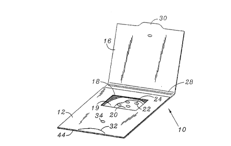
With reference to Figures 1 and 2, a device in
accordance with the present invention is in the form of
a specimen slide 10 and includes a front panel 12, back
panel 14, and a cover 16. A rectangular aperture 18 is
formed in the front panel. A thin sheet of mesh or
porous screening material 19 (shown partially cut-away
WO90/13802 2 0 ~ 2 4 8 2 PCT/US90/01g62
-5
in Figure 1) and a specimen sheet 20 are retained
between the front panel 12 and the back panel 14. The
screening material 19 is a high strength, high porosity
tissue composed, for example, of cellulosic fiber or
synthetic materials, such as polyester or nylon mesh.
Suitable materials include "Hollytex" brand material,
grade 3257, from Eaton-Dikeman Division of Filtration
Sciences Corporation, Mount Holly Springs, Pennsylvania,
and grade 785 tissue from the C. H. Dexter Division of
The Dexter Corporation, Windsor Locks, CT. The screen-
ing material 19 overlies the specimen sheet 20 and is
disposed between the aperture 18 and spt~ecimen sheet
20. The specimen sheet 20 is formed, for example, from
filter paper and includes a plurality of perforations 22
which define circular removeable portions 24 of the
specimen sheet 20 that can be easily removed as is
described hereinbelow. The cover 16 includes a tab 30
formed at the outer edge of the cover 16 and is adapted
to fold along a hinge line 28. The tab 30 is adapted to
engage a semi-circular cut 32 formed in the front panel
12 to thus close the specimen slide 10 once a specimen
has been applied through the aperture 18 and screening
material 19 to the specimen sheet 20.
A flap 36 (shown in its opened position in
Figure 2) is formed in the back panel 14 by an outline
of perforations 38 and a crease 40 defining a hinge.
The perforations 38 are spaced to define a plurality of
bridges 42, each comprising bridge portions 42a, 42b,
between the flap 36 and the surrounding portion of the
back panel 14. The bridges 42 hold the flap 36 in place
until the bridges 42 are broken as the flap 36 is opened
along the crease 40. When opened as illustrated in
Figure 2, the flap reveals the back of the specimen
sheet 20 and the removable portions 24.
WO90/13802 PCT/US90/01962
2 0 ~ 2 482 -6-
Preferably, the specimen slide 10 is assembled
using a single length of cardboard or paper into which
the aperture 18, cut 32 and perforations 38 are die-
cut. The specimen sheet 20, already including the
perforations 22, and the screening material 19 are
positioned against the inside of the back panel 14. A
bend is formed at edge 44 and the front and back panels
12, 14 are fixed together by means of a suitable
adhesive or glue. The cover 16 is folded along the
hinge line 28 and removably affixed to the front cover
by means of the drop or dot of glue 34.
In use, a patient opens the cover 16 breaking
the cover 16 away from the glue 34, revealing the
aperture 18. A specimen of fecal material or other
specimen is smeared with a suitable applicator through
the screening material 19 and onto the specimen sheet
20. The cover 16 is closed with the tab 30 beneath the
cut 32 and the specimen slide 10 is returned to the
physician's office or laboratory for analysis.
To remove a portion of the specimen from the
specimen slide 10, the flap 36 is freed from the back
panel 14 by breaking the bridges 42 and is opened.
Using a suitable implement such as tweezers, one or more
of the removable portions 24 to which a portion of the
specimen was applied is easily separated from the speci-
men sheet 20 and may be deposited in a microtiter plate
well or other suitable test vessel for subsequent
immunochemical assay.
With reference now to Figures 3 and 4, another
embodiment of a specimen slide 60 is illustrated. The
specimen slide 60 includes front panel 62, rear panel 64
and cover 66. Two apertures 68 and 70 are formed
through the front panel 62. A thin sheet of screening
WO9O/13802 2 0 3 2 4 8 2 PCT/Usgo/0l962
--7--
material 71 and a specimen sheet 72 are retained between
the front panel 62 and rear panel 64. The screening
material 71 overlies the specimen sheet 72 and is dis-
posed between the apertures 68 and 70 and the specimen
sheet 72. The specimen sheet 72 includes a plurality of
perforations 74 which define two removable portions 76
aligned with and visible through the aperture 68 and two
removable portions 78 aligned with and visible through
the aperture 70. As an alternative, the plurality of
perforations 74 may be arranged in a more densely packed
pattern such as illustrated in Figure 2. With such an
alternative pattern, the specimen sheet,72 may be
retained between the front and rear panels 62 and 64
without the need to carefully align particular perfor-
ations with the apertures 68 and 70 as is necessary withthe pattern illustrated in Figure 4.
A flap 82 is formed in the back panel 64. The
flap 82 is similar to the flap 36 of the slide 10 and
may be opened to reveal the removable portions 76 and
78. The specimen side 60 may be assembled in a fashion
similar to that described for the specimen slide 10 with
the cover 66 initially held in place by means of a glue
dot 84.
The use of the specimen slide 60 is similar to
that of the specimen slide 10. The cover 66 is separ-
ated from the glue dot 84 and is opened to reveal the
apertures 68, 70. However, two specimens from different
sites of the fecal material may be applied through the
two apertures to 68 and 70. The cover 66 is then closed
and is secured to the front panel 62 by means of a tab
86 and slit 88. To remove a specimen from the slide 60,
the flap 82 is opened and one or more of the removable
portions 76 or 78 carrying a portion of the respective
WOgO/13802 PCT/USgO/01962
2032482
specimens are removed and used for analysis of the
speclmen .
Various modifications to the present invention
will be readily apparent to those skilled in the art.
For example, the specimen slide 10 or 60 may be con-
structed without the screening material 19 or 71. The
shapes and sizes of the apertures 18, 68 and 70 may vary
according to, for example, the size of the specimen
slide or the amount of specimen that is to be applied to
the specimen sheet. For example, a smaller aperture may
have the effect of concentrating the specimen in a
smaller area, improving the reproducibility of the
specimen gathering technique. Also, the sizes of the
removeable portions 24, 76 and 78 may be varied to carry
more or less specimen to thereby accommodate differing
sensitivities of testing methodologies.
The specimen slides 10 and 60 advantageously
allow access to the fecal specimens without reopening
the portion of the slides to which the specimens were
originally applied. Also, the use of removable portions
of the specimen sheets for subsequent analysis is neat
and does not produce additional sub-parts or component
which may require separate disposal. Both of the
specimen slides 10 and 60 provide a convenient and
aesthetically improved means for collecting and handling
fecal specimens for immunochemical analysis.
The present invention is not to be limited to
the detailed description contained herein but is to be
afforded the full scope of the appended claims and all
equivalents thereto.