Note: Descriptions are shown in the official language in which they were submitted.
0~29~3i
INSTRUMENTATION CLAMP
Alan L. Grantz
David A. Gollnick
This patent application is a continuation-in-part of
5 patent application serial no. 07/463,741, filed
January 12, 1990 and now pending.
BACKGROUND O~ THE INVENTION
Field of the Invention
This invention relates to a means for clamping
10 instrumentation, especially cylindrical in shape, with
minimal slippage and deformation. In particular, this
invention relates to clamping optical fibers for
manipulation when using a la~er. One particular
application of the instrumentation clamp is in laser
15 surgery, such as percutaneous diskectomy.
DESCRIPTION OF THE PRIOR ART
Mechanically assisted percutaneous lumbar diskectomy
of the prior art is used as a treatment of leg pain
(sciatica) r~sulting from herniated discs of the lumbar
20 vertebral column. The lumbar vertebral column consists of
five vertebrae extending superiorly to the transitional
thoracic vertebrae (1) at a first end and extending
inferiorally to the sacrum (3) at a second end, as
illustrated in Figure 1. Between each lumbar vertebrae
25 and between the lumbar and sacrum are cartilaginous discs.
Each disc comprises an outer circular structure (annulus
fibrosus) 2 which surrounds and tightly binds an lnner
gelatinous material (nucleus pulposus) 4 in the center, as
illustrated in Figure 2. The annulus fibrosus 2 is made
30 up of concentric fibers which appear to cross each other
P~ 1217\1P\P\APP~)2.~L
- 2 ~ 81
obliquely. No blood vessels or nerves penetrate the
nucleus.
Usually with age, the fibers of the annulus begin to
degenerate. The degeneration results in the tearing of
5 individual fibers when the vertebral column is stressed.
Torn fibers can form fenestrations which allow the nucleus
pulposus to move through the fibers of the annulus and
bulge 5 outward away from the nucleus. If the bulged disc
presses upon an ad~acent nerve root 6, sciatica may
10 develop, as illustrated in Figure 3A.
It has been demonstrated that removing a portion of
the nucleus with grasping forceps through a small cannula
will produce good to excellent relief of pain in a
majority of patients having symptoms indicative of
15 sciatica. Once a portion of the nucleus is removed, the
pressure against the nerve root causing the pain is
relieved as the remaining nucleus contracts away from the
pressure point, as illustrated in Figure 3B. Hijikata S ,
Yamagishi M., Nakayama T., Oomori K., "Percutaneous
20 Diskectomy: A New Treatment for Lumbar Disc Herniation",
J. Toden Hospital 1975; 5:5-13. Since the Hijikata et al.
article, mechanical forceps for microlumbar and
percutaneous lumbar diskectomy procedures have been
developed related to relieving sciatica pain.
U.S. Patent No. 4,369,788, which issued in
January 25, 1983 to Goald discloses a forceps device
having an alligator jaw for microlumbar disc surgery. For
microlumbar disc surgery, a one-inch incision is made in
the patient into which the forceps are inserted and the
30 surgery is performed.
U.S. Patent No. 4,545,374, which issued on October 8,
1985 to Jacobson discloses a method and instrumentR for
performing percutaneous diskectomy using a knife to severe
the disc nucleus and Rongeur forceps to remove the severed
35 fragments of disc nucleus. The diskectomy tools are
inserted through a cannula to the injured disc area.
P:\M\1217\1P\P\APP002.SEL
3~ 9~
The instrumentation and procedure taught by Jacobson
require extensive manipulation of tools by the surgeon,
that a more streamlined procedure using fewer tools would
be desirableO
U.S. Patent No. 4,678,459 issued to Onik et al. on
July 7, 1987 discloses using an irrigating, cutting and
aspirating system for percutaneous surgery. Onik et al.
disclose using a system for removing nucleus pulposus
tissue which includes a probe and a guillotine type of
10 cutting means for cutting the nucleus pulposus. The
severed or cut fragments of nucleus pulposus are removed
from the cuttiny means using an internal fluid irrigation
system and a vacuum to aspirate the severed fragments out
of the disc area, through the system, and out of the
15 patient. This system provides for a relatively fast
diskectomy procedure compared to the other prior art
because nucleus pulposus can be fragmented and removed
without the need to manipulate many small blades, knives
and forceps, as described for the Jacobson patent
20 4,545,374. The probe and guillotine-type cutting means
disclosed by Onik et al. is sold on the market as a
Nucleotome Probe 7~, as illustrated in Figure 4. This
instrument is the most widely used instrument for
percutaneous diskectomy. The Nucleotome Probe 70 is
25 inserted into a cannula 73 and is locked into place on the
cannula 73, as illustrated in Figure 4. When the
Nucleotome Probe 70 is activated, the nucleus pulposus is
cut into fragments which are removed with irrigation
fluids and suction, all within the Nucleotome Probe 70.
30 The Nucleotome Probe 70 is activated until no further
material can be extracted. once complete, the Nucleotome
Probe 70 and cannula 73 are removed and the entry point is
covered with a sterile bandage. The cutting and
extracting process alone using ~he Nucleotome Probe 70
35 normally takes between 20 to 30 minutes.
P:\M\1217\1P\P\APP002.EEL
981
- 4 -
It would be desirable if a laser technique and laser
instrumentation were available to plerform percutaneous
diskectomies so that nucleus pulposus from herniated discs
could be vaporized using a laser in a safe and effective
5 way which is faster than cutting and irrigating using the
Nucleotome Probe and which would eliminate the need to cut
and remove fragmented debris from the patient.
SUMMARY OF THE INVENTION
A means for clamping is provided which clamps and
10 grips fragile instrumenta~ion with minimal slippage and
deformation. Each means for clamping can couple the
clamped instrument to a second instrument for manipulation
of both instruments simultaneously. In a first
embodiment, the clamping means is permanently attached to
15 a second instrument and in particular, the clamping means
attaches instrumentation to an introducer tube. In a
second embodiment, t~e clamping means can be temporarily
coupled to the second instrument. The instrumentation
clamp is self-contained and reusable. The means for
20 coupling of the second embodiment incorporates standard
temporary coupling means which makes the instrumentation
clamp readily adaptable for multiple uses. The means for
clamping works particularly well with the instrumentation
described herein for percutaneous diskectomy using a
25 laser.
According to the present invention, the means for
clamping comprises a resilient tube and means operatively
associated with the resilient tube for providing a
compressive force. The means for providing a compressive
30 force comprises a clamping portion having means for
holding said resilient tube and having means for coupling
to a second instrument; and a clamp housing operatively
secured to said means for holding said resilient tube,
said clamp housing selectively compressing said means for
35 holding against said resilient tube. The compressive
P:\M\1217\1P\P\APP002.EEL
- 5 ~ ~9~
force is distributed generally equally over the entire
length of the tube so that instrumentation disposed
through the tube is substantially secured with mini~al
deformation.
The means for clamping is especial'y adaptable to
clamping optical wave guides such as optical fibers, which
are typically clamped for manipulation. For example,
optical fibers are clamped and secured to instrumentation
which introduces the optical fiber into a patient for
10 surgical procedures using a laser. The means for clamping
according to the present invention can securely couple two
instruments together for applications such as percutaneous
diskectomy using a laser.
BRIEF DESCRIPTION OF THE DRAWINGS
Figure 1 is a posterior view of the lumbar vertebral
column.
Figure 2 is an oblique view o~ a lumbar disc and
inferior vertebrae.
Figure 3A is a sectional view of a herniated lumbar
20 vertebrae and an associated nerve root.
Figure 3B is a sectional view of the vertebrae in
Figure 3A after nucleus pulposus is removed.
Figure 4 is an oblique view illustrating the
Nucleotome Probe of the prior art.
Figure 5 is a posterior view of a patient in a
lateral decubitus position.
Figure 6 is a side view illustrating a probe used
with the present invention.
Figure 7 is an oblique view illustrating the probe
30 inserted into a disc according to the present invention.
Figure 8 is a sectional view of a herniated disc and
associated nerve root having the probe inserted thereinto
according to the present invention.
Figure 9 is a side view of a cannula having a dilator
35 inserted thereinto used with the present invention.
P:\M~1217\1P\P\APP002.EEL
2~ 9~31
-- 6 --
Figures lOA-lOF are sectional and plan view~ of a
bayonet type lock fitting used with the present invention.
Figure 11 is a side view illus;trating a curved
cannula used with the present invention.
5Figure 12 is a side view illustrating an introducer
means according to the present invention.
Figure 13 is a side view illustrating a stylet used
with the present invention.
Figure 14A~H are sectional views illustrating a
10 clamping means according to the present invention.
Figure 15 is a side view illustrating an introducer
means having a formed end according to the present
invention.
Figure 16 is an enlarged sectional view illustrating
15 an optical guiding means emanating from the formed end of
introducer means according to the present invention.
Figure 17 is a side view of an irrigation/aspiration
cannula used with the present invention.
Figures 18A-C are sectional views illustrating a
20 position indicator means according to the present
invention.
Figures l9A-B are plan views illustrating the first
line of a laser beam.
Figures 20A-C are plan views illustrating the second
25 line of a laser beam.
Figures 21A-21F illustrate sectional and plan views
of the clamping portion to the second embodiment of the
clamping means according to the present invention.
Figures 22A-22G illustrate sectional and plan views
30 of the clamp housing to the second embodiment of the
clamping means according to the present invention.
Figure 23A is a sectional view and Figure 23B is a
plan view illustrating the assembled in~trumentation clamp
of the second embodiment of the clamping means according
35 to the present invention.
P:\M~1217\lP\P\APP002.EEL
9B~
-- 7
Figure 24A illustrates the inst:rumentation clamp of
Figure 23B associated with both a first instrument and a
second instrument, according to the preferred embodiment
of the clamping means.
Figure 24s illustrates a cross-~sectional vi2w of
Figure 24A.
DETAILED DESCRIPTION OF THE INvENTION
A percutaneous diskectomy procedure using a laser is
designed for patients commonly showing evidence clinically
10 and radiologically of nerve root impingement. Conven-
tionally, physical examination of the patient sAould
reveal leg pain greater than back pain and signs of nerve
root irritation consistent with a herniated disc. Radio-
graphically, the patient should exhibit a focal herniation
15 or bulge that shows an impression on the thecal sac which
does not occupy more than fifty percent of the thecal sac.
Also, the radiographic results should correlate with the
patient's symptomatology.
Vaporization of nucleus pulposus material according
20 to the present invention suggests that the operative tools
be inserted at an entry site on the same side of the
patient's body that the herniation or other affliction is
evident. The path of entry to the afflicted disc should
avoid going through the psoas muscle since the lumbar
25 plexus has numerous fibers which traverse the muscle.
Conventionally, a computed tomograph (CT) scan slice of
the whole abdomen through the involved disc is quite
helpful for determining the entry path.
The safety of the procedure relies on radiologic
30 localization and guidance of the instruments into the disc
and a C-arm fluoroscope with image intensification, known
in the art, can provide clear and sharp images in antero-
posterior, lateral and oblique views.
Typically, the patient undergoing a percutaneous
35 diskectomy procedure is positioned on a fluoroscopic
P:\M\1217\1P\P\APP002.EBL
Z~3~98~
-- 8
table, which is known in the art, in a lateral decubitus
position, as illustrated in Figure 5. The patient must be
stabilized to prevent rotation of t:he patient's shoulders
and hips during the procedure. Using a fluoroscope, the
s sacrum is identified and located, the afflicted disc is
located, and as illustrated in Figure 5, a posterolateral
entry point is selected. The entry point typically is
8-12 centimeters from the midline and hoth parallel and
midway between the end plates of the afflicted disc, as
10 determined using a measuring scale. Local anesthetic is
used to anesthetize the area to be operated on which is
administered typically with a long spinal needle.
At this point in the patient preparation procedure,
the percutaneous diskectomy procedure using a laser and
15 means for inserting instrumentation according to the
present invention is described.
First, one end of a semi-rigid trocar or probe 100,
which is preferably 18 gauge Birmingham Wire Gauge (BWG),
is inserted at the entry point once the anesthetic has
20 taken effect. Probe 100 has an elongated body lOOa and
has a standard tube clamp lOOb with a threaded lock lOOc
connected to probe 100, as illustrated in Figure 6.
Clamp lOOb is removable from body lOOa by loosening
lock lOOc and sliding clamp lOOb in either direction
25 beyond the end of probe 100. Clamp lOOb serves as a
handle to hold probe 100 while it is inserted into the
patient~ Clamp lOOb is removed for subsequent steps
described below. Clamp lOOb may be made of plastic, for
example acrylonitrile butadiene-styrene (ABS) plastic or
30 preferably polycarbonate plastic, or metal, preferably
stainless steel.
Probe 100 is preferably made of stainless steel, for
example type 304 or an equivalent, No. 3 temper. The
inserted end of probe 100 has a sharp tip and is guided
35 into the damaged or herniated disc area 18c with
radiologic localization and guidance, preferably using the
P:\M~1217~1P~P~APP002.EEL
29~
g
C-arm fluoroscope with image intensification, as described
above. Probe 100 is inserted until the inserted end
punctures through the annulus fibrosus 18a of the disc
18b, as illustrated in Figures 7 and 8. While probe 100
5 is in place, extending from the disc area to outside the
patient's body, a cannula 104 having a dilator 102
inserted thereinto is inserted over probe 100 at the
exterior end and into the probed disc area 18c. One end
of dilator 102 (102b) and cannula 104 (104b) remain on the
10 exterior of the patient.
Cannula 104 and dilator 102 are preferably 12 gauge
stainless steel tubing, for example type 304, No. 3 temper
(full hard). Stainless steel tubing may be purchased at
any stainless steel tubing supplier, for example Pop~r and
15 Sons, N.Y. Dilator 102 preferably is longer than cannula
104 and has a tapered end 102a which extends beyond the
end 104a of cannula 104, as illustrated in Figure 9, for
ease of insertion over probe 100 through the patient's
skin. Dilator 102 has bore 102d which extends through the
20 center of dilator 102 along its length. Probe lGO fits
within bore 102d of dilator 102. Cannula 104 is
preferably a straight tubular member having a central bore
104d, which extends along its length. Dilator 102 and
probe 100 fit within bore 104d of cannula 104.
Cannula 104 and dilator 102 have locking means 103
(103 mechanism not shown in Figure 9) for locking
dilator 102 to cannula 104 at end 104b and 102b,
respectively, and a locking stabilizer 105, as illustrated
in Figure 9. Locking means 103 is preferably a bayonet-
30 fitting locking mechanism, as illustrated in
Figures lOA-lOF as portions 103a and 103b. Dilator 102
has portion 103a and cannula 104 has portion 103b of
bayonet-fitting locking mechanis~ 103. Portion 103b on
cannula 104 has a segmented body and flared legs for
35 gripping anA preferably projection 103b-1 having two
laterally extending flanges 103b-2 which oppose one
P:\M~1217\lP\P\APP002.EEL
2C~298~L
-- 10 --
another. Portion 103a on dilator 102 preferably has
aperture 103a-1 and sockets 103a-2. Aperture 103a-1 is at
least as deep as the distance projection 103b-1 projects.
Sockets 103a-2 oppositely extend oif aperture 103a-1 and
5 are at least as deep as flanges 103b-2 are thick.
Projection 103b-1 and flanges 103b--2 fit within aperture
103a-1 and sockets 103a-2, respectively. Once fitted
together, end 102b of dilator 102 is turned clockwise
thereby rotating dilator 102 within cannula 104 to lock
10 locking portion 103a to locking portion 103b and thereby
lock dilator 102 to cannula 104 using locking means 103.
Locking means 103 can also be a luer lock, threaded screw
lock, snap lock or a friction fitted lock, which are known
in the art. Locking means 103 and stabilizer 105 can be
lS made of plastic, ABS or preferably polycarbonate plastic,
or metal, preferably stainless steel. In the preferred
embodiment, means 103 and stabilizer 105 are made of a
plastic which can withstand at least the stresses
associated with gamma sterilization techniques without
20 distortion. The plastic may also withstand the stresses
associated with autoclaving and usage of ethylene oxide
gas sterilization methods. Locking stabilizer 105 is
adjustably located along the length of cannula 104 and
serves to rest against the patient's skin when the cannula
25 is properly placed.
In another embodiment, curved cannula 106 may be
inserted into the patient instead of straight cannula 104.
Cannula 106 is a curved tubular member having a locking
dilator 107 and locking stabilizer 108, as illustrated in
30 Figure 11. Curved cannula 106 ic used in situations where
the patient's afflicted area is within the lumbar 5-
sacrum 1 region of the vertebral column, as shown in
Figure 1.
Once dilator 102 and cannula 104 are confirmed,
35 preferably fluoroscopically, to be embedded in the annulus
fibrosus, dilator 102 is unlocked from locking mechanism
P:\M\1217\1P\P\APP002.EEL
Z~3X981
103 and removed. Dilator 102 is unlocked by turning
portion 103a counterclockwise while holding portion 103b
on cannula 104 stationary. As dilator 102 is withdrawn,
cannula 104 is advanced forward to embed in the wall of
5 the annulus approximately the distance equal to the
difference in length of dilator 102 and cannula 104.
Cannula 104 is secured by stabilizer 105 by unlocking the
screw mechanism, sliding stabilizer 105 up against the
patient's skin, and locking the screw. Probe 100 is
10 removed once cannula 104 is secured.
Second, one end of a first introducer means or tube
110 for inserting instrumentation according to this
invention is inserted into central bore 104d at the
exterior end 104b of cannula 104. First introducer means
15 110 is a substantially straight elongated member
preferably 14 gauge along most of its length and having a
17 gauge tip llOa at one end, as illustrated in Figure 12,
and having a clamping means 111 at an opposite end for
clamping to an optical guiding means, as is described
20 below. In another embodiment, the first introducer means
and means for clamping are separate devices. First
introducer means 110 is metal, preferably type 304
stainless steel, No. 3 temper (full hard). Clamping means
111 can be plastic, preferably polycarbonate plastic or
25 metal, preferably stainless steel. First introducer means
110 haq a bore llOd which extends through the center of
first introducer means 110 along its length.
When the one end llOa of first introducer means 110
is inserted into bore 104d of cannula 104, first
30 introducer means 110 preferably has a stylet 112 extending
therethrough. Stylet 112 is a long straight member,
preferably 18 gauge stainless steel, having a sharp tip
112a at one end and a handle means 112b for handling
stylet 112 at the opposite end, as illustrated in
35 Figure 13. The sharp end 112a can be a conical-shaped
tip, diamond shaped tip or beveled, for example. Sharp
P:~M\1217\1P\P\APP002.EEL
- 12 - ~ 981
end 112a extends out of the inserted end llOa of the first
introducer means and stylet 112 is clamped by clamping
means lll on first introducer means 110 at handle means
end 112b. The clamping mechanism for clamping means 111
5 will be descri~ed below. Stylet 112 is longer than and
narrower in diameter than first introducer means 110 and
fits within bore llOd of first introducer means 110.
Stylet 112 can be adapted to also lock with the means for
clamping at handle means 112b either with a luer lock,
10 threaded screw lock, snap lock, or friction fit lock,
depending on the embodiment of the means for clamping.
Because the sharp tip extends out of the inserted end
llOa of first introducer means 110, stylet 112 contacts
the outer wall of the nucleus 18d and enters into the
15 nucleus with its sharp tip 112a, leaving a small opening.
Since the nucleus is a soft gelatinous material,
stylet 112 enters the nucleus with ~inimal resistance and
the inserted end llOa of first introducer means 110 is
placed in the nucleus. Stylet 112 is removed from the
20 nucleus through the first introducer means 110, and the
first introducer means or tube 110 is left in place for
introducing instrumentation into the nucleus.
Third, one end of a first optical guiding means 116
for guiding laser light is inserted through bore llOd of
25 first introducer means 110 after stylet 112 is removed.
First optical guiding means 116 is inserted until it
emanates from end llOa of first introducer means into the
small opening made by stylet 112.
First optical guiding means 116 is preferably an
30 optical fiber or a hollow optical wave guide (both not
shown), depending on the embodiment. In one embodiment,
an optical fiber can be used which is preferably 400
misrometers in inner diameter and 600 micrometers in outer
diameter and is made of quartz. In another embodiment, a
35 hollow optical wave guide can be used which can be made
P:\M\1217\1P\P\APP002.EEL
r~9~
- 13 -
from metal or preferably ceramic. The hsllow waveguide is
rigid compared to the optical fiber.
First optical guiding means 1~6 passes through first
introducer means 110 and into the nucleus 18d at a first
5 end and is connected to a first la~;er means for producing
laser light at a second end outside of the patient. First
optical guiding means 116 can have a position indicator
means lllh for indicating a preset distance optical
guiding means 116 must extend out of first introducer
10 means 110 at end llOa, depending on the embodiment.
Positioning indicator means lllh, which is described
below, is illustrated in Figures 18A-18C and serves to
prevent optical guiding means 116 from being inserted
beyond the preset distance by contacting clamping means
15 111 from one end.
Clamping means 111 then clamps optical guiding means
116 in place in first introducer means 110, according to
the first embodiment. Clamping means 111 serves to ensure
that optical guiding means 116 moves with first introducer
20 means 110 as first introducer means 110 is manipulated
during the diskectomy procedure for example.
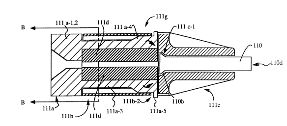
As illustrated in Figure 14A, the first embodiment of
a means for clamping, clamping means 111, has a clamping
end llla, midsection lllb and an introducer end lllc.
25 Midsection lllb and introducer end lllc comprise clamp
housing lllg. Clamping means 111 can be made of a metal,
for example stainless steel, but is preferably made of
molded plastic, preferably polycarbonate plastic.
Clamping end llla comprises a clamp llla-1 having a
30 clamping head llla-2, two integrally connected compression
legs llla-3, and side members llla-7, as illustrated in
Figures 14A-14C. Side members llla-7 are wider than
compression legs llla-3. Clamping head llla-2, side
members llla-7, and compression legs llla-3 are preferably
35 molded as one piece. Legs llla-3 and side members llla-7
P:\~\1217\1P\P\APP002.EEL
8~
are integrally connected to head llla-2 at one end while
the opposite ends are free~
Compression legs llla-3 and side members llla-7 of
clamp llla-1 fit within midsection lllb of housing lllg,
5 and each compression leg llla-3 has a cylindrical boss
llla-4 which projects laterally therefrom, as illustrated
in Figures 14A-14F. Bosses llla-4 fit within internal
curved recesses lllb-1 of midsection lllb, as illustrated
in Figure 14G, when clamp llla-l is fully inserted into
10 housing lllg. Curved recesses lllb-1 serve as cam
surfaces while tAe cylindrical bosses llla-4 serve as cam
followers. Compression legs llla-3 also each have an
engagement ear llla-5 at the free ends thereof.
Engagement ears llla-5 project laterally out and fit
15 within retention slots lllb-2 in outer housing lllg, as
illustrated in Figures 14A-14F and 14H. Retention slots
lllb-2 are located in midsection lllb near where
midsection lllb and introducer end lllc meet. As clamp
llla-l is in~erted into housing lllg, bosses llla-4 slide
20 along recesses lllb-1 until engagement ears llla-5 snap
into retention slots lllb-2, thereby fixing the assembly
together, as illustrated in Figure 14A. Then ~ousing lllg
is rotated while clamping end llla is held stationary,
causing midsection lllb to press in on compression legs
25 llla-3 with cam and follower action. Engagement ears
llla-5 also move out of retention slots lllb-2 and within
midsection lllb as midsection lllb is rotated.
An elastomer llld is retained by the inner radius of
compression legs llla-3 and side members llla-7 and is
30 preferably tubular in shape, extending from head llla-2 to
engagement ears llla-5, a~ illustrated in Figure 14A.
Elastomer llld is a resilient material, preferably
silicone rubber. Elastomer llld grips or clamps to
optical guiding means 116 when housing lllg is rotated.
35 Optical guiding means 116 is inserted into first
introducer means 110 through bore 110d to a position
P:\M\1217\1P\P\APP002.EEL
981
- 15 -
indicated by its position indicator means lllh, as
illustrated in Figures 18A-18C. Optical guiding means 116
is intended to extend out of end 110a for a distance which
is determined by the surgeon to be within the nucleus 18d
5 of the afflicted disc 18b. Compression legs llla-3
compress elastomer llld against optical guiding means 116
in the fully rotated, clamped position. The side members
llla-7 prevent radial expansion of the elastomer llld
during the compression. Elastomer llld grips and prevents
10 axial slippage of optical guiding means 116. The
compressed elastomer llld distributes the clamping force
on the optical guiding means in such a manner that the
optical transmission characteristics of optical guiding
means 116 are not degraded. In addition, elastomer llld
15 exhibits a large coefficient of friction against optical
guiding means 116. This large coefficient of friction
minimizes the clamping force required to sustain a given
degree of restraining force. Because of the
characteristics of elastomer llld, clamping means 111 is
20 also removable by rotating housing lllg in the opposite
direction to release the compression forces without
degrading the optical guiding means 116 optical
characteristics.
Both clamping means 111 and position indicator means
25 lllh of the first embodiment clamp and grip onto optical
guiding means 116 in the same way and clamping means 111
also clamps to stylet 112 in the same fashion. The shapes
of housing lllg and position indicator means (lllh)
housing lllh-l differ although they comprise similar
30 components. The differences in housing lllg and the
housing lllh-1 of position indicator means lllh relate to
introducer end lllc. Introducer end lllc is shaped to fit
and grip end 110b of first introducer means 110. End 110b
is flared as illustrated in Figure 14A and flare grip
35 lllc-1 holds end 110b in place. In the first embodiment
of the means for clamping, flared end 110b is bonded into
P:\M\1217\1P\P\APP002.EEL
~X9~3~
- 16 -
introducer end lllc using an organic adhesive, for example
fast bondinq adheslves which are compatible with both
plastics and metal, like cyanoacry:Late adhesives.
Clamping means 111 is permanently attached to first
5 introducer means 110 with the adhesiYe. On the other
hand, position indicator means lllh is shaped to
facilitate the insertion of the optical guiding means 116,
which does not have flared ends, as illustrated in
Figures 18A-18C, and is not permanently attached to other
10 instruments.
Clamping means 111 is assembled as follows: First,
end 110a of first introducer means 110 is inserted into
housing lllg from midsection 111~ end until flared end
110b contacts with flared grip lllc-1. End 110b of first
15 introducer means 110 is held in place until bonded with a
pre-applied adhesive. Second, elastomer llld is then
inserted within the inner radius of compression legs
llla-3. Third, clamp llla-1 is inserted into midsection
lllb until engagement ears llla-5 engage with retention
20 slots lllb-2. Housing lllg is not rotated into the
clamping position until optical guiding means 116 or
stylet 112 is inserted and clamping is necessary.
A second embodiment of the clamping means, instrumen-
tation clamp 700, according to the invention is
25 illustrated in Figures 21A-21F, 22A-22G, 23A, 23B, 24A and
24B. Figure 23B illustrates the assembled instrumentation
clamp 700, while Figure 23A is a cross-sectional view of
the assembled instrumentation clamp 700 illustrating the
internal components of the clamp assembly.
Figure 21A is a cross-sectional view of clamping
portion 500. Clamping portion 500 comprises compression
legs 501 attached to clamp head 502. Compression legs 501
are preferably integrally connected to clamp head 502 at
one end and clamping portion 500 is typically molded as
35 one part. Compression legs 501 each have a projecting ear
503 and a cylindrical-shaped raised portion 504 formed
P:\M\1217\1P\P\APP002.~EL
981
- 17 -
near the free end thereof. The raised portion 504 is
located adjacent to projecting ear 503. Clamping portion
500 further comprises extension 505 which projects from
the interior of clamp head 502. Extension 505 has a
5 tapered bore which has a larger diameter d1 at the end
505a which protrudes from clamp head 502 than diameter d2
at end 505b where compression legs 501 emanate. The
tapered bore facilitates threading a first instrument 800,
such as an optical fiber, through bore 505b diameter d2
10 from end 505a of extension 505.
According the second embodiment, clamp head 502
provides a means for coupling 508 a second instrument 801
to instrumentation clamp 700. Means for coupling 508 can
utilize any locking means including a threaded screw lock,
15 a snap lock, bayonet-type lock, or a friction fitted lock,
which are known in the art, or preferably a luer lock.
Second instrument 801 can have the complementary fitting
to clamp 700. In the preferred embodiment, the means for
coupling 508 incorporates thread receiving grooves 508-1
20 on the interior surface of clamp head 502. The thread
receiving grooves 508-1 receive the threads 802-1 of a
female luer fitting 802 of a second instrument 801, as
illustrated in Figure 23A. Instrumentation clamp 700 can
be made of engineering plastic, such as ABS or
25 polycarbonate plastic. For the preferred embodiment,
instrumentation clamp 700 is made of a polycarbonate
plastic. For the invention, the means for coupling 50
can be either the male lock fitting or the female lock
fitting, while the male lock fitting is preferred for
30 instrumentation clamp 700.
Figure 21C and 21E are side views of clamping portion
500 of the second embodiment. Figure 21C illustrates a
side view of compression legs 501. In Figure 21~, a front
view of one compression leg 501 is illustrated along with
35 a side view of compression extensions 506. Compression
extensions 506 facilitate holding resilient tube 507 along
P:\M\1217\1P\P\APP002.EEL
981
- 18 -
with compression legs 501. Compression legs 501 and
compression extensions 506 have internal and external
curvature, as illustrated in Figure 21B. Although Figure
21B illustrates only two compression legs 501, in other
5 embodiments, there can be more than two. As the number of
compression legs 501 increases, the siæe of the
compression extensions 506 decreases. Compression legs
501 are designed to flex inwardly, as shown by arrows in
Figure 21A, toward a resilient tube 507 (not shown).
10 Resilient tube 507 is illustrated in cross section in
Figure 23A and is similar to elastomer llld of the first
embodiment of the means for clamping. Resilient tube 507
has a central bore 507a which is aligned with bore end
505b having diameter d2 in clamp head 502.
Figure 21D illustrates an end view of clamp head 502
wherein the tapered bore having diameters d1 and d2 through
projection 505 are shown in relation to clamp head 502.
Also shown are a plurality of ribs 509 on the surface of
clamp head 502. Ribs 509 serve as gripping and handling
20 members of clamp 700.
Figure 22D illustrates a cross sectional view of
clamp housing 600 of the second embodiment. Clamp housing
600 is essentially a cylindrical tube having a bore 603
and comprises a clamp receiving end 601 and a clamp
25 securing end 602. Clamp receiving end is illustrated in
Figure 22F. Bore 603 having diameter d3 extends along the
length of clamp receiving end 601 and narrows to diameter
d4 in clamp securing end 602. Bore 603 flares out to
diameter d5 at the other end of clamp securing end 602
30 (see Figures 22D, 22F and 22G). The tapering of bore 603
facilitates threading a first instrument into clamp 700
from clamp securing end 602. The end view of clamping end
601, as illustrated in Figure 22F, also illustrates
recesses 604 for receiving raised portions 504 and
35 projecting ears 503 on compression legs 501. Recesses 604
extend along the length of the interior surface of clamp
P:\M\1217\1P\P\APP002.EEL
~ 19 - ~03Z98~
housing 600 and have a radius of cllrvature designed to
match the radius of curvatur~ of raised portions 504.
Figure 22G illustrates an end view of clamp securing
end 602. Clamp securing end 602 has cutout portions 605
5 which extend approximately from an edge of each recess 604
for a distance around the perimeter of end 602 and along
the length of clamp securing end 602. Recesses 604 are
generally aligned with one edge of cutouts 605 and each
respective recess 604 and cutout 605 are directly opposite
10 the other in the preferred embodiment. Through each
cutout 605 is a projecting ear receiving slot 606 which
extends from edge to edge of cutout 605 near where clamp
securing end 602 ends and clamp receiving end 601 begins.
Receiving slots 606 are illustrated in Figures 22D and
15 22E.
Figure 22B is a cross sectional view of clamp housing
600 cut along line ~-B indicated Figure 22D. Cutouts 605
are not cross-hatched and are shown in relation to
recesses 604 in Figure 22B.
Figure 22A is a cross sectional view of clamp housing
600 cut along line A-A in Figure 22D. Figure 22A
illustrates the cross section of clamp housing 600
directly opposite to that of the cross section of clamp
housing 600 in Figure 22B where recesses 604 end in clamp
25 receiving end 601 and cutouts 605 begin in clamp securing
end 602.
Figure 22C is a cross-sectional view of clamp
securing end 602 cut along line C-C of Figure 22D.
Figure 22C illustrates the tapering of bore 603 from
30 diameter d5 to diameter d4 in clamp securing end 602. Also
shown are cutouts 605 and recesses 604.
To assemble clamp 700, resilient tube 507, made from
a resilient elastomer, such as silicone rubber, for
example, is placed within the interior curvature of
35 compression legs 501 and compression extensions 506 so
that bore 507a in resilient tube 507 is aligned with bore
P:\~\1217\lP\P\APP002.E~L
- 20 - ~ ~ ~X981
505b of clamping portion 500. Clamp housing 600 is snap
coupled to clamping portion 500, after resilient tube 507
is installed. First, projecting ears 503 of compression
legs 501 are aligned with recesses 604 of clamp housing
5 600. Compression legs 501 are compressed inwardly until
projecting ears 503 fit into clamp housing 600 within
recesses 604. Second, while holding clamping portion 500
by clamp head 502 and holding clamp housing 600 by clamp
securing end 602, clamp housing 600 is slid over
10 compression legs 501 until projecting ears 503 are
received by receiving slots 606. Projecting ears 503
emanate from receiving slots 606 and curved portions 504
are received by recesses 604 when instrumentation clamp
700 is in an open position.
To clamp to a first instrument, preferably an optical
fiber 800, one end of optical fiber 800 is inserted into
bore 603 from clamp securing end 602. Optical fiber 800
is threaded through bore 603 diameter d5 and through end
505b of clamping portion 500 to emanate out end 505a of
20 extension 505 in clamping portion 500. When the optical
fiber 800 is inserted to the desired distance through
clamp 700, optical fiber 800 is clamped in place by
rotating clamp housing 600 while clamping portion 500 is
held stationary until projecting ears 503 slide from one
25 end of receiving slots 606 to the other end of receiving
slots 606. When clamp housing 600 is rotated, raised
portions 504 exit recesses 604, thereby causing
compression legs 501 to compress against resilient tube
507 by cam and follower action. Instrumentation clamp 700
30 is now in a closed position. The compressive force
generated when clamp housing 600 is rotated closed is
sufficient to hold optical fiber 800 securely with minimal
slippage and minimal deformation of the optical fiber's
transmission characteristics.
To temporarily couple a second instrument 801 to
clamp 700, second instrument 801, having a central bore
P:\M\1217\1P\P\APP002.EEL
- 21 - 2~3298~
803, preferably has a threaded luer female fitting 802 at
one end with bore 803a therethrough to receive extension
505 from clamping portion 500 as threads 802~1 are
received by thread receiving grooves 508-1. Moreover,
5 second instrument 801 preferably has bore 803b which is
narrower than bore 803a and is generally aligned
therewith. A first instrument 800, such as an optical
fiber, can be threaded through clamp 700 and through bore
803 of second instrument 801 for simultaneous
10 manipulation.
Second instrument 801 can be coupled to clamp 700
using means for coupling 508 prior to the insertion and
clamping of first instrument 800. Once second instrument
801 is coupled, optical fiber 800 is threaded through bore
15 603 at end 602 until it emanates from bore 803b of second
instrument 801 to a desired distance. Once threaded,
clamp housing 600 is rotated relative to clamping portion
500 and second instrument 801 to a closed position, as
described above, wherein projecting ears 503 slide to
20 opposite ends of receiving slots 606 and raised portions
504 exit recesses 604 within clamp housing 600. At this
point, optical fiber 800 is securely clamped with minimal
slippage and deformation within second instrument 801 for
simultaneous manipulation. Figure 24A illustrates the
25 simultaneous clamping of optical fiber 800 and coupling of
second instrument 801 to clamp 700 according to the
preferred embodiment. Figure 24B illustrates the clamping
of the preferred embodiment in cross section. Gripping
handle 804 is attached to the periphery of second
30 instrument 801 and facilitates manipulation of second
instrument 801.
According to a preferred implementation of
instrumentation clamp 700, a position indicator means is
no longer necessary. Once the desired length of optical
35 guiding means 116 is determined, clamp 700 clamps optical
guiding means 116 at a predetermined position in the same
P: \M\ 1217 \ lP\ P\APP002 . EEL
981
- 22 -
way that position indicator means lllh was used. The
clamped optical guiding means 116 is threaded through an
introducer means to extend a predetermined distance out of
the introducer means. In this embodiment, clamp 700 is
5 not permanently attached to the introducer means and is a
self-contained means for clamping according to the
preferred embodiment. The introducer means is adapted to
couple to clamp 700 by coupling means 508, as described
above, using the preferred luer lock coupling mechanism
10 508, as illustrated in Figures 23A, 24A and 24B. The
instrumentation clamp 700 provides a temporary and secure
coupling to the introducer means and clamp 700 can be
readily disconnected and reused. The introducer means of
the invention can be any fiber introducing instrument and
15 the instrument and clamp are not limited to use in
percutaneous diskectomy. For example, the introducer
means and means for clamping of the first and second
embodiments can be used in orthopedic surgery.
Fourth, using laser energy from the first laser means
20 through first optical guiding means 116, some of the
nucleus pulposus within nucleus 18d is vaporized to create
a first vaporized area in the nucleus 18d of the herniated
disc 18b. The first vaporized area provides a space or
cavity in the nucleus pulposus into which nucleus pulposus
25 from the herniated area 18c can fill and thereby contract
away from nerve root 18e. First optical guiding means 116
along with first introducer means 110 are removed from
cannula 104 when the vaporization step is complete.
According to the invention, a second vaporization
30 step is included. According to the preferred embodiment,
a second introducer means 130 is inserted into cannula 104
to contact the first vaporized area.
Second introducer means 130 is preferably 14 gauge
along its length and has a 17 gauge tip 130a. Second
35 introducer means 130 is metal, preferably type 304
stainless steel, No. 3 temper (full hard). Moreover, the
P:~\1217\1P\P\APP002.EEL
9~31
- 23 -
opening in tip 130a of second introducer means 130 is
formed differently from first introducer means 110, as
illustrated in Figure 15 and in an enlarged view
illustrated in Figure 16. Rather t:han opening 130a-1
5 being perpendicular to the longituclinal axis of the
tubular member as is shown for first introducer means,
opening 130a-1 at end 130a is curved relative to the
longitudinal axis. Curved end 130a is not flared out nor
wider than the 14 gauge portion of the tubular member. As
10 a result, curved end 130a of second introducer means 130
need not be wider in diameter than first introducer means
110. In the preferred embodiment, second introducer means
130 has the same inner and outer diameter as first
introducer means 110 and has a curvature at end 130a
15 within that outer diameter. Therefore, second introducer
means 130 fits within cannula 104 in the same way firs~
introducer means 110 fits within cannula 104. Cannula 104
remains in the patient's body to receive second introducer
means 130 for the second vaporization step according to
20 the preferred embodiment, as described below.
Fifth, the curved end 130a of second introducer
means 130 enters the nucleus 18d and contacts the first
vaporized area when second introducer means 130 is
inserted into cannula 104. One end of a second optical
25 guiding means 132 is inserted through a central bore 130d,
of second introducer means 130 to emanate from opening
130a-1 into the first vaporized area at the formed end
130a of second introducer means 130, as illustrated in the
enlarged view in Figure 16. Second optical guiding means
30 132 has a position indicator means which is the same as
position indicator means lllh on first optical guiding
means 116 according to the first embodiment. The position
indicator means on second optical guiding means 132
contacts with clamping means 131 in the same way as
35 described above for first introducer means 110 and
position indicator means lllh. Clamping means 131 and 111
P:\M\1217\1P\P\APP002 .EEL
- 24 - 2~32981
are essentially the same and clamping means 131 is
illustrated in Figure 15. Alternatively, instrumentation
clamp 700 of the second embodiment can be used for
clamping the optical guiding means 116, 132 for both
5 vaporization steps. Clamp 700 eliminates the need for two
clamping means 111 and 131, permanently associated with
first and second introducer means 110 and 130, and two
position indicator means. The first and second introducer
tubes only need to be adapted to accommodate the means for
10 coupling 508 of the second embodiment instrumentation
clamp 700.
When end 132a of second optical guiding means 132
emanates from opening 130a-1 of curved end 130a on second
introducer means 130, end 132a of second optical guiding
15 means 132 is deflected off the longitudinal axis of the
second optical guiding means 132. The amount which second
optical guiding means 132 is deflected is dependent upon
the radius of curvature of end 130a of second introducer
means 130.
The considerations made when determining what radius
of curvature to use at least depended on several factors,
according to the invention. First, the minimum radius of
curvature should be so formed at the tip of an introducer
means so that the curved introducer means still fits
25 within cannula 104. Second, optical guiding means 116 or
132, for example an optical fiber, should deflect with
uniform curvature to achieve a minimal loss of laser light
guiding efficiency. Third, the radius of curvature of the
introducer means allowable and the diameter of the optical
30 guiding means allowable are mutually dependent. According
to the preferred embodiment, the radius of curvature is
0.45 which deflects second optical guiding means 132 ahout
17 from the longitudinal axis when second optical guiding
means 132 is a 400~m optical fiber. Second optical
35 guiding means 132 can be deflected between the range of 1
P:\M\1217~lP\P\APP002.EEL
- 25 - 2~ 81
to 30 by curved end 130a of second introducer means 130,
for the preferred embodiment.
Once second optical guiding means 132 is in place and
positioned so that it extends out of curved end 130a of
5 second introducer means 130 for a distance, as
predetermined by the surgeon, optical guiding means 132 is
clamped in place by clamping means 131 in much the same
way as described previously for clamping means 111 of the
first embodiment, or alternatively, can be clamped in
10 place as described for instrumentation clamp 700 of the
second embodiment. Therefore, clamping of second optical
guiding means 132 to second introducer means 130 allows
second optical guiding means to be manipulated as second
introducer means 130 is manipulated. An end of second
15 optical guidiny means 132 opposite to the deflected end is
attached to a second laser means. Light energy from the
second laser means is guided by second optical guiding
means 132 into the nucleus 18d to vaporize nucleus
pulposus and create a second vaporized area within nucleus
20 18d. The second vaporized area of the preferred
embodiment is larger than the first vaporized area and the
larger area is created by the deflected beam emanating
from deflected end 132a of second optical guiding
means 132 during this vaporization step. Manipulation of
25 second introducer means 130 with second optical guiding
means 132 clamped thereto will cause manipulation of the
deflected beam as well.
According to the invention, when the laser beam is
applied generally along a line 30-1 to a herniated disc
30 area, the line or path that the laser beam takes ic
illustrated by example in Figure l9A. Line 30-1 is
obtained by moving first introducer means 110 having ~irst
optical guiding means 116 disposed therethrouqh axially
within cannula 104. Since laser beams according to the
35 invention are divergent and emanate in a 15 cone from the
guiding means, the line or path defined by the divergent
P:\M\1217\1P\P\APP002.EEL
- 26 -
beam is described as a single overall direction the laser
beam travels, as illustrated by arrow A in Figure l9A.
Line 30-2 to a herniated disc area can also be the path of
the laser beam, as illustrated in Figure l9B. Line 30-2
5 to a herniated disc area is obtained with second
intrcducer means 130 having second optical guiding
means 132 disposed therethrough, being deflected off the
longitudinal axis by curved end 130a. The laser beam
guided through deflected second optical guiding means 132
10 is applied along line 30-2.
The laser beam can be applied along a line 31-1, as
illustrated in Figure 20~. Line 31-1 is different from
line 30-1, as illustrated in Figures l9A and 20A, and the
difference is at least due to shape of straight first
15 introducer means 110 relative to the shape of curved
second introducer means 130. Line 31-1 is obtained by
guiding a laser beam along second optical guiding means
32 while second optical guiding means 132 is disposed in
curved second introducer means 130.
When the laser beam is applied along line 31-2, line
31-2 is different from line 30-1 and line 30-2, as
illustrated in Figures 20A and 20B. Line 31-2 is obtained
by guiding a laser beam along second optical guiding
means 132 while second optical guiding means is disposed
25 in curved second introducer means 130. Moreover, curved
end 130a of second introducer means 130 is inserted into
cannula lQ4 at a position rotated a distance from line
30-2. Line 31-2 is at an angle to both line 30-1 and line
30-2.
When the laser beam is applied along a line 31-3,
line 31-3 is different from line 30-1 and line 30-2, as
illustrated in Figure 20C. Line 31-3 is at an angle to
line 30-1 and parallel to line 30-2. Line 31-3 is
obtained by guiding a laser beam along second optical
35 guiding means 132 while second optical guiding means 132
iB disposed in curved second introducer means 130 and
P:\M\1217\1P\P\APP002.EEL
- 27 - Z~981
second introducer means 130 is moved axially a distance
within cannula 104 along the path followed by second
introducer means 130 for llne 30-2.
According to the preferred embodiment, second
5 introducer means 130 having curved tip 130a can be moved
axially within cannula 104 while the laser beam applied to
the nucleus from deflected end 132a of second optical
guiding means is at an angle to the direction of movement.
Moreover, second introducer means 130 having second
10 optical guiding means 132 disposed therethrough can be
rotated to any distance through 360 degrees to apply the
laser beam in an arc up to 360 degrees. The laser beam
from second introducer means 130 can be applied along a
plurality of lines through 360 degrees or less and each
15 line would be at an angle to the previous line. Second
introducer means 130 can be moved axially within cannula
lQ4 while being rotated through 360 degrees at least one
time and preferably several times during the second
vaporization step. The deflected beam from second optical
20 guiding means 132 and the movement increase the amount of
nucleus pulposus vaporized in the second vaporized area.
Second introducer means 130 having curved end 130a
articulates second optical quiding means 132 to increase
the amount of nucleus pulposus which can be vaporized.
25 Second introducer means 130 articulates the second optical
guiding means 132 in a static way because second
introducer means 130 has one predetermined curved end 130a
which will deflect second optical guiding means 132 in one
way and to a fixed degree. Different introducer means
30 having different radii of curvature can be used in
addition to second introducer means 130 and still be
within the scope of the invention.
On the other hand, variable articulators are known in
the art which articulate optical fibers in numerous ways
35 and to different degrees in endoscopic procedures.
Variable or dynamic articulators of the relevant art are
P: \M\ 1217 \ lP\ P\APPOO 2 . EEL
X~ 9~3~
- 28 -
much larger in diameter and require much larger paths
along which they are manipulated. As a result, variable
articulators are used in endoscopic surgery through
preexisting body cavities. Second introducer means 130 is
5 a static articulator which can be manipulated within much
smaller paths than the variable articulators because of
second introducer means 130 design and construction.
Therefore, static articulator or second introducer means
130 of the present invention works well in percutaneous
10 procedures while variable articulators do not. Also,
second introducer means 130 can vaporize a larger given
area than straight first introducer means 110 when each is
manipulated along the same small path or cannula 104, as
described above.
Second optical guiding means 132 may be of the same
construction as first optical guiding means 116 or may be
different. In the preferred embodiment, second optical
guiding means 132 has the same construction as first
optical guiding means 116, and first optical guiding means
20 116 preferably is reused as second optical guiding means
132 during the second vaporization step in a single
diskectomy procedure. Reuse of an optical fiber in
multiple diskectomy procedures is not recommended. In one
embodiment, an optical fiber is used as first optical
25 guiding means 116. The optical fiber is preferably used
as second optical guiding means 132, or another optical
fiber may be used. The optical fibers preferably have the
same construction but they can have different
constructions, depending upon the application. For the
30 invention, the optical fibers are for single use and
disposable. In another embodiment, a hollow optical
waveguide is used as first optical guiding means 116. A
hollow optical waveguide can be used as second optical
guiding means 132, as well, with slight modification to
35 one end of the optical waveguide to adapt it to formed end
130a of second introducer means 130. The optical fibers
P:\M\1217\lP\P\APP002.EEL
2C~329~31
- 29 -
are preferred over the hollow optical waveguides for the
present invention. Alternatively, one optical guiding
means can be an optical fiber, while the other optical
guiding means can be an optical wave guide in still
5 another embodiment. The particular optical guiding means
used for the different embodiments will depend on the
application and the laser means which is also used.
The first and second laser means are typically the
same laser which emits one wavelength of light.
10 Alternatively, two wavelengths of laser light or two
different lasers may be used to produce a laser beam for
vaporizing nucleus pulposus. According to the present
invention, only one laser is necessary. The laser system
used for percutaneous diskectomy according to the present
15 invention can emit energy in the temporal continuous mode
or pulse mode in the ultraviolet, visible and infrared
ranges of the electromagnetic spectrum. Table I lists the
lasers and the associated wavelength~ for use in
percutaneous diskectomies according to the invention. For
20 example, a Nd:YAG laser which emits energy at 1064 nm can
be modified by second harmonic generation to create a
laser beam at another wavelength. In the preferred
embodiment, a Nd:YAG laser ~hich emits light at 1064 nm is
coupled with a frequency doubler to generate a laser beam
25 at 532 nm. For the preferred embodiment, a solid state
media is used as a frequency doubler, in particular a
potassium, titanyl phosphate crystal (KTP), to create a
laser system according to the present invention which is
usable with either the first or second laser means, or
30 both. The laser system of the preferred embodiment, has
been used for other percutaneous surgical procedures in
the areas of gynecology, urology, dermatology,
gastroenterology, otorhinolaryngology, and other
neurosurgeries, but has not been used for applying a laser
35 beam in percutaneous diskectomies, according to the
present invention. The laser system of the preferred
P:~M\1217\1P\P\APP002.EEL
_ 30 _ ~3~81
embodiment is known in the art as KTP/532Tm Surgical Laser
System.
TABLE I
LIST OF LASERS FOR USE IN PERCUTANEOUS DISKECTOMY
Laser TYPe Wavelenqth
(Nanometers or Micrometers)
C2 10.6 ~m
CO 5, 7 ~m
Erbium:YAG 2.94 ~m
Holmium:YAG 1950 nm, 2150 nm
Krypton 647 nm
Argon 488 nm, 514.5 nm
Dye Lasers 350 nm, 1000 nm
Nd:YAG 1320 nm
Nd:YAG (frequency doubled) 532 nm, 660 nm
Nd:YAG (frequency tripled) 354.7 nm, 440 nm
Nd:YAG (frequency quadrupled) 266 nm, 330 nm
Tunable Lasers:
Co:MgF2 1. 75 ~m, 2.5 um
Ti:Sapphire 660 nm, 990 nm
Ti:Sapphire (frequency doubled) 330 nm, 495 nm
Alexandrite 730 nm, 780 nm
Alexandrite (frequency doubled) 365 nm, 390 nm
Excimer Lasers:
Xenon Chloride 308 nm
Xenon Fluoride 248 nm
Argon Fluoride 193 nm
Krypton Fluoride 248 nm
P:\M\1217\1P\P\APP002.EEL
- 31 ~ 2
The laser system according to the present invention
should be any laser which emits lase.r energy that is
absorbed by body tissue. The first and second laser means
are preferably one laser system which is used in both the
5 first and second vaporization steps.
Any laser system used in accordance with the present
invention that emits a laser beam in the ultraviolet or
visible range of the electromagnetic spectrum can be used
in conjunction with optical guiding means 116 and 132 of
10 the preferred embodiment, namely an optical fiber. Any
laser system that emits a laser beam in the infrared range
of the electromagnetic spectrum can be used in conjunction
with a hollow optical waveguide. Therefore, the Argon
laser for example, or preferably Nd:YAG laser modified by
15 second harmonic generation will emit a laser beam that is
conducted by an optical fiber, according to the present
invention. The C02 laser will emit a laser beam that is
conducted by a hollow optical wa~eguide, according to the
present invention. In another embodiment, two different
20 wavelengths of laser light or two different lasers are
used, one laser which typically uses an optical fiber to
conduct it laser beam and one laser which typically uses
a hollow optical waveguide to conduct its laser beam, as
described above.
After the second vaporization step according to the
preferred embodiment, second optical guiding means 132 and
second introducer means 130 are removed from cannula 104.
In the preferred embodiment, cannula 104 is also removed
and the entry point through the skin is covered with a
30 sterile bandage. Th~ patient is then allowed to leave the
hospital and recuperate at home under minimal restrictions
or reguirements.
Alternatively, in still another embodiment an
irrigation/aspiration cannula 150, is inserted into
35 cannula 104 after second introducer means 130 and second
optical guiding means 132 are removed. Irrigation/
P:\M\1217\1P\P\APP002.EEL
3~3~
- 32 -
aspiration cannula 150 is preferably 15 gauge along its
length and has a 17 gauge tip 150a, as illustrated in
Figure 17. Cannula 150 is used to levacuate the second
vaporization area so that the second vaporization area can
5 be further cleansed in the unlikely event that loose
fragments or debris might be present. A vacuum suction
device is attached to end 150a of cannula 150 and the area
is aspirated, before cannula 104 is removed.
The means for inserting instrumentation necessary
10 for percutaneous diskectomy using a laser can be packaged
in a kit and sold, for example, for single use or multiple
use. The kit may contain probe 100, straight cannula 104,
curved cannula 106, first introducer means 110, second
introducer means 130, stylet 112, cannula 150 and tools
15 such as a marking pen, scalpel with blade, measuring scale
and a locking stabilizer 105. The kit may contain all
these items or some of them. For example, the kit may
contain instrumentation clamp 700, accordinq to the second
embodiment. Furthermore, optical guiding means 116 and
20 132 may be included. The laser system according to the
preferred and exemplary embodiments may be supplied
separately also.
While the invention has been described in connection
with several exemplary embodiments, it will be understood
25 that many modifications will be apparent to those of
ordinary skill in the art, while still being within the
intended scope of the present invention.
P: ~M\ 1217 \ lP\P\APP002 . EEL