Note: Descriptions are shown in the official language in which they were submitted.
8 2 8
BACKGROUND OF THE INVENTION
In cataract surgery, the clouded natural lens is
normally removed. An artificial lens known as an intraocular
lens, or IOL, is implanted within either the posterior cham~er or
the anterior chamber of the eye. The IOL comprises an optic lens
portion and a portion to retain and support the lens within the
eye. The supporting and retaining portion usually employs one or
more elongated strands, referred to as loops or haptics, which
are resiliently deformable to facilitate insertion of the IOL
into the eye, and expansion of the portion bearing against the
interior surface of the eye, once implanted. The present
invention relates in general to such an intraocular lens
apparatus, and in particular, to a posterior chamber intraocular
lens apparatus having loop-shaped haptics having areas of varying
transverse cross-sectional area, so as to minimize the effective
maximum width dimension or transverse profile of the IOL upon
insertion through the smallest possible incision, and maximize
th~ region of contact by the haptics with the interior surface of
the eye upon implantation for greater stability.
In order to effectively utilize an intraocular lens
within the eye, the clouded natural lens must first be removed
prior to insertion of the IOL. Such removal can be achieved by a
num~er of different processes. One such process is
phacoemulsification, wherein a micro-needle is vibrated
approximately fourty thousand times per second so as to
effectively liquify the nucleus of the natural lens for
facilitated removal. Once this occurs, the remainder of the
natural lens is removed from the eye by a finely requlated
suction process.
In cataract surgery, it is extremely important to keep
the incision made in the eye, which provides access to the
posterior chamber, as small as possible in order to speed the
healing process. While several prior art IOLs have employed
haptics configured so as to provide the narrowest transverse
2t~3~28
profile and thereby enable insertion of the IOL through the
smallest possible incision in the eye, few, if any such IOLs,
have been designed so as to allow for a maximum region of contact
with the interior surface of the eye after implantation and
positioning within the eye. Likewise, such narrow profile prior
art IOL's tend to have relatively rigid haptics that tend to
increase the risk of puncturing or scratching the interior of the
eye. Furthermore, no known prior art IOL incorporates a haptic
which is configured so as to have alternating regions of varying
transverse cross-sectional areas along the length of the haptic
which serve to combine a narrow insertion profile with great
flexibility for maximum stability upon implantation within the
eye.
Historically, conventional IOLs have been of either
the C-loop type or the J-loop type.
J-loop haptics have a thin insertion profile and
relatively stiff legs extending outwardly in a nearly straight,
tangential fashion from the lens, and have a small, sharply
curved open end. ~ence, J-loop haptics provide a higher
potential for damaging (by scratching or puncturing) the interior
of the eye, and at times are less stable, once implanted, due to
their increased, almost column-like rigidity.
C-loop haptics provide improved stability upon
insertion, in light of their prolonged, smoother curved regions
of co~tact with the interior of the eye, but require larger
incisions and usually more manipulation upon insertion, as a
result of their wide profile. Examples of prior art J-loop
haptics are U.S. Pat. Nos. 4,159,546; 4,581,031; and 4,636,210,
while example~ of prior art C-loop haptics are U.S. Pat. Nos.
4,535,896; 4,585,456: 4,601,722; and 4,629,461.
It is thus an object of the present invention to
provide a posterior chamber intraocular lens apparatus which can
be inserted through a relatively small axial incision in the eye,
that is smaller than the incision required for a normal C-type
~i J~28
haptic, much like a J-type haptic, while retaining the retention
and increasing stability capabilities of a C-type haptic, once
implanted.
It is another object of the present invention to
provide a posterior chamber intraocular lens apparatus which
employs regions of varying cross-sectional area along the length
o~ its haptics to maximize flexibility and, in turn, the region
of contact with the interior of the eye once inserted.
Another object of the present invention is to provide a
posterior chamber intraocular lens apparatus which can be easily
manipulated into proper position once it has been inserted into
the eye, while also reducing the risk of damaging the interior of
the eye during such manipulation.
It is another object of the invention to provide a new
and improved posterior chamber intraocular lens that assumes the
shape of the intraocular confines, such as the capsular bag or
ciliary sulcus, because of the increased flexibility of the
haptic design.
It is another object of the invention to provide a new
and improved posterior chamber intraocular lens apparatus which
avoids one or more of the above-mentioned limitations and
disadvantages of prior art intraocular lenses.
These and other objects of the present invention, shall
become apparent from the description of the drawings and claims
that follow.
~?J ~.~28
SUMMARY OF THE INVENTION
The present invention relates to an intraocular lens
apparatus for implantation in the posterior chamber of an eye, in
which the apparatus is to be inserted into the eye along a
longitudinal or radial direction with respect to the center of
the eye through a relatively small incision which is made in the
eye along the outer periphery of the corneal-scleral junction.
The apparatus is configured in such a way as to have strong, yet
flexible support means which are used for positioning, and
maintaining the position, of the apparatus once it has been
inserted within the interior of the eye, and to further enable
ease of manipulation, rotation and positioning of the apparatus
after it has been inserted into the posterior chamber of the eye.
The intraocular lens apparatus includes a substantially
circular transparent optical, light-focusing lens means which
includes an anterior side, and an opposite posterior side, as
well as a peripheral edge. The apparatus may or may not also
include positioning means which are operably attached to the lens
means, and which serve to enable facilitated manipulation of the
intraocular lens apparatus once it has been inserted within the
eye, thereby enabling proper positioning therewithin. Resilient
support means are also a structural feature of the intraocular
lens apparatus. These resilient support means are operably
attached to the lens means, and they extend outwardly therefrom,
and serve to retain and stabilize the intraocular lens apparatus
once it has been appropriately located within the eye by
contacting the eye interior along a region of contact. ~he
resilient support means additionally comprises at least one
flexible loop member which has a plura~ity of regions of
increased cross-section which are each positioned next to a
region of narrowed cross-section, along its length, so as to
maximize the length of the region of contact within the interior
of the eye.
3~ 2 8
In the preferred embodiment of the invention, the
support means further comprises loop members which are operably
_attached at a first end to the peripheral edge of the lens means
and end at a second end. These regions of narrowed cross-section
of the loop member have successively smaller transverse cross-
sectional areas than the next preceding region of the narrowed
cross-section. Furthermore, each of the regions of narrowed
cross-sections decrease in transverse cross-sectional area from
the next preceeding narrowed region along the length of the loop
member from the first end to the second end, so as to provide for
greater flexibility of the loop member proximate to its second
end.
In another embodiment of the invention, the suppoxt
means further comprises loop members which are operably attached
at a first end to the peripheral edge of the lens means, and
which end at a second end. Each of the regions of narrowed
cross-section of the loop member has a successively smaller,
constant transverse cross-sectional area than the next preceding
region of narrowed cross-section as one moves from the first end
to the second end. Furthermore, each of the regions of narrowed
cross-section progressively increase in transverse cross-
sectional area along the length of the loop member from the next
preceeding narrowed region, from the first end to the second end,
so as to provide for greater flexibility of the loop member
proximate to its first end.
In yet another embodiment of the invention, the support
means further comprises loop members which are operably attached
at a first end to the peripheral edge of the lens means, and
which end at a second end. Each of the regions of increased
cross-scction of the loop member has successively smaller
transverse cross-sectional areas than the next preceding region
of increased cross-section. In addition, each of the regions of
increased cross-section increase in transverse cross-sectional
area from the next preceeding narrowed region along the length of
2 ~ 8
the loop member, from the first end to the second end, so as to
provide for greater flexibility of the loop member proximate to
its first end.
The support means in another embodiment of the
invention, further comprises loop members being operably attached
at a first end to the-peripheral edge of the lens means and
ending at a second end. The reqions of increased cross-section
of the loop member have successively smaller transverse cross-
sectional areas than the next preceding region of the increased
cross-section, as one moves from the first end to the second end.
In addition, each of the regions of the increased cross-section
decrease in transverse cross-sectional area from the one
preceeding it along the length of the loop member, from the first
end to the second end, so as to provide for greater flexibility
of the loop member proximate to its second end.
In one embodiment of the invention, the support means
comprises a first and a second loop member. Each of these loop
members are generally J-shaped and are diametrically opposed to
each other. Furthermore, each has a first end which is operably
secured to the lens means.
In another embodiment of the invention, the support
means comprises a first and a second loop member. Each of these
loop members are generally C-shaped and are diametrically opposed
to each other. Each of these C-shaped loop members have a first
end thereof which is operably secured to the lens means.
In yet another embodiment of the invention, the support
means comprises two or more elliptically shaped loop members.
Each of these loop members have both of their ends operably
affixed to the peripheral edge of the lens means.
3Q In the preferred embodiment of the invention, the
sl~pport means comprise loop members which are constructPd so as
to be resiliently compressible, and which have a pre-insertion
maximum transverse width which is substantially equal to that of
the lens means. Such a construction enables the apparatus itself
,'~3 ~3 ~
to be inserted into the eye through a relatively small arc-shaped
incision. Once the intraocular lens apparatus is actually
inserted into the eye, these resiliently compressed loop members
will expand in a transverse direction. Once this expansion has
occurred, the resiliently compressed loop members provide maximum
retention and stability of the intraocular lens apparatus within
the eye.
In another embodiment of the invention, the support
means comprise a plurality of loop members which are operably
attached to the peripheral edge of the lens means. Each of these
loop members are diametrically opposed and arranged radially
symmetrically in relation to each other, so as to ensure that the
loop me~bers are substantially co-planar with the peripheral edge
of the lens means.
In yet another embodiment of the invention, these
support means further comprise a plurality of loop members which
emanate angularly upwardly with respect to the posterior side of
the lens means.
The lens means preferably comprises an optic lens which
has an anterior side which is substantially convex. In addition,
the lens means is configured in such a way where the posterior
side is also substantially convex so as to comprise a bi-convex
lens.
In another embodiment of the invention, the lens means
comprises an optic lens in which the anterior side is
substantially convex. Furthermore, the posterior side of the
optic lens has a substantially flat configuration.
Alternatively, the posterior side can be substantially concave.
In one embodiment of the invention, the posit-ioning
means comprises one or more elliptically shaped portions which
are contiguously positioned along the peripheral edge of the lens
means. These elliptically shaped portions may comprise aperture
means which are integrally positioned within the one or more
elliptically shaped portions. The aperture means enable
20~4~28
receptive cooperation with a surgical instrument during
manipulation of the intraocula-; lens apparatus.
- In another embodiment of the invention, the positioning
means further comprise a plurality of slot means which are
S operably and distally positioned juxtaposed to the peripheral
edge of the lens means. Accordingly, these slot means allow for
the receptive cooperation of the intraocular lens apparatus with
a surgical instrument during manipulation of the intraocular lens
apparatus.
- .......... . . . ...
2Q3~28
BRIEF DESCRIPTION OF THE DRAWINGS
Fig. l of the drawings is a top plan view showing, in
particular, the inventive posterior chamber IOL apparatus and,
the haptics, positioning means and optic lens;
Fig. 2 of the drawings is an elevated side view of one
embodiment of the postPrior chamber intraocular lens apparatus
wherein the haptics are angled upwardly with respect to the
posterior side of the optic lens;
Fig. 3 of the drawings is an elevated side view of
another embodiment of the posterior chamber intraocular lens
apparatus which shows the haptics being co-planar with the
posterior side of the optic lens;
Fig. 4 of the drawings is a top plan view of another
e~bodiment of the posterior chamber intraocular lens apparatus
particularly showing the adaptation of a single, oval haptic
which is attached at diametrically opposed sides of the optic
lens and concentrically positioned about the optic lens itself;
Fig. 5 of the drawings is an elevated cross-sectional
side view of a human eye and showing, in particular, the anterior
chamber of the eye, the pupilary cavity and the posterior chamber
of the eye;
Fig. 6 of the drawings is an elevated side view of the
posterior chamber intraocular lens apparatus operably positioned
within the posterior chamber of a human eye, and particularly
showing the positioning of the opt;~ lens in relation to the
pupilary cavity tparticularly showing the convex surface of the
optic lens3;
Fig. 7 of the drawings is a top plan view of one
embodiment of the posterior chamber intraocular lens apparatus
particularly showing the expanded position of the haptics in
phantom prior to manipulation and implantation into the eye, and
_,
.
2 8
then showing the same haptics after insertion and manipulation
with a surgical instrument, through a small axial incision in the
eye;
Fig. 8 of the drawings is a top plan view of another
embodiment of the poste~ior chamber intraocular lens apparatus
and showing, in particular, the oval, single haptic attached to
the outer periphery of the optic lens at diametrically opposed
points thereon, with the expanded position, prior to insertion
being shown in phantom and the compressed position once
manipulated and implarted in the eye through a small incision in
solid:
Fig. 9 of the drawings is a top cutaway view of one
embodiment of the present invention having a haptic construction
with a number of regions of narrowed cross-sectional area wherein
the transverse cross-sectional area of each such region
successively increases from that of the next preceeding region as
one moves in the direction of the free end of the haptic;
Fig. 10 of the drawings is a top cutaway view of
another embodiment of the invention having a haptic construction
wherein the transverse cross-sectional area of each narrowed
region successively decreases in the direction of the free end of
the haptic;
Fig. 11 of the drawings is a top cutaway view of
anothQr cm~odiment of the posterior chamber intraocular lens
apparatus having a haptic construc~ion with a number of regions
of increased cross-sectional area wherein the transverse cross
sectional area of each such increased region successively
decreases in the direction of the free end of the haptic; and
Fig. 12 of the drawings is top cutaway view of another
embodiment of the posterior chamber intraocular lens apparatus
having a haptic construction wherein the transverse cross-
11 '
. ,, . ........ , .. . , . .................... . ........ . _ . .. . .
. .
2034828
sectional area of each increased region successively increases in
the direction of the free end of the haptic.
2034828
DETAILED DESCRIPTION OF THE DRAWINGS
While this invention is susceptible of embodiment inmany different forms, there is shown in the drawings and will
herein be described in detail, several specific embodiments with
the understanding that the present disclosure is to be considered
as an exemplification of the principles of the invention and is
not intended to limit the invention to the embodiment
illustrated.
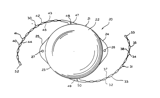
A first preferred embodiment of posterior chamber
intraocular lens apparatus 20 is shown in Fig. 1 as including
optic lens 21 and its posterior side 22, as well as haptics 30
and 31 which are used to secure posterior chamber intraocular
lens apparatus 20 within the eye. Implantation of an intraocular
lens apparatus requires resilient deformation of the free end of
the haptic substantially radially inwardly towards the lens.
Optic lens 21 is also shown having optional elliptically shaped
positioning means 24 and 25 emanating from the peripheral edge 23
of the light-focusing optic lens 21. Lens 21 should be made of a
suitable transparent biocompatible material for optical
correction such as polymethylmethacrylate ~PMNA), silicon,
hydrogyl or other soft polymers, acrylate or ophthalmic glass,
while haptics 30 and 31 may be constructed by biocompatible
material such as polypropylene or polymethylmethacrylate.
Haptics 30 and 31 are normally rather fine and have an
approximately circular cross-section with a diameter of
approximately 0.005 inches. The haptics may also be formed of
either single or multiple filaments. The positioning means 24
and 25 further include apertures 26 and 27 respectively, which
when used in cooperation with a surgical instrument serve to
efficiently allow for the manipulating and positioning of the
posterior chamber intraocular lens apparatus 20 once inserted
within the eye. The positioning means could also comprise
apertures (not shown) formed in the outer periphery of the lens
21, as well. Haptics 30 and 31 attach to lens 21 at shoulders 47
.. . . . ,, , . . , ~ . ... . .. .. .
~ 0 ~ 8
and 49 respectively, attached to the peripheral edge 23 of optic
lens 21. Haptics 30 and 31 can have optional apertures 48 and 50
respectively, which are formed in shoulders 47 and 49 to further
facilitate manipulation of intraocular lens apparatus 20 with a
surgical instrument. Haptic 30 is shown having alternating
regions of increased, transverse cross-sectional area 40 through
43 and adjacent regions of narrowed cross-sectional area such as
44 and ~5, along the length of haptic 30. Haptic 31 is also
shown as having alternating regions of increased and reduced
transverse cross-sectional area, such as regions of increased
cross-sectional areas 32 through 35, and regions of narrowed
cross-sectional area 37 and 38. These alternating narrow and
wide portions of haptic 30 and 31 serve to allow for greater
flexibility, and the maximum length of contact with the interior
surface of the eye, which is needed for the aforementioned
resilient deformation of the free end of the haptic radially
inwardly towards the lens while retaining the requisite rigidity
necessary to facilitate proper securement within the eye. Free
ends 52 and 53 of haptics 30 and 31, respectively, are also shown
in Fig. 1.
A preferred embodiment of the invention 60 employing
angled haptics 64 and 65 is shown in Fig. 2. These haptics 64
and 65, each have one end affixed to peripheral edge 63 of optic
lens 61, and extend upwardly at an angle of approximately 5-20
degrees to the plane of the posterior side 77 of lens 61, and
toward anterior side 62 of lens 61 as shown in Fig. 2. Haptics
64 and 65, as shown in Fig. 2, are provided with the above-
described alterna~ing regions of increased cross-sectional area
such as 67, 68, 73 and 74, and regions of narrowed or decreased
cross-sectional area such as 69, 70, 75 and 76. ~his
configuration coupled with the angular positioning of haptics 64
and 65 enable posterior chamber intraocular lens apparatus 60 to
be secured within the capsular bag, and in turn, the posterior
chamber of the eye 149, as shown in Fig. 6, while allowing each
14
203~28
of said haptics to be extremely resiliently compressible.
Fig. 3 shows another preferred embodiment of the
posterior chamber intraocular lens apparatus 90 as having haptics
95 and 96 being aligned on a plane that is coplanar with the
posterior peripheral edge 94 and posterior side 93 of optic lens
91. Anterior side 92 and posterior side 93 of optic lens 91 is
also shown in Fig. 3. Haptics 95 and 96 of embodiment 90 of the
intraocular apparatus likewise has alternating regions of wide,
transverse cross-sectional area, such as 97 and 99 immediately
adjacent to narrowed regions such as 98 and 100, respectively.
In Fig. 4, another preferred embodiment of posterior
chamber intraocular lens apparatus 110 is shown as having a
single oval haptic 117 positioned around peripheral edge 127 of
optic lens 111. Haptic 117 is attached to optic lens 111 by
shoulders 129 and 130 serving as attachment means, which are
affixed to peripheral edge 127 of optic lens 111. This single
haptic configuration allows intraocular lens apparatus 110 to be
inserted within the eye 145, as shown in Figs. 5 and 6, as a
single unit without the need to separately manipulate the
haptics. Intraocular lens apparatus 110 is relatively easy to
insert, and may further reduce the risk of scratching, and
potentially damaging any part of the eye, during such insertion
or the subsequent positioning of the lens within the eye.
Single haptic 117 is also configured with a series of
wider regions, such as wider regions 118 through 120, and
narrower regions, such as 123 through 125, which are positioned
adjacent to said wider regions. Accordingly, this alternating
sequence of wider regions 118 through 120, and narrower regions
123 through 125, enable maximum flexibility of posterior chamber
intraocular lens apparatus 110 prior to and during insertion into
the eye, and further allows for maximum spreading of end regions
132 and 133 of haptic 117 along the inside surface of the eye
once intraocular lens apparatus 110 is operably positioned
therewithin. Varying numbers of narrower and wider regions
20~828
should also be considered as being contemplated by the present
invention.
Also shown in Fig. 4 are elliptically shaped portions
113 and 114 comprising positioning means and their respective
apertures 115 and 116, which allow for maneuvering intraocular
lens apparatus 110 with surgical instruments to properly align
it, once it is actually inserted within the capsular bag of the
posterior chamber 149 of the eye 145, as shown in Figs. S and 6.
In the preferred embodiment of intraocular lens apparatus 110,
narrowed regions such as 123 and 125 of haptic 117 are
symmetrically arranged along its length. Moreover, in a
preferred embodiment, both wider regions such as 118, 119 and 120
and narrowed regions such as 123 and 125 are each of a
substantially constant diameter and substantially circular cross-
lS section, so as to result in substantially uniform stresses being
distributed throughout haptic 117, and in turn distributed in a
substantially uniform fashion to the interior of the eye once
implanted.
In Figs. 5 and 6, a human eye 145 is shown prior to,
and after posterior chamber intraocular lens apparatus 140 has
been inserted into the eye 145. In particular, there is shown in
Fig. 5, a human eye 145 wherein the natural lens has been
extracted because of a cataract condition. Typically, a small
incision is made in the front wall of the scleral-corneal
iunction or interior capsule through the cornea 151. A process
such as phacoemulsification is then used to liquify the natural
nucleus of the lens prior to removal by suction, or the nucleus
can be expressed through a slightly larger incision. The length
of the insertion incision should be minimized so as to speed the
healing process. Left behind are posterior capsule 160, iris 161
and ciliary sulcus 162. The pupil lS3 is ordinarily dilated to
facilitate centered positioning of the lens 140 in the posterior
chamber 149, behind it.
16
2~3~28
In order to insert posterior chamber intraocular lens
apparatus 140 into eye 145, a small arc-shaped incision (not
~shown) needs to be made upon the outer periphery of cornea 151.
After the incision is made, posterior chamber intraocular lens
S apparatus 140 is maneuvered and inserted through pupilary cavity
lS3 until it has entered into posterior chamber 149 of eye 145.
Once inserted into posterior chamber 149, posterior chamber
intraocular lens apparatus 140 is maneuvered, as shown in Fig. 6,
until its convex surface 146 is centered behind pupilary cavity
153, through the use of surgical instruments (not shown).
~osterior side 146a is also a substantially convex surface so as
to comprise a bi-convex lens together with anterior convex side
146. As shown in Fig. 6, once implanted in the eye, haptics 142
and 143 resiliently expand to contact ciliary sulcus 162 or the
capsular bag, so as to assume the shape of the intraocular
confines in order to maintain lens apparatus 140 within the
interior of the eye 145. Once this has occurred, posterior
chamber intraocular lens apparatus 140 will have been
appropriately seated and the surgical instruments withdrawn from
the interior of the eye 145.
Fig. 7 shows intraocular lens apparatus 160 in the
compressed position, as in the interior of the eye, as well as in
its expanded position, prior to insertion, in phantom. Haptics
161 and 162 are attached to lens 169 at shoulders 165 and 166.
The maximum, pre-insertion longitudinal length dimension is shown
as tl. As further shown in Fig. 7, the maximum transverse width
dimension of lens apparatus 160 is kept to a minimum to provide
the narrowest possible transverse profile for insertion purposes.
However, upon implantation, the longitudinal length dimension t2
3Q is shorter than original length dimension Tl, due to the
compression of haptics 161a and 162a along the longitudinal axis
to arrive at post-insertion haptic positions 161 and 162. At the
same time, upon insertion of lens apparatus 160, the maximum
transverse width dimension increases from Wl, to W2, due to the
?Q v~ Q ~28
resilient spreading of haptics 161a and 162a in the transverse
direction. The alternating regions of wider and narrower haptic
~ross-section enable the maximum amount of such spreading of the
haptics in the transverse direction so as to maximize the region
of contact Wc of haptics 161 and 162 with the interior of the
eye, while still providing a transverse profile substantially
equal to the width of the lens portion. This combines to enable
ease of implantation through the smallest possible incision, and
greater stability once implanted, through maximized region of
contact Wc along the interior of the eye.
Another embodiment of intraocular lens apparatus 170 is
shown in Fig. 8. In this embodiment, both ends of haptics 171
and 172 remain attached to lens 179 at shoulders 175 and 176.
The pre-insertion configuration is shown in phantom. Once
implanted in the interior of the eye, longitudinal length
dimension of apparatus 170 decreases from Tl to T2, while
transverse width dimension increases from Wl to W2. The region
of contact Wc of the haptics with the interior of the eye is
likewise maximized with the present embodiment of the invention.
In Fig. 9, one embodiment of haptic 180 is shown having
wide portions 181 through 184, and narrow portions 185 through
187. Each narrow portion, such as 185 through 187, has a
constant and progressively larger transverse cross-sectional area
than the preceding narrow portion preceding it, as one moves
25 toward the free end A of haptic 180. Accordingly, haptic 180
will be less flexible near its free end A, than at its point of
attachment to the lens, so as to spread less against the interior
of the eye near the free end A than ~4Rr the attached end of the
haptic.
In Fig. 10, another embodiment of haptic 190 is shown
having regions of wider cross-sectional area 191 through 195 and
regions of narrowed cross-sectional area 196 through 199 which
are adjacently positioned thereto. In this particular
embodiment, each individual region of narrowed constant cross-
18
.. . . . ..
203~28
sectional area, such as narrowed regions 19~ through 199, have a
smaller transverse cross-sectional area than each respective
~receding region of narrowed cross-sectional area, as one moves
toward the free end A of haptic 190. This configuration allows
for greater rigidity and therefore lens flexibility closer the
attached end of the hapt ~ and greater deflection and flexibility
near the free end A of haptic 190.
Another embodiment of haptic 215 is shown in Fig. 11
wherein regions of enlarged transverse cross-sectional area 202
through 207 each have constant transverse cross-sectional areas
that are smaller than the next preceding regions of enlarged
cross-sectional area. The transverse cross-sectional areas of
enlarged regions 202 through 207, each decrease as one moves to
free end 214 of haptic 215 and away from attachment 201 on lens
200. In this embodiment, regions of narrowed transverse cross-
sectional area 208 through 213 have substantially uniform
transverse cross-sectional areas throughout the entire length of
haptic 215, however, in other embodiments, these narrowed regions
208 through 213 can also decrease or increase in cross-sectional
20 area as one moves toward free end 214 of haptic 215. This
particular configuration allows for greater rigidity closest to
securement point 201 of haptic 215 while allowing greater
flexi~ility near tip 214 of haptic 215.
In Fig. 12, another embodiment of haptic 240 is shown
having regions of enlarged transverse cross-sectional area 224
through 229 interposed between regions of narrow portions 218
through 223. In this embodiment, while each narrowed transverse
cross-sectional area 218 through 223 has a substantially uniform
transverse cross-sectional area, regions of enlarged~ cross
30 section 224 through 229 each have increasingly larger transverse
cross-sectional areas than the next preceding enlarged region as
one moves towards free end A of haptic 290. This particular
configuration allows greater flexibility near securement point
231 of optic lens 210, while providing for greater rigidity and
~034~28
stability at the free end of haptic 240.
The foregoing description and drawings merely explain
- and illustrate the invention and the invention is not limited
thereto except insofar as the appended claims are so limited as
S those skilled in the art who have the disclosure before them will
be able to make modifications and variations therein without
departing from the scope of the invention.