Note: Descriptions are shown in the official language in which they were submitted.
~~3~~i~~:~.
SURGICAL INSTRUMENT GUIDANCE SYSTEM
The present invention relates to surgical
instruments. More particularly the present invention
relates to medical, operating room power drills which
are typically used in conjuction with X-ray machines
~or drilling precisely-located holes in bone for
receiving and anchoring prosthetic implants and
especially to a radiolucent, eg. X-ray transparent,
drill assembly and targeting drill bit for allowing the
holes to be drilled more precisely.
Certain surgical procedures require that a
surgeon drill and/or place wires, pins or screws (or
some other component) through bones or implants which
cannot be completely seen without the use of 3C-ray
equipment. For example, when an intramedullary nail is
inserted in the medullary canal of a bone, it may be
necessary to lock the end of the nail in place by
inserting retaining pins, also called transfixion
screws or transverse locking screws, through
interlocking holes at the distal end of the nail. It
is important that in the interlocking of intramedullary
nails that the orthopaedic surgeon know the precise
position of the interlocking holes in the nail when
drilling through a bone to avoid unnecessary damage to
the bone or the nail.
~~9~~~~:~.
- 2 -
There are several methods currently being used
by orthopaedic surgeons when drilling through a bone to
anchor an intramedullary nail. The methods have been
called the "free-hand" method and the distal aiming
device method.
In the free-hand method, the surgeon uses a sharp
awl, or drill bit, to locate the starting point under
X-ray imaging. The surgeon then rotates the drill
point parallel to the line of x-ray and forces the
sharp pointed instrument through the bone and through
the intramedullary nail. This method, although quick
and relatively accurate for surgeons who frequently use
it, can increase the amount of radiation the surgeon
receives because his or her hand must remain in the
path of the X-rays fox a period of time.
In the distal aiming device method, a distal
aiming device, such as an X-ray transparent target, is
used to assist in locating the correct path of
transfixion screws for anchoring the intramedullary
nail so that the drill bit goes through the hole in the
nail and opens a path in the bone for the locking
screw. There are distal aiming devices in which a
drill template is adjusted and ultimately fixed in the
desired position by means of an X-ray image amplifier.
- 3 -
For example, Swiss Patent No. CH-A5635998 discloses an
aiming device which has an aiming head with a hole for
the insertion of a directional socket. the aiming head
mounting is positioned in a holder that is connected to
the x-ray machine and is suspended form the X-ray
machine. This arrangement makes it difficult to
position the apparatus and to fix it in position, with
resulting unsatisfactory target precision. In
addition, the stationary arrangement limits operation.
There is an aiming device that can be used
independently of an X-ray machine, described in German
Industrial Design Patent 018417438. A device with a
receiving head rests in a holder and is permeable to
X-rays, and which accepts a drill bit or a drill wire.
Even this improved device, however, has major
disadvantages. In particular, the aiming process takes
place during the drilling of the bone, which causes
considerable darkening of the working field and low
image resolution.
US Patent No. 4803976 describes another aiming
device which may be held between a radiation source and
radiation receiver, the position of which can be
represented visibly by means of an image converter,
allowing continuous adjustment of its orientation. The
device has a socket for the drill bit and a direction
- 4 -
finder which must be maintained in a defined position
relative to one another during the drilling process.
This device still has the advantage that it does not
permit direct observation of the drill bit as it drills
through the bone and nail because of the obstruction in
the radiation field resulting from the power drill
which is in the same line of sight as the drill bit.
The present invention seeks to overcome the
drawbacks of the prior art by providing a device which
allows the surgeon to observe the drill bit on a
monitor of an %-ray image converter as the bit drills
through the bone and intrameduallary nail opening.
In an embodiment of the invention there is
included a radiolucent offset drill assembly with a
drive mechanism for transmitting rotational energy from
a drill to a drill bit where the drill is out of line
of sight of the axis of the drill bit so that only the
drill bit can be seen on a standard %-ray image
monitor. The drill bit is formed with a metal bit
portion mounted in a holder formed of an %-ray
transparent material so that the bit can be targeted
through the use of an %-ray image monitor.
In accordance with the present invention there is
provided a guide for aligning a rotatable elongate
CA 02035801 2000-03-22
-5-
member during a surgical procedure comprising:
(a) means for holding the elongate member
(b) drive means for rotating the elongate member
(c) coupling means for operatively connecting a drive
source to the drive means and
wherein at least the holding means and the drive means
are formed from an X-ray transparent or translucent
material and the coupling means is adapted to be coupled
to a remote drive source and the guide is adapted to move
along a path of alignment.
In accordance with a second aspect of the invention
there is provided a guidance system for aligning a
rotatable elongated member during a surgical procedure
comprising:
(a) means for holding and for guiding the elongated
member
(b) drive means for rotating the elongated member
(c) means for moving the guiding means along an
alignment path
(d) means for generating an X-ray field
(e) imaging means for viewing X-ray opaque objects
within a generated X-ray field
(f) the means for holding and the drive means
comprising X-ray transparent or X-ray translucent material
(g) and drive source means coupled to said drive means
wherein said drive source means is remotely located such
that an aligned path indicated on the imaging means is not
obscured by X-ray opaque objects.
In accordance with a third aspect of the invention
there is provided An elongated member for drilling a hole
or inserting a pin adapted to engage a drive means,
comprising:
CA 02035801 2000-03-22
-6-
(a) an elongated shaft,
(b) means for engaging the drive means connected at
one end of the elongated shaft included a tapered holder,
and comprised of an X-ray transparent or translucent
material.
The guide is a medical, X-ray transparent adapter for
aligning a drill bit, pin or other elongated member during
a surgical procedure and comprises:
(a) drive means;
(b) means for holding the elongated member and means
for engaging a drive source which is connected to the
drive means and located and oriented relative to each
other so that the drive source is out of a line of sight
in the direction of the elongated member, the drive means
being adapted to rotate the elongated member when driven
by the drive source; and
(c) at least the drive means, and means for holding
the elongated member being formed of X-ray transparent
material so that an operator can align the drill bit with
an internal predetermined direction in an X-ray
transparent medium.
The guidance system is a medical X-ray system for
aligning a drill bit, pin or other elongated member with
an internal line of sight during a surgical procedure and
comprises:
(a) X-ray generating means for creating an X-ray
field;
(b) imaging means for observing an internal
configuration in a patient's body with the X-ray field;
(c) an elongated member for drilling a hole or
inserting a pin within the internal configuration;
(d) a power source for rotating the elongated
- 7 -
member; and
(e) an adapter substantially formed of an X-ray
transparent material for operatively connecting the
power source to the elongated member, the adapter being
shaped and dimensioned so that the power source is out
of the line of sight of the elongated meatier when a
surgeon is aligning the elongated member in a
predetermined direction toward the internal
configuration in the X-ray field.
In a preferred embodiment, the assembly includes
a gear box housing which contains two shafts connected
to each other through appropriate gears or the like.
One of the shafts is connected to a drive source such
as a power drill. The other shaft exte~ls through the
gear box housing and is adapted to receive a drill bit.
As the one shaft is rotated by the power drill, the
other shaft through the gears causes the drill bit to
rotate.
The entire offset drill assembly is formed of
X-ray transparent or radiolucent materials with the
exception of the drill bit and an adapter for
connecting the one shaft to the power source. The
gears allow the drill bit to be offset from the power
source, preferably by 90°, so the plane of the drill
bit is perpendicular to the plane of the power drill or
CA 02035801 2000-03-22
_8_
other power source. The drill bit is formed so that only
the bit and not the bit holder shows up on the X-ray image
monitor. In this way the power drill and other non-X-ray
transparent parts are removed from the radiation field and
the precise orientation of the drill bit can be observed
on the monitor of the X-ray image converter as the drill
bit drills through the bone to the target interlocking
hole in, for example, an intra medullary nail.
Embodiments of the invention will be further described
with reference to the accompanying drawings, in which:
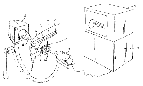
Figure 1 is a perspective view of the radiolucent
offset drill assembly embodying the subject invention and
a power drill and associated X-ray equipment;
Figure 2 is a perspective view of the drill assembly
of fig. 1, rotated 90° to show the drill bit relative to
the drill
Figure 3 is a sectional view looking along section
lines 3-3 of Fig. 2;
Figure 4 is an exploded view of the drill assembly of
Figs. 2 and 3; and
Figure 5 is an enlarged plan view of a drill bit shown
in Fig. 4.
An exemplary embodiment of a radiolucent offset
.. __... . . .. _. ..... . _._ _ _. . . . . .. .. .. . ...... .. . . .. .. ..
. ... ... . .. . . .. . . . .. . . . .
- 9 -
drill assembly which is the subject of the invention is
shown in Fig. 1 and designated generally by reference
numeral 10. The drill assembly 10 is dziven by a power
drill 1, in this case a standard medicall operating room
power drill, for rotating a drill bit 12. The drill
assembly 10 is used in conjunction with an X-ray source
2, an X-ray receiver 3, and an image converter 4 having
a monitor 5 connected to the X-ray recenver 3.
In a radiation field 8 generated by the X-ray
source 2 and receiver 3, a patient's thigh T is shown
positioned with an intramedullary nail ~ inserted in
the medulla of a femur F. The orientation of the drill
bit 12 with regard to an interlocking hole 6 in the
intramedullary nail 7 can be observed on the monitor 5
of the image converter 4 because the drill assembly 10
and all of its component parts described below are
transparent to X-rays and the drill 1 is not in the
path of the X-rays.
Referring to Figs. 2, 3 and 4 the drill assembly
includes a gear box housing 14 with two halves, an
upper half 16 and a lower half i8. The gear box
housing 14 is made of a material that is transparent to
X-rays and is autoclavable so that the drill assembly
can be sterilised and reused and at the same time be
strong enough to perform as described. Such a material
CA 02035801 2000-03-22
-10-
is a polyetherimide known as ULTEM~, which is a
thermoplastic material sold by General Electric Company.
The two halves 16, 18 are held together with bolts 20
which are inserted through apertures 22 in the upper half
16. The bolts 20 engage internally threaded apertures 24
in the lower half 18 of the gear box housing 14. The bolts
20 are made of a nylon or other X-ray transparent
material.
The drill assembly 10 is driven by a standard
operating rrom power drill 1 shown in Figs. 1 and 2.
Preferably the drill 1 is offset 90° from the drill bit 12
as best shown in Figs. 1 and 2. Other angles may be used.
Instead of a gear assembly as described, the offset
drill assembly can be formed of a flexible shaft (not
shown), universal connection (not shown) or other assembly
for allowing a standard drill to drive a drill bit 12 and
at the same time be out of the line of site along the axis
of the drill bit 12. The important characteristic of the
invention is to remove the drill from the path of the X-
rays so that the orientation and location of the drill bit
can be observed at all times by the operating physician.
CA 02035801 2000-03-22
-11-
The power drill 1 engages an adapter rod 26 which
extends from one face 28 of the gear box housing 14. The
adapter rod 26 is generally irregular in cross-section,
preferably triangular, to provide a firm engagement with
the power drill 1. The adapter rod 26 is preferably formed
of metal because of the strength requirements for
engagement with the power drill 1.
The adapter rod 26 is connected to a drive shaft 32
which is made of the ULTEM~ resin material mentioned
above. A bevel gear 34 formed of DELRIN~ or other X-ray
transparent material, is mounted on the drive shaft 32. A
rear face 36 of the gear 34 has a pair of raised surfaces
38 which abut a pair of cooperating notched surfaces 40 of
the drive shaft 32 which prevent rotational slippage of
the gear 34 on the drive shaft 32. A drive shaft aperture
42 is formed in the face 28 of the housing 14. A bearing
race 44 is located immediately behind the aperture 42 to
receive a bearing 46 which fits onto the drive shaft 32
and includes plastic ball bearing formed of a suitable
resin such as DELRIN~, a thermoplastic material of DuPont,
Inc. The inner end 48 of the drive shaft 32 extends beyond
the gear 34 and a bushing 50 with a flange 52 is inserted
over the inner end 48. A larger bushing 56 is inserted
over the bushing 50 and is positioned between mating half
CA 02035801 2000-03-22
-12-
saddle supports 54 which are formed in the halves 16, 18
of the gear box housing 14. The small and large bushings
50 and 56 can be formed of teflon or other suitable
radiolucent material.
The gear 34 meshes with a bevel gear 58 formed of
DELRIN~ which is mounted on a drill bit shaft 60 formed of
ULTEM~ plastic. The drill shaft gear 58 has a rear face 62
with a pair of raised surfaces 65 engage corresponding
receive surfaces 66 of the drill shaft 60 to lock the gear
58 to the drill shaft 60.
The drill shaft 60 is oriented perpendicular to the
drive shaft 32 and extends through the front face 68 of
the gear box housing 14. The front face 68 has a front
aperture 72 through which a drill bit receiving end 74 of
the drill drive shaft 60 extends. The rear face 70 has a
rear aperture 76 for exposing a hollow rear end 78 of the
drill drive shaft member 60. A bearing race 80 is formed
immediately behind the front aperture 72 to receive a
bearing 82 which fits onto the drill shaft 60. A rear
drill shaft bearing race 84 is formed immediately adjacent
to the rear aperture 76 to receive a bearing 86 which fits
onto the drill shaft member 60. The bearings 82,86 include
plastic ball bearings (not shown) formed of DELRIN~.
i,
~~~'~0~~. ',
- 13 --
The drill bit 12 is a standard operating room
drill bit formed of stainless steel. One end of the
drill bit 12 has flutes 87 formed on it while the other
end is a drill shank 88 that is fixed to a bit holder
92 with a tapered outer surface 96, as shown in Figure
5, formed of an X-ray transparent material such as a
suitable high strength plastic as an acetal copolymer.
The bit holder 92 can be moulded or mounted through an
adhesive to the shank 88 which is irregular in shape to
resist rotation of the holder 92 relative to the shank
88. By forming the holder 88 of an X-ray transparent
material a more precise targeting can be achieved
because only the drill bit will show up on the X-ray
image monitor. A series of numbered depth indicators
generally designated by reference numeral 89 are formed
on the outer surface on the drill bit 12 to indicate to
the surgeon the depth of the drilled hole.
A cooperating, tapered receiving opening 97 is
formed in the drill bit receiving end 74 of the drill
drive shaft 60 (Fig. 4). After the bit holder 92 is
inserted in the drill bit receiving end 74 of the drill
drive shaft 60, a locking knob 98 having a central
aperture 100 and internal threads snot shown) is placed
over the drill bit 12 so that the drill shank 88
extends through the central aperture 100 of the locking
2~~ ~ ~~.
- 14 -
knob 98. The locking knob 98 is threaded onto the
external threads 102 on the drill bit receiving end 74
of the drill drive shaft 60. The locking knob 98 holds
the bit holder 92 against the receiving opening 97 to
eliminate any slippage between the two surfaces. The
bit holder 92 can also be modified to accept other
types of instruments or tools requiring a rotational
torque.as, for example, locking screws, bolts or
guidewires for spiral pedicle screws, sacral screw
fixation, pelvic screw-fixation, or any other surgery
requiring precisely-placed guidewires or screws.
The upper half 16 of the gear box housing 14 has
a recessed threaded aperture 106 for receiving a handle
108 having a threaded end 110. A similar recessed
threaded aperture 112 can also be provided in the lower
half 18 of the gear box housing 14 to provide the
operator with maximum versatility in the use of the
offset drill assembly 10.
In use, the surgeon holds the power drill 1 in
one hand and the handle 108 in the other hand, with the
offset drill assembly 10 positioned in the radiation
field 8 and the drill 1 outside the radiation field
(Fig. 1). The drill bit 12 is aligned, for example,
with the interlocking hole 6 in the intrameduallary
nail 7 by the surgeon viewing the image monitor 5.
- 15 -
Once properly aligned, the bone is drilled. Continuous
observation on the monitor 5 during the drilling
process permits the surgeon to accurately and precisely
align the targeted interlocking hole 6 with the
corresponding opening in the intramedullary nail 7.
The radiolucent offset drill assembly 10 permits
high image resolution on the monitor 5. The high image
resolution results not onlyfr.om the fact that the
drill assembly 10 is made primarily of radiolucent
materials and the nearest metal around the drill bit 12
is approximately 1.5" away (the adapter rod 26), but
also due to the fact that the drill bit drive shaft
member 60 is substantially hollow with the exception of
the sonically shaped receiving area 97 of the drill bit
drive shaft member 60. Thus, minimal darkening occurs
within the immediate area surrounding the targeted
interlocking hole 6. Additionally, the offset drill
assembly 10 reduces the amount of radiation that the
surgeon would receive since the surgeon's hands are
outside the radiation field 8.
It is intended that the foregoing description of
the exemplary embodiment of the invention is not to be
limiting by improvements and modifications can be made
without departure from the spirit and scope of the
invention, which is defined in the appended claims.