Note: Descriptions are shown in the official language in which they were submitted.
2~3~87
NON-BIOD~6R~UUUaLB TWO P~AS~ CORN~AL IHPLANT
AND MæTUOD FOR P~EPARrNG SAM~
~he present invention relates to a non-biodegradable
corneal implant and a method of preparing such an implant.
~ore particularly, this invention relates to a non-biodegrad-
able corneal Lmplant comprising (13 a polymerized soluble
transparent collagenous core having acylated amine groups or
esterified carboxyl gro~ps, and (2) a polymerized opaque peri-
phery surrounding said core, ~aid periphery comprising
polymerized fibrous collagen, said collagen being in the form
of fibrils under suitable physiological conditions. The
corneal Lmplant is useful in human corneal transplantation, for
example, replacement of damaged cornea, corneal inlays or
corneal onlays.
Various materials have been described for use as
transplanta~le corneal implants.
In U.S. Patent Nos. 4,505,855 and 4,581,030 Bruns et
al. disclose native, non-fibrilized transparent collagen
~ ~ r ~ ; r,'
material that can be fixed, i.e., cross-linked to form a
prosthetic cornea replacement. The collagen material employed
by Bruns et al. i8 soluble or rendered soluble by treatment
with dilute acids, e.g., acetic acid; base, e.g., NaOH; and in
dilute aqueous salts, e.q., NaCl. Bruns et al.'s pelleted
collagen material is described as exhibiting strand like
structures upon electron microscopic examination (see U.S.
4,505,855 and U.S. 4,581,030, col. 6, Example 3). Bowever, the
materials and corneal prosthesis disclosed in the Bruns et al.
patents are not suitable for promoting cell ingrowth and
enhancing the adhesion of the prosthesis to surrounding
recipient tissue after transplantation.
In U.S. Patent No. 4,772,283, White discloses a corneal
Implant prosthesis having a transparent lenticula to which is
attached a carrier that is constructed from preserved biologi-
cal tiQsue, e.g., cornea, sclera, fascia. The transparent
lenticula can be made from a number of ~non-biological
materials~, such as polymethylmethacrylate (PMMA), polycar-
bonates, polyhydroxyethylmethacrylate (~EMA), polysulfones and
silicones. White's implant prosthesis is difficult to con-
struct, however, because in order to attach ~uch n non-biologi-
cal" materials to the carrier, it is necessary to carry out
ela~orate and time consuming mechanical procedures, such as
driving stakes into place where the lenticula and carrier are
to be joined and heat fusing the tisQue. Alternatively, the
carrier tis~ue may be retained in the peripheral groove of the
lenticula by crimping flanges which have been provided on the
latter.
In U.S. Serial No. 157, 638 and ~uropean Patent
Application Publication No. 330,389, there is disclosed a
chemically modified, crosslinkable, solubilized collagenous
substance obtained from autogenic intact human tissue, i.e.,
the donor and recipient are the ~ame individual. This colla-
genous substance is useful as a corneal implant among others.
3S It would be highly desirable, therefore, to discover a
corneal implant which would overcome the disadvantages of prior
art materials, prosthesis, implants, and the like, such as the
2 ~ 3 ~
aforementioned construction problem and the difficulty in
incorporating the implant into neighboring endogenous tissue of
the recipient following tran~plantation.
It is among the object~ of the present invention to
prepare a non-biodegradable corneal implant that is easily
incorporated into endogenous recipient ti6sue following
transplantation without adver~e Lmmunologic reaction.
These and other object~ of the present invention will
be apparent to those skilled in the art, in light of the
accompanying description, drawings and appended claims.
The pre6ent inventor has discovered that a non-
biodegradable two-phase corneal implant can be prepared using
different but compatible collagenous materials as the lens core
and as the peripheral opaque carrier or attachment portion.
The core comprises a polymerized soluble transparent collage-
nous core having acylated amine groups or esterified carboxyl
groups. The opaque periphery surrounding the core comprises
fibrous polymeri~ed collagen, ~aid collagen being in the form
of fibrils under suitable physiological conditions xo as to
attach to endogenous tissue following transplantation of the
implant into a subject recipient.
The present inventor has discovered that the just-
described non-biodeqradable corneal Lmplant can be prepared by
following the steps of either incubating neutralized acid
soluble collagen under conditions sufficient so that the
collagen forms fibrils and then recovering the collagen fibrils
that are formed, or preparing collagen fibrils from intact
dermis by treating dermis with acylating or esterifying agents
and recovering the fibrous fraction by centrifugation. The
fibrous fraction i~ then suspended in sterile water and added
to physiological solution to form fibrils which are subsequent-
ly recovered by centrifugation. The recovered ccllagen fibrils
are treated or contacted with a binding agent to bind the
fibrils followed by a polymerizing or cross-linking step. At
this point, a core of the polymerized (cross-linked) collagen
fibrils is excised (cut out) and replaced with a ~oluble
collagen material that is next subjected to polymerizing
S conditions to cross-link the core material and thereby yield a
non-biodegradable corneal implant suitable for human corneal
transplantation and other corrective ophthalmic procedures. A
preferable alternate procedure involves forming the fibrous
ring around a clear central core of soluble collagen and then
polymerizing or cro6clinking tbe two phase lens as one unit.
In the accompanying drawings:
Figure 1 represents three embodiments of the two phase
corneal implants of the present invention.
Figure 2 illustrates a non-limiting range of dimensions
of the two phase corneal Lmplant.
All literature references, patents and patent publica-
tions cited in this specification are hereby incorporated by
reference in their entirety.
The present invention provides a non-biodegradable
corneal implant comprising: (1) a polymerized transparent
core, (lenticula), which comprises a polymerized ~oluble
transparent collagenous core having acylated amine groups or
esterified carboxyl groups, and (2) a polymerized opague
periphery surrounding said core, the periphery comprising
fibrous collagen in the form of fibrils under suitable physio-
logical conditions.
In order to minimize the risk of graft rejection and
other adverse immunologic reactions, it `i6 preferred that the
ti~sue from which the implant core and periphery are con-
structed, be autogeneic, i.e., obtained from the individual
recipient. This is par~icularly the case with the material
used to construct the periphery of the implant.
In the case of the collagenous core, this component can
be prepared from autogeneic Type I collagen (i.e., obtained
2 ~ . 7
from the individual recipient) or from Type IV collagen (e.~.,
human umbilical tissue). The transplant core may alternately
be composed of known synthetic polymers such as PMMA, ~EMA,
sulfones, etc. In preferred emhodiments of this invention, the
collagenou~ core is derived from at least one member ~elected
from the group consisting of purified Type I collagen, purified
Type IV collagen, predominantly Type I collagenous preparation
obtained from human tissue, e.g., dermis. Preferably the core
(1) comprifie~ a Type I collagenous preparation or material
obtained from human tisfiue. A~ used herein, the purity of the
Type I collagen or Type IV collagen comprises from about 70 to
about 100 weight percent, preferably from about 95 to about 100
weight percent. In a further preferred embodiment, the -
predominantly Type I collaqenous material compri~es fibril
forming collagen, the preparation of which i8 de~cribed in
detail below and in the examples which follow. As used herein,
the term ~predominantly" refers to a high concentration of Type
I collagen in the collagenous preparation, e.g., from about 70
about to 95 weight percent, preferably from about 80 to about
95 weight percent.
Methods for preparing the soluble collagenous core
material having acylated amine or e6terified carboxyl groups
are described in the aforementioned U.S. Serial No. 157,638 and
European Patent Application Publication No. 330,389.
These methods entail the following procedures.
Attendant noncollagenous protein contaminates including lipid
constituents are desirably removed from telopeptide (non-
helical extension peptides) collaqen-containing intact
autogenic tissue, i.e., tissue that has been obtained from a
sole human donor, to form essentially purified telopeptide
collagen-containing tissue material, and extracting and
chemically modifying the purified telopeptide collagen to form
an autoimplantable, crosslinkable substance useful as the
periphery material in the present invention.
The contaminates may be removed by contacting the
tissue with a substantially neutral liquid which is capable of
7~ solubilizing contaminates without solubilizing the collagen, or
by utilizing specific enzymes to solubilize noncollagen tissue
components.
At this stage in the processing, a fibrous collagenous
matrix has been prepared which i8 capable of forming fibrils
under suitable physiological conditions and is therefore,
useful as the periphery of the corneal implant of this inven-
tion. This human fibril formung collagen (h~lm~n FF collagen)
is not soluble in organic acid~ as iB bovine FF collagen.
human FF collagen is dispersed by chemical treatment into a
form that will undergo rapid fibril orga~ization when mixed or
contacted with physiological fluid. As used herein, "suitable
physiological conditions" include neutral p~, e.g. 6.8, human
body temperature and the presence of buffer, e.g., phosphate
buffer.
When contacted in this way, i.e., phosphate buffer, p~
6.8 and approximately 37-C temperature, fibril formztion of the
human FF collagen from 6uspensions in water will begin to occur
in about 30 minutes.
In order to obtain the collagenous core material, the
contaminate-free telopeptide collagen i8 then extracted by
reaction of the tissue directly with a chemical modifying
agent, e.g., acylating agent or esterifying agent.
The acylating agent is amine reactive. The acylation
reaction is carried out in a solubilizing aqueous medium of
substantially neutral to basic pH sufficiently to solubilize at
least partially the telopeptide collagen in the aqueous medium,
with the at least partially solubilized collagen thereafter
being recovered and purified to form the autoimplantable
telopeptide-containing collagenous substance as product.
The esterifying agent is carboxylic acid reactive, and
in this instance, the esterifying reaction is carried out in a
solubilizing nonaqueous organic medium at acidic p~ ~ufficient-
ly to solubilize at least partially the telopeptide collagen
therein, with the at least partially solubilized collagen
thereafter being recovered and purified to form the autoim-
plantable telopeptide-containing collagenous core substance as
product. It should be understood that while both acylation and
233~7
esterification of the collagen can be carried out in accordance
with the present i~vention, the yield of clear, transparent
modified collagen from the ecterification of soluble, mam-
malian, e.g., bovine, collagen is generally less than that
obtained by acylation. Alternatively, the chemical modifica-
tion may be carried out using both the amine acylating and
carboxylic acid esterifying steps.
For the amine modifying reaction, the noncollagenous
protein contaminate-free, and lipid-free extracted, tissue
powder is resuspended in aqueous medium. The suspension may be
in any appropriate aqueous medium such as water, deionized
water, balanced salt solution, saline solution, etc., preferab-
ly 0.05 to 0.5 M buffer at pH 9.0, i.e., Tris ~uffer, bicar-
bonate, etc.
Although the amine modifying reaction will proceed at a
p~ of from about 7 to about 11, it is preferably e~fected at
mildly basic p~ to increase the reaction speed and reduce the
processing time. The reaction is desirably effected at about
pH 8.0-10.0, and especially at about pH 8.5-9Ø
The amine reactive modifying agent used as a solubiliz-
ing agent may be an acylating agent, such as a carboxylic acid
anhydride, e.g., ~uccinic anhydride, glutaric anhydride,
benzoic anhydride, 1,2,4,5-benzene tetracar~oxylic acid
dianhydride; carboxylic acid ester, e.g., monophenyl terephtha-
late, ethyl benzoate, alpha-naphthoic acid ethyl ester;
carboxylic acid halide, e.g., succinic acid chloride; ~ulfonic
acid, e.g., 1,3-ben 2 ene-disulfonic acid, aniline-2-~ulfonic
acid, 3-nitro-benzene-sulfonic acid, 2-formylbenzene-~ulfonic
acid, 4-amino-naphthalene-sulfonic acid; or ~ulfonic acid
halide, e.g~, 4,4'-biphenyl-disulfonyl chloride, benzene
sulfonyl chloride; and mixtures thereof. Preferred as the
acylating agent is glutaric anhydride, and combinations of
glutaric anhydride with methacrylic anhydride, ~thylene/maleic
anhydride; B-sulfonyl chloride.
In general, the acylating agent may be an aliphatic or
aromatic, mono-, di- or higher functional, carboxylic acid
anhydride, ester or halide, or sulfonic acid or halide, such as
8 20359~7
a lower alkanoic, lower alkane-dioic or higher functional lower
alkane carboxylic, or aryl mono-, di- or higher functional
carboxylic (e.g., benzoic or naphthoic), acid anhydride, ester
or halide, or lower alkyl, or aryl (e.g., phenyl or naphthyl),
mono-, di- or higher functional sulfonic acid or halide, to
provide the corresponding acyl (carbonyl or ~ulfonyl) moiety on
the amine group, e.g., lower alkanoyl, aroyl (e.g., phenoyl or
naphthoyl), alkyl sulfonyl, or ~ryl (e.g., phenyl or naphthyl)
sulfonyl, substituted amino (amido or sulfon~m;do).
The acylating agent may be added directly as a solid
material, e.g., powder, or dissolved in a suitable org~nic
solvent ~uch as acetone, N,N-dimethylformamide (DMF), ethanol,
or methyl pyrrolidone.
The total guantity of acylating agent added depend~ on
the extent of disruption, modifying and extrscting of the
telopeptide collagen desired. For instance, one addition at
150 mg agent per gram of wet tissue may not be ~ufficient to
disperse and solubilize totally the collagen content of the
ti~sue; as many as four such additions may be required.
The quantity required should generally ~atisfy the
weiqht ratio of acylating agent to wet ti~sue of broadly 0.005-
O.S:1, and preferably O.OS-0.1:1.
The reaction time for achieving complete fiolubilizing
of the collagenouQ ti~sue may range from about 30 minutes to 2
hours. The time depends on the quantity of solubilizing agent,
specific solubilizing agent used, rate of agitation or ~tirr- -
ing, temperature, pH, and degree to which the tissue wa~
initially pulverized or dispersed in the preliminary homogeniz-
ation treatment.
For the carboxylic acid modifying reaction, the
noncollagenous protein contaminate-free, and lipid-free
extracted, tissue powder is desirably dried, e.g., n vacuo or
by freeze drying, and combined with a carboxylic acid reactive
esterifying agent in a nonagueous organic medium at acid~ic pH,
preferably no more Shan about pH 3.2, such as about pH 0.1-3.2.
The quantity required should generally ~atisfy the
~,
2 ~ ~ Q~ I
weight ratio of esterifying agent to dry tissue of broadly 1-
30:1, preferably 1-20:1, and more preferably 5-20:1.
In particular, the medium i~ advantageously a large
excess of the esterifying agent in the form of ~n acidified
S liquid, such as an Acidified alcohol, especially nn aliphstic
alcohol, such as a water soluble lower alkanol, e.g., methanol
and ethanol. The esterification reaction which forms the ester
and water is favored by use of an excess of the alcohol to
assure efficient formation of the ester product, in the
presence of a catalytic amount of an acid such as 0.1 ~ ~Cl as
acidifying agent, thereby providing a system pH of about 0.1-
3.2.
The reaction is desirably ef f ected under anhydrous
conditions using dehydrated starting materials for optLmum
result6, although acceptable results are still obtainable with
starting materials which have not been dehydrated, such as wet
tissue powder.
In general, the e~terifying agent may be an aliphatic
or aromatic alcohol, such as a lower al~anol or an aryl alcohol
(e.g., a phenol or a naphthol), to provide the corresponding
aliphatic or aromatic, e.g., alkyl or aryl (e.g., phenyl or
naphthyl), ester.
Where the esterifying agent i8 a solid at room tempera-
ture, it may be dissolved in a suitable nonaqueous organic
solvent such as acetone, N,N-dimethylformamide (DMF), ethanol,
or methyl pyrrolidone, as the organic medium.
The esterification reaction is conducted at the 6ame
temperature and for the same reaction tine as the acylation
reaction, for the same reasons, but since the e~terifying agent
is advantageously used in large excess a~ nonaqueous organic
reaction medium, the esterifying agent amount will preferably
be several times larger than that of the dry starting tissue,
e.g. in a weight ratio thereto of about 2-20:1, although the
ratio may be 1-30:1, and preferably 1-20:1, in general,
especially where the esterifyir.g agent is a solid and an
organic solvent is used as the reaction medium.
2 ~ 7
In the Aqueous medium, the completely ~olubilized
material intended for use as the collaqenous core, is a
transparent, viscous telopeptide-containing collagen ~solution"
product.
The solubilized product constitutes chemically
modified, crosslinkable, telopeptide-containing, naturally
crosslinked, collagen, in which the individual helical 6trands
of the triple helix molecules remain in interconnected ~ide by
side helical disposition ~long the corresponding collagen
polypeptide backbone, with the terminal ~m; no group-containing
site of each given strand still linked to it~ adjacent non-
helical telopeptide end moiety, and with the tenminal car-
boxylic acid group-containing ~ite of the same strand ~till
linked to its adjacent non-helical telopeptide end moiety.
Thus, all three helical strands of one tropocollagen
molecule remain linked at their ends to their respective
telopeptide moieties, telopeptide moietie may remain cross-
linked to adjacent tropocollagen molecules, and ~djacent
helical strands may remain crosslinked to each other along
their central regions, and to telopeptide regions of adjacent
tropocollagen molecules, to retain the original polypeptide
backbone arrangement and to retain some order of the original
intermolecular configuration. However, these strands now
contain acylated (~uccinylated) amino groups which render the
collagen soluble at neutral to basic p~, while still preserving
the integrity of the intermolecular arrangement.
It will be understood that these individual chemically
modified tropocollagen molecules, consequent their solubiliza-
tion, are no longer in packed staggered arrangement in fibrils
of fiber bundles as in the starting tis~ue, but rather con-
stitute substantially intact separate units, which are com-
pletely dissolved in the reaction medium where complete
solubilization is carried out. If partial ~olubilization is
carried out, the suspension contains a mixture of intact
separate units and various degrees of fiber units sized in
dependence upon the extent of solubilization, which are
11 2 ~ 3 ~ 1 3 7
suspended or dispersed in the reaction medium as fine particle
material.
Where the tissue powder has already been solubilized in
the amine modifying reaction, the recovered and purified
acylated product may be dried, e.g., Ln v~cuo or by freeze
drying, and then combined with the acidified esterifying agent
and reacted to form the corresponding acylated and esterified
product. Alternatively, the tissue may fir~t be subjected to
the esterification step and the esterified solubilized product
is then subjected to the acylation step.
It will be readily apparent to those skilled in the art
that in order to provide an implant suitable for example, in
the replacement of damaged cornea, the collagenous core must be
optically clear and not infiltrated with fibroblasts. In most
applications, the collagenous core has a refraction index of
preferably from about 1.283 to about 1.545 when the transparent
collagenous material i6 modified with an agent that exhibits an
index of refraction in this range. This range can be modified
further as necessary, as tescribed next.
For specific ophthalmic applications, it i~ preferable
that the chemical modifying agent be employed that i5 capable
of modifying the collagenous core to provide a solubilized
collagen with a high index of refraction. This is most
~ffective for correcting sight. The solubilized collagen i8
recovered, purified and com~ined with aqueous liquid to form a
telopeptide collagen solution, of a selective index of refrac-
tion for correcting sight, as product.
The agent u~ed to achieve such selective index of
refraction (nD) i~ ~uitably an amine modifying acylating agent
which is capable of achieving complete solubilization of the
collagen to provide a product that is essentially completely
~oluble at physiological pH conditions, such as glutaric
anhydride, aniline-2-sulfonic acid (nD = 1.586)l 3-nitroben-
zene-sulfonic acid ~nD c 1.550), 2-formylbenzene-sulfonic acid
~aD = 1. 544), 1,3-benzene-disulfonic acid, 1,2,4,5-benzene-
tetracarboxylic acid dianhydride, and B-styrene ~ulfonyl
t chloride or like reagents whose particular constituent reactive
12 2v~
group or functional group exhibits a high index of refraction
or Lmparts a resultant high index of refraction to the so
modified collaqenouc core substance.
Preferred as a chemical modifying agent to provide a
solubilized collagen suitable a8 the coll~genous core of high
refractive index in the corneal implant of thi~ invention i6 B-
styrene sulfonyl chloride. Styrene exhibitfi a refractive index
of about 1.545.
Thus, 6uch an agent will generally possess an index of
refraction of at least about nD 1.500, such as an index of
refraction of from about ND 1.500 to about nD 1.600.
~ he refractive index of the collagenous core may be
modified as well, e.g., reduced, by modifying the collagenous
material wit~ an acylating agent such as trifluoroacetic
anhydride. Trifluoroacetic acid exhibits a refractive index of
about 1.283. The normal cornea provides a refractive index of
about 1.370. Common Hbiologically acceptable" materials
exhibit the following nD's: PMMA: about 1.495; and hydrogel
intercorneal lenses: about 1.375. It may be possible to
combine modified collagen with hydrogel to form a composite
lens with varying indices of refraction, all higher than the
cornea.
Thus, a biologically acceptable material can be
incorporated into the collagenous core (1) of the implant
provided by thi~ invention. The "biologically acceptable"
material is one that will not impair or diminish the com-
patibility of acceptance of the implant following transplanta-
tion into a suitable recipient. The material is selected from
the group consisting of polyhydroxyethylmethacrylate, poly-
methylmethac~ylate, hydrogel, or a combination of any of thefcregoing.
Because the collagenous ccre can be prepared to provide
a range of refractive indice~, the implant of the present
invention will reduce the need for human donor cornea in
keratoplasty. By providing such a range of refractive indices,
this implant is also useful for correcting refractive errors
~j3~
13
without the need for corrective devices, e.g., eye glasses and
contact lens.
In one embodiment of this invention, the collagenous
core (1) of the implant can be derived ~y cbemically modifying
pulverized human dermal tissue, using conventional technigue~.
Such ch~ical modification can be carried out by contacting the
tissue containing the collagen with an acylating or an esteri-
fying agent as described hereinabove.
The periphery t2) of the implant provided by thi~
invention comprises predominantly Type I collagenous material
that can be obtained from mammalian tissue, e.g., human tissue
or bovine tissue. For purposes of enhancing the non-biodegrad-
ability or immunologically-acceptable features of the implant,
it is important that the tissue from the Type I collagenous
material be autogeneic, i.e., the donor and recipient be the
same individual.
In another aspect of this invention, the core (1) and
the periphery (2) ~n the Lmplant are polymerized by means of
exposure to polymerizing ayents, e.g., ultraviolet irradiation,
chemical agents, or a combination of polymerizing agents.
The non-biodegradable corneal implant of this invention
can be prepared from the aforementioned collagenous core and
periphery materials in a method provided by this invention.
This method has two ~eparate polymerizing steps ("two step
polymerizing method") and comprises the steps of (a~ incubating
neutralized fibrous collagen under conditions sufficient to
form fibrils; (b) recovering ~aid formed fibrils; (c)
contacting said fibrils with a binding agent to bind ~aid
fibrils; (d) polymerizing said bound fibrils; (e) replacing a
core of said bound fibrils with a soluble collagenous material
having acylated P~;ne or esterified carboxyl groups; and (f)
polymerizing said soluble collagenou~ material.
Alternately, th~ lens may be formed by polymerizing the
soluble collaqenous core and the fibrous periphery as one unit,
3S i.e., using a single polymerizing step. In this case, the
fibrous collagen is placed in the mold to provide a homogeneous
l~yer, the central zone is removed and replaced with soluble
14
collagenous material and the entire unit is polymerized. Thu~,
the present invention provides an alternative method of forming
the non-biodegradable corneal implant de6cribed. A prepoly-
merized implant i6 formed which comprises a collagenous core
having acylated amine or esterified carboxyl groupq, and a
fibrous collagen periphery, the periphery having been treated
under conditions ~ufficient to form fibrils. The pre-poly-
merized implant is then polymerized by expo6ing, e.g., to W
irradiation and/or chemical agents, or both, to form a non-
biodegradable corneal implant.
The ~teps of thi6 method are described in more detail
hereinbelow and in the examples which follow.
The fibrou6 collagen compri~es Type I collagen derived
from mammalian ti~sue, e.g., bovine or human ti~sue. In a
preferred aspect of the method, the mammalian tissue comprises
autogeneic human ti~sue.
FF collagen is neutralized (i.e., removing charged
particles) by mlxing with a buffer solution, e.g., phosphate
buffer, at p~ 6.0 to about 8.0, preferably from about p~ 6.8 to
about 7.4.
The neutralized FF collagen i~ next incubated under
conditions sufficient to cause the formation of fibrils. Such
conditions comprise a temperature in the range of from about
25-C to about 40C, preferably about 37-C, for a period of time
of from about 10 to about 45, preferably about 30 minute6 or
so .
After fibril formation, the fibrils are recovered by
conventional recovery techniques, e.g., centrifugation at
12,000 RPM. A fibril peliet is recovered. The recovered
pelleted fibril material is then treated by contacting with a
binding agent to bind the fibrils. By way of example, ~uitable
binding agents include the above described soluble collagen or
collagenous material which has been obtained by treating
fibrous Type I collagen with a chemical modifying agent (e.g.,
an acylating or esterifying agent, or a combination of an
acylating and esterifying agent).
5 v ., i
Next, the fibril containing collagen mixture i8 cast
upon a sui~able support surface, such as a glass or ceramic
mold, in order for polymerization or cross-linking to be
carried out, e.g., by exposure to ultraviolet irradiation (or
g~mma irradiation). In order for polymerization or cro~slink-
ing to be effectively, the FF collagen must be dispersed prior
to polymerizing or cro6slinking. ~his can be done for example,
by placing the FF collagen in deionized water. Short wave-
length W light is preferred, such as 7.5 cm from W 60urce of
8 watts, for about 25 minutes in nitrogen. Polymerization or
cross-linking can be also carried out by exposing the molded
collagen to chemical polymerizing or cro~s-linking agent6 auch
as isocyanate, aldehyde, e.g., glutaraldehyde, epoxy compounds
such as polyglycerol polyglycidyl ether, diglycerol polygly-
cidyl ether, and such other compounds, or a combination
thereof.
From the polymerized fibrous ~button~ that has been
made, a central core zone of suitable area, e.g., 3-4 mm
diameter may be excised or cut out by conventional methods,
e.g., using a cork bore. The core zone is filled with the
soluble collagenous core material, described above, which has
acylated amine groups or esterified carboxyl groups or both.
Following replacement of the core zone with such
soluble collagenou~ core material, the entire "button" i8
exposed to polymerizing or cross-linking steps, e.g., W
irradiation for at least about 20 minutes. Then the "button"
is removed from the support surface and allowed to air dry
overnight.
After air drying, the button is exposed once again to
polymerization or crocs-linking conditions. The polymerization
or cross-linking of th~ collagenous core and the fibrous
collagen containing periphery is important to provide a corneal
implant that is non-biodegradable so as to resist breakdown
following transplantation.
Alternately, the entire unit may be polymerized as
"one". It has also been found desirable to melt the central
core or soluble collagenous core at about 35-380C before sir
~ J
16
drying and subsequent crosslinking. In addition, melting the
soluble, binding collagen, in the fibrous component before
crosslinkinq has been found to improve the strength and
elasticity of the implant.
S Accordingly, the present invention provides a method of
forming the aforesaid non-biodesradable corneal implant
comprising the steps of forming a pre-polymerized Lmplant
comprising a collagenou6 core having acylated amine or es-
terified carboxyl groups, and a fibrou~ collagen periphery, the
periphery having been treated under conditions sufficient to
form bound fibrils; and polymerizing said collagenous core and
caid bound fibrils to form the non-biodegradable corneal
implant.
The corneal implant thus prepared can be stored for
long periods of time, up to 6 months or even longer in saline
solution (0.2M).
Those skilled in the art will appreciate that the
corneal implant of the present invention can be used in various
ophthalmic applications. One particularly useful application
is as an autoimplant for correcting sight in which the implant
is formed into a ma6s of selective shape and ize corresponding
to an effective implant device. ~he implant may be so formed
from a mold having a concave surface of selective ~ize and
shape corresponding to an effective shape and size for the
outer surface of the damaged cornea to be replaced or reshaped.
Other uses of the corneal ~mplant described herein include
corneal onlay, corneal inlay and intraorbital lens (e.g.,
behind the iris). Onlay lends will be placed on the surface of
the existing cornea, after removal of epithelial cells which
~hould migrate over the lens. Inlay lens will be placed in the
corneal lamellae and should not retard diffusion of oxygen and
nutrients. Glutaric lens, hydrated, contain approximately B5-
90~ water.
Again, it is preferable to melt the soluble, central
core collagen prior to drying and subsequent polymerization.
It has also been recently discovered that a strong oxidizer,
such as sodium persulfate, available from Aldrich Chemical
'~ 3 ~ ,3 3 r~
17
Company, Milwaukee, Wiscon~in, will dramatically accelerate the
polymerization reaction using W -irradiation, even in the
presence of oxygen. This is particularly evident if the
soluble collagen contains some methacrylic moieties. Other
~uitable oxidizing Agents include ~odium bisulfite, ferrous
chloride tetrahydrate, sodium thiosulfate, all available from
Aldrich Chemical Company, Milwaukee, Wisconsin.
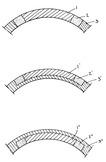
Referring to Figure 1, three embodiments of the two
phase corneal implant of the present invention. In lA, there
is shown a full thickness corneal graft comprising (1) a clear
central zone; (2) a collagen fiber periphery; and (3) the
surrounding corneal tissue. Figure lB and lC are illustrations
of partial thickness corneal implants. lB is an on-lay lens
and lC is an intrastromal lens.
It is preferable that the lenses be grafted or im-
planted 80 that the fibrous periphery is attached, i.e.
sutured, to the scleral tissue, i.e. at the limbus.
Figure 2 are front view illustrations of the two phase
corneal implant of the pre~ent invention within various
dimensions that are not to be construed as lLmiting. The total
diameter of the implant can be from a~out 6 mm to about 20 mm.
The thickness of the fibrous periphery can range from about 2
mm to about 6 mm. The diameter of the clear optical core can
be from about 2 mm to about 8 mm. The implant thickness can
vary from about 0.025 mm to about 2.0 mm. The curvature of the
concave can also vary as follows: top diameter from about 6 mm
to about 20 mm; bottom diameter from about 3 mm to about 12 mm.
EXAMPLES
The iollowing examples are ~et forth by way of il-
lustration and not limitation of the present invention.
I~XAHPI~ 1~ PR~3PARATION OF TWO PllASE AND
CORNEAL IMPI~NT FROM BOVI~3F TISSUE
A. Fibrous Type I collagen was prepared from bovine
material (calf hide) using the following procedure:
.,
2~9~7
18
1. Clean, dehaired split hides, which are
commercially available from the Andre Manufacturing Co.,
Newark, New Jersey, and 6tore frozen in sealed plastic bags
until ready for use.
2. Thaw approximately 200g of cow hide at room
temperature.
3. Cut the hide into 6mall pieces, approximately
1 cm3 using a scalpel and tweezerR. Weigh the wet tis6ue and
record its weight.
4. Place the cow hide in 15 liters of 0.5M
acetic acid and stir at room temperature usinq a lightning
mixer for at least one hour. The cow hide will swell.
5. Add 2% or 3.9g of pepsin from porcine gtomach
mucosa (manufactured by Sigma Chemicals, St. Louis, Missouri)
to the cow hide solution, after disRolving it in approximately
10 mls of 0.5M acetic acid. Continue stirring with mixer over-
night.
6. Add 1% or 1.96g of the above pepsin to the
cow hide solution dissolved in approximately 10 mls of 0.5M
acetic acid. Continue stirring with mixer overnight.
7. Refrigerate the dissolved cow hide ~olution
until it is uniformly at a temperature of about 4C. This may
take until overnight.
8. Remove the solution from the cooler and begin
~tirring with the lightning mixer. Increase the p~ of the
~olution to 9.0 using lON NaO~ to denature the pepsin. Ice
cubes may be added during the process to keep the solution
cold. (Collagen will precipitate at p~ 9.0 if the temperature
is higher than 6-C.) Quickly return the ~olution to 4C. The
aolution must remain in the cooler for at least 4 hours.
9. Remove the solution from the cooler and
centrifuge at 4C for 30 minutes at 9 rpm. Save the super-
natant, which contains the collagen and discard the precipitate
which containC the pepsin.
10. Add enough NaCl to the solution to bring up
the concentration to 2.5M. This will precipitate the desired
~ 3 ~ `. 7
19
collagen. Stir with the lightning mixer for at least two
hours.
11. Centrifuge for 30 minutes at 9 rpm to recover
precipitate. The resultant collagen precipitate is collected
and then reconstituted in 15 liters of 0.5M acetic ~cid (at
least two hours).
12. The collagen solution is precipitated again
by adding enough NaCl to the solution to bring up the con-
centration to 0.8M. It is stirred well for at least two hours
then centrifuged for 30 minute~ at 9 rpm.
13. The precipitate is collected and then
reconstituted in 15 liters of 0.5M acetic acid (at least two
hours).
14. Enough NaCl is added to the collagen solution
to brinq up the concentration to 0.8M. The precipitate is
formed by mixing for at least two hours. Centrifugation at 9
rpm for 30 minutes will recover the precipitate.
15. For the final tLme the precipitate i8
collected and then reconstituted in O.lM acetic acid to provide
a high purity of approximately 0.3 percent wt/wt collagen Type
I ~olution having a p~ of about 3.
16. The collsgen solution is filtered first
through a prefilter which has a pore size of a~out 0.3 um and
then through a final filter which has a pore fiize of 0.22 um
for sterilization. This material can now be uced in the
modification procedure or for preparation of fi~ril~.
Bovine Type I collagen is soluble in organic acid and
undergoes fibril formation under physiological conditions,
e.g., neutral pH, body temperature, in buffer. The periphery
of the two part Lmplant was composed of this bovine fibril-
forming (FF) collagen. Bovine physiological soluble (PS-
collagen) was prepared by chemically acylating acid solutions
of bovine FF-collagen~ The bovine FF-collagen was treated with
a monofunctional acylating agent, such as glutaric anhydride.
~he glutaric treated collagen was clear and viscous in buffer
at pH 6.8. The central zone was composed of this PS-collagen.
,3 ,~ ,~ ,",; .' r)
1~ J t~
The corneal implant was made as follows: The bovine FF-
collagen formed above was mixed with phosphate buffer at p~ 6.8
to neutralize the preparation. This was incubated ~t 37-C for
30 minutes to allow fibril formation to occur. The fibrils
- S were recovered by centrifugation at 12,000 RPM. The fibril
pellet was recovered and mixed with approxLmately 0.1 ml of PS-
collagen to bind the material. The mixture was then cast onto
a concave microscope slide of about 14 mm in diameter and dis-
persed in deionized water. Thi~ fibrous collagen material was
cross-linked by exposure to short wavelength UV light t7.5 cm
from source of 8 watts) for 25 minutes, in nitroqen. The
polymerized fibrous button was trimmed and a central zone of 9
mm was cut at with a cork bore. This zone was then filled with
PS-collagen and the button was again exposed to W irradiation
for 20 minutes. The button was removed and allowed to air dry
overnight. The dried button was then exposed to W irradiation
once again. ~he button was then stored in 0.2M saline solu-
tion. The button had a clear central zone and a white, fibrous
periphery, as shown in Figure 1.
EXAMPLE 2: PREPARATION OF TWO P~ASE
CORNEAL INPL~N~ FROM ~UMAN TISSUE
Human skin biopsy tissue (or human skin tissue obtained
from recon~tructive surgery, or the like), of the donor
patient, is immediately frozen. Specimens of the frozen tissue
are di~sected to remove the attendant epidermal Pnd sub-
cutaneous layers, and the remaining dermal layer is sectioned.
The following steps and procedures are carried out.
STBP 1 - Dissection
1) Remove skin sample from the ~reezer and equi-
librate at room temperature for no more than four hours.
2) Place the ~kin on a clean, dry cutting board.
Only one specimen can be dissected on a cutting bofird at any
~iven time. The dermal layer of the skin is dissected using a
scalpel with a fresh hlade, tweezers, and scissors that have
been soaked in alcohol. The epidermal layer of the skin and
21
any hair on the skin i8 removed by scraping the outer portion
of the skin with the scalpel while holding the skin in place
with the tweezers. The inner portion of the f2kinl which may
contain a lot of fat c~ n be cut off with 8ci8sors ~nd then
scraped with the ~calpel until the white dermal layer remains.
3) The wet dermis i8 then cut into very 6mall pieces
with s~issors and placed into a pre-weighed labelled ~terile
50ml centrifuge tube. The weight of the centrifuge tube is
subtracted from the weight of the centrifuge tube and dermis.
The wei~ht of the dermis is then recorded.
,STEP 2 - Purification Dnd Sterili~ation
4) Add to the dermis 10 ml8 of Rterile filtered 0.lN
HCl. Cap the centrifuge tube nnd place it on a shaker for two
hours.
5) Centrifuge the tube for 15 minutes at 8 revolu-
tions per minute. Using a f~terile transfer pipet, remove the
supernatant which is the excess HCl being careful not to remove
any dermis.
6) To the dermis add 10 mls of reagent alcohol
(formula 3A - denatured). Cap the centrifuge tube and place on
a shaker for two hours.
7) The tube i8 then centrifuged for 15 minutes at
8,000 rpms. Following centrifugation, the tube is ~prayed down
with alcohol and placed in the sterile hood.
NOTE: From this point on the specimens are to be processed
using aseptic techniques in the sterile laminar flow
hood.
8) Using a sterile transfer pipet the alcohol/super-
natant is removed and discarded. Fifteen mls of ~terile water
i6 added to the dermi~. The tube iB capped and shaken well.
The ~entrifuge tube i~ removed from the hood and centr,ifuged
for 15 minutes at 8 rpm. The tube is sprayed with alcohol and
returned to the sterile hood.
9) The dermis is washed two more times with sterile
water, repeating step 9. On the last wash after the water is
removed add 10 mls of sterile filtered 0.5M Tris ~uffer. The
2 t~ 3 ~
22
dermis pieces will be equilibrated for one hour. Check the pH
of the solution, it ~hould be between 8.5 and 9Ø If it i8
not then adjust with ~terile filtered lN acl or lN NaOH.
S ST~P 3 - Modification of the Collagen
10) The ~ample is placed in the small mixer attachment
of the Waring blender. An aliquot of 1 part glutaric anhydride
per 10 parts of dermi~ wet weight in 1 ml of DMF is added to
the solution. ~he top i8 placed on the blender and blending
begins. After approximstely seven minutes into the blending, a
second aliquot of glutaric anhydride and DMF exactly ~Lmilar to
the fir~t one will be added. Each sample will receive 5 - 1
minute blendings over a period of 15 minutes. The blender
~hould not be allowed to build up heat because heat will break
down the collagen.
11) After blending, the pH should be between 6.8 and
7.4. If it is not then ~terile filtered lN ~Cl can be added to
make this adjustment.
12) The sample is removed from the hood and centri-
fuged at 8 rpm for 30 minutes. The tube is sprayed down with
70% alcohol and then returned to the hood.
13) The supernatant i8 removed with a sterile transfer
pipet and then di~carded. The layer on top of the residue
IdisPerse fraction~ i~ scr~ped off using a ~terile ~patula and
placed into a labelled 15 ml ~terile centrifuge tube.
14~ The disperse fraction is washed three times with
sterile water ~imilar to the procedure in step 9.
15) The disperse fraction is redispersed in sterile
phosphate buffered saline. Fibers will form.
The aboYe procedures yielded fibrous collagen.
Preparation of ~oluble collagen from human dermis was
carried out as follows:
Processed dermis is incubated in pH 9.0 buffer for at
least 2 hours. This is then homogenized in a small container
using a commercial Waring blender. To the homogenate is added
th~ amine reactive modifying agent, preferable glutaric
anhydride, at about 1 part to 10 parts of wet tissue. This
~ v `~ v ~
23
mixture is blended about 5 times for one minute each. Care i8
taken to avoid reaching an exce6sive temperature during the
blending. A second aliquot of ~mine reactive modifying agent
is ad~d and the mixture is blended 5 more times. The p~ of
the mixture is then decreased to 6.7-7.4 u6ing lN hydrochloric
acid and the mixture centrifuged at about 8,000 rpm for 20
minutes. The soluble collagenous component appears as a
gelatinous ma~s covering the dispersed tissue. This i~ removed
and placed in alcohol. The material immediately precipitates
10 and i6 washed 3 times in alcohol. At this point it i~ prefer-
able to dry the alcohol precipitate in a laminar flow hood.
The dried precipitate is then dissolved in buffer at p~ 6.8-7.4
to a viscous consistency of approximately 40,000 centipoise.
This solution can then used as the clear core for the human two
15 phase corneal implant.
An alternate method has been found to obtain additional
soluble collagen. This alternate method involves taking the
supernatant from the above mentioned centrifugation. This
solution i~ adjusted to pH 4.3 and stirred for a~out 1 hour.
20 The solution is then adjusted to pH 6.8-7.4. After about 10
minute~, clear, gelatinous material forms in the solution.
This material appears to be solubilized dermal collagenous
material which can also be used for the clear core of the human
two phase corneal implant. This material may be placed in
25 alcohol (e.g., ethanol) for storage.
In the case of all human material, skin spe~imen can be
processed as described in the aforementioned U.S. Application
Serial No. 157,638 and European Patent Application Publication
No. 330,389. Using these procedures, human physiologically
30 ~oluble (PS) collagen was prepared as was human fibril-forming
(FF) collagen dispersions. Unlike the bovine FF-collagen, the
human FF-collagenous fraction is not soluble in organic acids.
Instead, human FF-collagen is dispersed, by chemical treatment,
into a form that will undergo rapid fibril organization when
35 mixed with physiological fluid. An aliquot of h~man FF-
collagen was mixed with a drop of human PS collagen. This
mixture was placed onto a concave microscope slide as descri~ed
~ .
24
~bove. It i8 important that the human FF-collagen be dispersed
in deionized water. The material was irradiated with W , after
which a central zone of about 4-8 mm was removed and then
filled with human PS-collagen., The button was again exposed
S to W irradiation. The button was removed and mix dried and
again exposed to W irradiation. The final product button had
a slightly opaque periphery and a clear central core. When
immersed in 0.2m saline solution, the periphery immediately
became white and opaque and the center remained clear.
Two phase corneal implants were prepared using fibrous
collagenous material prepared from human dermis and soluble
collagen obtained from bovine collagen. Since the cornea is
avascular, such an implant is efficacious as a full thickness
corneal graft. It ~hould be noted that previous attempts with
collagen corneal implants (Type IV in particular) have failed
due to degeneration at the periphery of the implants. The two
phase implant, even with a bovine collagen core, may be more
stable because the periphery is fibrous and composed of
autologous collagenous material.
In Example 3 which follows, the n YiVo efficacy of the
corneal Lmplant of the present invention was evaluated in one
animal model.
EXAMPL~ 3: IN VIRO ~FFICACY OF TE~ CORNEAL
IHPLANT IN RABBIT HODEL
Two corneal grafts were prepared from rabbit skin.
Rabbit skin wa~ dissected to remove fur, epidermal layer, and
underlying subcutaneous tis~ue. Sections of resulting dermal
layer were minced, weighed and placed in 70% alcohol for 16-18
hours. The tissue was removed from the alcohol and treated
with 0.lN hydrochloric acid for 2 hours. The tissue was
recovered following centrifugation at 8,000 rpm for 30 minutes,
washed with sterile water and placed in 10 volumes of 0.5M Tris
buffer at pH 8.7. After equilibration for 2 hours, the tissue
3~ was placed in the small blender container (12-37 ml) and
blended using a Commercial Waring blender. The tissue was
homogenized 2 times for about 1 minute each time. At this
point the tiscue pieces were still intact. The tissue was ~
dispersed by adding 1 part of glutaric anhydride per 10 parts
of wet dermis. ~he anhydride was dissolved in O.2-1.0 ml of
dimethyl forma~ide (DI~F). The mixture was blended 4 times for
about 1 minute e~ch time. After 1 minute of continuous
blending, the temperature of the blender container began to
increase and the homogenization was stopped until the container
cooled. A second aliquot of glutaric anhydride was added and
the mixture blended 4 times more, as described above.
The pP of the mixture was adjusted to 6.8-7.4 by
addition of l~ON sodium hydroxide or hydrochloric acid. The
mixture was then centrifuged at 8,000 rpm for 30 minutes to
separate the dispersed fractions. The supernatant was removed
and stored at 4C. The gelatinous fraction was removed and
placed in 70% alcohol an~ the dispersed fraction removed and
washed three times with sterile water.
Rabbit corneal grafts were formed as follows:
Graft 1. The disperGed fraction, in sterile water, was mixed
in sterile phosphate buffered saline, p~ 7.3, to form fibers.
The fibers were recovered by centrifugation and mixed with a
small aliquot sf glutaric modified, soluble bovine collagen.
The mixture was then placed in a glass mold 15 mm in diameter
and 2 mm in depth. A thin layer was applied to a diameter of
approximately 12 mm and the material exposed to ultraviolet
radiation (25~ nm) for 20 minutes in a nitrogen atmosphere.
The polymerize~ fibrous disc was removed and a center core of
about 5 mm removed. The center was filled with an aliquot of
glutaric mod,fied, soluble, bovine collagen and again ~ubjected
to W irradiat~on. The final graft appeared as a 12 mm concave
disc with a 5 mm clear central core. This was placed in a
clear pouch, ealed and gamma sterilized.
Graft 2. The fibrous portion was prepared as described above.
The solu~le f~action was again composed of glutaric modified,
soluble, bovine collagen. Soluble fractions of glutaric
modified, rab~it collagen were available and made into two-
26
phase corneal grafts, but were not implanted. The graft wasmade in one step. Fibers were isolated as di~cussed above,
mixed with a fimall aliquot of soluble collagen ~nd placed in a
concave glass mold, 22 mm in diameter and 5 mm in depth. A
center core of about 5 mm was removed and filled with glutaric
modified, soluble, bovine collagen. The mixture with clear
center and fibrous periphery was placed in an oven at 38C for
3 minutes to ~melt~ the soluble fraction. The mold was then
placed in a ~terile, l~minar-flow hood to dry the graft. After
about 4 hours, the mold was removed from the hood and subjected
to W -irradiation, in nitrogen, for 20 minutes. ~he implant
was trimmed to about 12 mm diameter and contained a 5 mm clear
center core. It was placed in ~0% alcohol, washed with ~terile
water, and placed in a sterile pouch for storage.
The first graft was used as a full thic~ne~6 corneal
graft in the rabbit model. The rsbbit was anesthetized and a 7
mm trephine was used to remove a core of the natural cornea.
The graft was trephined to 7 mm and sutured into the rabbit
eye. The graft appeared clear but was difficult to suture. At
one point, the sutures tore through the graft. The eye was
treated with antibiotics and closed using sutures. The rabbit
remained alive for about 24 hours at which time the grafted eye
was enucleated and prepared for histopat~ological examination.
Results indicated the beginning of hea~ing at the margin
between the graft and the host tissue. A few inflammatory
cells were present near the wound margin and there wa~ a hint
of reepithelialization. The graft seemed to be well tolerated
and on its way towards proper healing.
The second graft was also used as a full thickness
corneal graft in the rabbit model. The graft was sutured into
the rabbit eye as discussed above. In this case, the graft
~utured extremely well. The fibrous periphery exhibited
unexpected strength and flexibility. After implantation, the
central core was clear. Slit-lamp examination of the graft
indicated excellent approximation to the host ti6sue. There
was no adverse tissue reaction and the graft remained intact.
There was, however, cloudir.g of the central core and upon
27 ~ ~ 3 ~ ~9 ~, ~
enucleation, at approximately 4 weeks, minute erosion at the
apex of the graft. ~he enucleated eye was again prepared for
hiRtopathological evaluation.
?~