Note: Descriptions are shown in the official language in which they were submitted.
, 2038170
Chemical luminescence-detectinq apparatus
The present invention relates to a chemical luminescence-
detecting apparatus in which the intensity of chemical
luminescence generated in a photometric cell is detected by
means of an optical detector.
Since the drawings are referred to in the description
below, these drawings are first briefly introduced as follows:
Fig. 1 is a cross-sectional view showing one example of a
chemical luminescence-detecting apparatus according to the
present invention;
Fig. 2 is a block diagram showing a basic electrical
circuit for the apparatus shown in Fig. 1;
Figs. 3 to 5 show one example of an enzyme immuno assay
system provided with a chemical luminescence-detecting
apparatus according to the invention; in particular:
Fig. 3 is a perspective view showing the inside of the
entire system;
Fig. 4 is a partially cut away partial side elevational
view; and
Fig. 5 is a plan view showing the main components;
Fig. 6 is a sectional view showing a chemical
luminescence-detecting apparatus according to another
preferred embodiment of the present invention;
Figs. 7 and 8 show still another preferred embodiment of
the present invention, in which:
Fig. 7 is a sectional view showing a chemical
luminescence-detecting apparatus; and
Fig. 8 is a perspective view showing the main components
of the apparatus shown in Fig. 7;
Fig. 9 is a graph showing the relationships among the
-
2038170
_~ 2
output from a high sensitivity photomultiplier tube, the
output from a low sensitivity photomultiplier tube and the
concentration of luminous substance;
Fig. 10 is a graph showing the results measured by means
S of the chemical luminescence-detecting apparatus according to
the present invention; and
Fig. 11 is a block diagram showing a conventional
chemical luminescence-detecting apparatus.
As shown in Fig. 11, in order to detect the intensity of
chemical luminescence of a solution in a prior art procedure
referred to as a batch-type measuring method, a cylindrical
photometric cell 91 made of glass or plastics is mounted in a
spherical cell holder 92. A photomultiplier tube 94, acting
as an optical detector and provided with a high-voltage power
source 95, is positioned so as to face the photometric cell 91
to detect the quantity of chemical luminescence generated
within the photometric cell 91. Light from the cell passes
through a shutter 93 and is detected by the photomultiplier
tube 94 and the output is amplified by an amplifier 96.
The conventional apparatus shown in Fig. 11 has only one
photomultiplier tube 94, so that the range of intensity over
which said quantity of chemical luminescence can be measured
is limited in those cases where the measurement is carried out
under essentially the same conditions, and thus it has been
necessary to carry out the measurements by varying the
measuring conditions if a greater range of sensitivity is
required, for example by varying the supply voltage from the
high-voltage power source 95 of the photomultiplier tube 94,
the value of feedback resistance in the amplifier 96 and the
like, which requires the provision of additional circuitry and
equipment.
Enzyme immuno assays have recently been carried out on
the basis of the chemical luminescence method but it has been
quite difficult to use the above described conventional
chemical luminescence-detecting apparatus as it is. Because a
large number of items must be randomly measured in the enzyme
immuno measurement procedure, the quantity of light to be
3 2038170
measured varies over a very wide range and thus the
measurement can not be carried out by means of a single
optical detector having a limited range of sensitivity.
In addition, this problem occurs not only in the so-
called batch-type measuring method but also in the so-called
flow through-type measuring method using a spiral flow
through-type of photometric cell.
It is accordingly an object of the present invention to
provide a chemical luminescence-detecting apparatus capable of
accurately detecting a wide range of quantities of
luminescence.
In order to achieve the above described object, the
invention provides a chemical luminescence-detecting
apparatus, comprising: a photometric cell for containing
luminescent materials to be measured; a plurality of optical
detectors of different measurement sensitivities associated
with said cell, said detectors generating different output
signals for the same intensity of luminescence and two said
detectors being provided on opposite sides of said cell; means
for converting some or all of said output signals so that the
output signals from each of the detectors, following said
conversion, have the same values for the same intensity of
luminescence; and display means for receiving an output signal
from one or other of said detectors and displaying a value
corresponding to said measured luminescence.
In the usual case, there are two detectors, one of low
sensitivity and one of high sénsitivity. The output signal
from the low sensitivity detector is converted into a value
which is the same as the output from the high sensitivity
detector for the same concentrations of sample to be detected.
A chemical luminescence-detecting apparatus having the
above described characteristics according to the present
invention is provided with a plurality of optical detectors of
different light measurement sensitivities in the vicinity of
the photometric cell, so that the measuring range of
sensitivities of the apparatus is the sum total of the
individual ranges of sensitivities of each of the optical
detectors. Thus a wider range of measurements is possible
203817û
than when using a single optical detector. In order to make
the use of more than one optical detector feasible, the ratio
of outputs from the optical detectors based on the intensity
of luminescence is first determined using solutions of known
concentration in order to find the factors by which the
outputs of the individual detectors should be multiplied in
order to make the outputs of all of the detectors the same for
the same concentrations of material to be detected. For
example, the output values of low sensitivity optical
detectors may be multiplied by a factor determined from
standard solutions so that a converted value corresponding to
the outputs from high sensitivity optical detectors may be
obtained. This permits the detectors of low sensitivity to be
used when the sensitivity ranges of the high sensitivity
detectors have been exceeded (i.e. when the detectors have
been saturated) while permitting the converted output signals
from the low sensitivity detectors to be processed in the same
way as the output signals from the high sensitivity detectors,
so that a highly accurate measurement can be achieved ranging
widely from a low sensitivity zone to a high sensitivity zone.
The way in which this can be achieved will be apparent
from the description below of preferred embodiments of the
present invention in which reference is made to the
accompanying drawings.
Figs. 1 to 5 show one preferred embodiment of the present
invention. However, before the chemical luminescence-
detecting apparatus according to the present invention is
described in detail, an enzyme immuno assay system provided
with a chemical luminescence-detecting apparatus incorporated
therein is first described with reference to Figs. 3 to 5 in
order to show the context in which the present invention is
intended to operate.
Referring first to Fig. 3, reference numerals 1 and 2
designate horizontal partition plates dividing the internal
volume of an apparatus case 3 into three spaces P1, P2, P3
arranged one above the other in the vertical direction. As
shown in Fig. 4, a tube-conveying elevator 4 is provided to
203817û
_ 5
convey tubes from the central space Pl to the upper space P2.
Reference numeral 5 designates an immobilized antibody tube
cooling device comprising a suction-exhaust portion 6
communicating with a cooler (not shown) provided in the lower
space P3 and a cooling case 7 communicating with said suction-
- exhaust portion 6. The cooling case 7 can be freely withdrawn
at the front of the apparatus case 3.
Referring again to Fig. 3, reference numeral 8 designates
immobilized antibody tubes provided with an antibody
immobilized on an inner surface at the bottom of each tube and
an aluminum foil cap sealing the upper open end of each tube.
Reference numeral 9 designates dilution tubes. The tubes 8
and 9 are each supported by tube-supporting cases 10 provided
with open lower sides removably positioned on an upper surface
portion of the cooling case 7 so as to form cooling ducts
around the tubes.
Reference numeral 11 designates a tube-conveying
mechanism movable horizontally in two-directions and provided
with a freely elevatable vessel chuck 12 (as shown in Fig. 4)
for conveying an immobilized antibody tube 8 (or a dilution
tube 9 as the case may be) to the lower end of the elevator 4.
Referring to Fig. 5, reference numeral 13 designates a
constant temperature shaker provided with a plurality of tube
holding portions 14 and first to third rotors 16 to 18
arranged in front of the shaker 13. The rotors are provided
with a plurality of receiving holes 15 for receiving the
immobilized antibody tubes 8. A washer 19 and a diluent
dispenser 20 are arranged around the first rotor 16; a washer
21 and a substrate solution dispenser 22 are arranged around
the second rotor 17; and a washer 23 and an enzyme conjugated
antibody reagent dispenser 24 are arranged around the third
rotor 18. The rotors 16, 17 and 18 may be freely rotated in
the direction of the arrows so that receiving holes 15 may be
moved beneath the various dispensers as required.
Reference numeral 25 designates a tube-conveying
mechanism provided with a tube chuck 26 (refer to Fig. 4)
freely movable in three orthogonal directions for conveying an
203~170
_ 6
immobilized antibody tube 8, delivered to the space Pz by the
elevator 4, from the constant temperature shaker 13 to a
sample station 27 via the first to third rotors 16 to 18.
Reference numeral 28 designates a sample tube-housing
region in which sample tube-housing cases 30, each housing a
plurality of sample tubes 29 containing a sample (for example
serum) therein, are provided in line in the right and left
direction (refer to Figs. 4 and 5). Referring to Fig. 5,
reference numeral 31 designates cover members closing the
upper openings of the sample tube-housing cases 30 and
reference numeral 32 indicates a cover member-closing
mechanism provided at one end of the row of said cover
members 31.
Reference numeral 33 designates a stock region of pipette
tips 34 and reference numeral 35 designates a sample dispenser
mechanism horizontally movable in two orthogonal directions.
The sample dispenser mechanism 35 is provided with a freely
elevatable probe 37 (refer to Fig. 4) communicating with a
suction-exhaust pipe 36 at an upper end thereof and a pipette
tip 34 at the lower end thereof. A descending movement of the
probe 37 within the stock region 33 loads a pipette tip 34 and
then the probe is moved to the sample to be tested in the
housing region 28 so that a sample may be sucked into the
pipette tip 34 from a sample tube 29 by vacuum and then
discharged into an immobilized antibody tube 8 held in the
first rotor 16 by an exhausting action.
Reference numeral 38 (refer to Fig. 5) designates a stock
region of reagent bottles 39 containing enzyme conjugated
antibody reagents.
Referring to Fig. 3 and Fig. 5, reference numeral 40
designates a photometric detection station provided with a
photometric cell 41 in the form of a glass tube, reference
numeral 42 designates a reactant dispenser for transferring a
reactant from an immobilized antibody tube 8 conveyed to said
sample station 27 into the photometric cell 41, reference
numeral 43 designating a reagent dispenser for pouring a
luminescent reagent (for example a luminol solution) into the
`203~17~
_ 7
photometric cell 41, and reference numeral 44 designating a
washer for the photometric cell 41. Reference numeral 45
designates a recovery station for the immobilized antibody
tube 8 and reference numeral 46 designates a discard station
5 for receiving the used pipette tip 34.
With the enzyme immuno assay system having the above
described construction, an enzyme immuno assay may be carried
as follows by, for example, the so-called two-step sandwich
method.
An immobilized antibody tube 8 with an antibody
corresponding to the item to be measured immobilized thereon
is received in a tube-receiving hole 15 of the first rotor 16
by means of the tube-conveying mechanism 11 on the lower space
P1, the elevator 4 and said tube-conveying mechanism 25 in the
15 upper space P2. The aluminum foil sealing the upper opening of
the immobilized antibody tube 8 is broken during the process
of moving the tube.
The probe 37 is provided with a pipette tip 34 at a lower
end thereof so that the sample may be sucked in the pipette
20 tip 34 from a sample tube 29 and then poured into the
immobilized antibody tube 8 positioned in the first rotor 16
followed by ejecting the pipette tip 34 into the discard
station 46.
Upon rotating the first rotor 16 through a predetermined
25 angle, a diluent is poured into the immobilized antibody tube
8, into which the sample has previously been poured, and then
the immobilized antibody tube 8 is set in the constant
temperature shaker 13 and is shaken for a predetermined time
at a constant temperature of about body heat to carry out a
30 first immuno reaction.
The immobilized antibody tube 8 is moved to the second
rotor 17 to be washed and then subjected to a so-called B/F
separation followed by pouring in an appointed dose of enzyme
conjugated antibody reagent corresponding to the agent to be
35 measured and is set in the constant temperature shaker 13
again to carry out a second immuno reaction.
Subsequently, the immobilized antibody tube 8 is moved to
. 2o3gl7o
_ 8
- the rotor 18 to be washed and then an appointed quantity of
substrate solution is poured into the immobilized antibody
tube 8 followed by setting it in the constant temperature
shaker 13 again to carry out a further enzyme reaction for an
appointed time. Hydrogen peroxide is generated in the
- immobilized antibody tube 8 in a quantity corresponding to the
quantity of substance to be measured by this reaction.
After the enzyme reactions have taken place, the
immobilized antibody tube 8 is conveyed to the sample region
27 where the reaction solution containing hydrogen peroxide is
added to the photometric cell 41, into which the luminescent
reagent has been previously poured, to bring about a
luminescent reaction. The immobilized antibody tube 8 is then
ejected into the discard station 45.
Light generated during the above described luminescent
reaction in the photometric cell 41 is electrically measured
and the result is processed by means of a computer to display
an analytical result (concentration of luminescent substance)
on a monitor 47 and the result is recorded by means of a
printer 48.
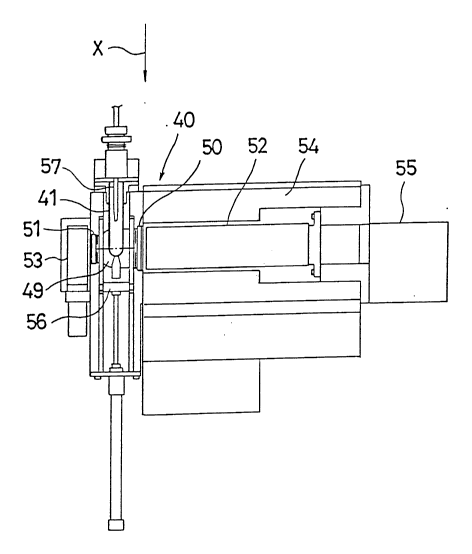
In the photometric region 40 of the above described
enzyme immuno assay system, as shown in Fig. 1, the
photometric cell 41 is held by a cell holder 49 of spherical
shape and a high sensitivity photomultiplier tube 52
(hereinafter referred to as HPMT) and a low sensitivity
photomultiplier tube 53 (hereinafter referred to as LPMT) are
disposed via interference filters 50, 51, respectively, on
opposite sides of the photometric cell 41 so that they are
aligned when viewed from the direction of arrow X. Reference
numeral 54 designates a housing for the HPMT 52 and the
housing 54 is provided with a cooler (not shown) for reducing
the dark current of the HPMT 52. In addition, reference
numeral 55 designates an amplifier for the HPMT 52, reference
numeral 56 designating a shutter, and reference numeral 57
designates a reactant-pouring nozzle.
In the case where PMTs 52 and 53 of different sensitivity
are used to detect the chemical luminescent quantity, as above
20~170
- 9
described, the signal from HPMT 52 is greatly different in
level from the signal from LPMT 53, so that equivalent signals
must be obtained by converting by varying one or both of the
signals so that the same output signal is obtained from each
detector for any given sample concentration. In order to
achieve this, the present invention employs an arrangement as
shown in Fig. 2.
Fig. 2 shows the interconnections of the HPMT 52 and the
LPMT 53 and other components of the equipment. Reference
numerals 55 and 58 designate amplifiers, reference numerals 59
and 60 designate log amplifiers, reference numeral 61
designates a changeover switch, reference numeral 62
designates an A/D converter, reference numeral 63 designates
an inverse log converter, reference numeral 64 designates an
integrator, reference numeral 65 designates a display, and
reference numeral 66 designates a memory. The log amplifiers
59 and 60 and the inverse log converter 63 are not always
required, depending upon the measuring range and on the
capability of the A/D converter 62. Furthermore, the
changeover switch 61 is not limited to the location shown.
That is to say, it may be disposed on the output sides of two
A/D converters (for use individually with the HPMT and the
LPMT) or the output sides of two integrators, in addition to
the input portion of the A/D converter 62 as shown.
The changeover switch 61 is an analog switch for
alternately directing the output signal from the HPMT 52 and
the output signal from the LPMT 53 to the A/D converter 62
every 50 m sec to enter the two sets of data.
In this preferred embodiment, the radiant life is usually
about 10 seconds, and, as above described, the output from the
detector is alternately taken out one by one every 50 m sec,
so that, after all, the output from the respective detectors
is divided into 200 pieces to be put in the computer. (The
integral value of the respective outputs becomes the datum
adopted in the operation of concentration.) The signal from
the HPMT 52 and the signal from the LPMT 53, which have been
analogized in the A/D converter 62, are subjected to the
~20~8~170
-- 10
inverse analog operation in the computer to be memorized.
In this time, the signal from the HPMT 52 is preferentially
adopted as the datum for the operation of concentration and,
in the case where the signal from the HPMT 52 exceeds the
regulation current, the signal from the LPMT 53 is adopted,
and then the output from the LPMT 53 is multiplied by a factor
determined by the ratio of the output from the HPMT 52 to the
output from the LPMT 53 based on previously determined
luminescent intensities from standard sample solutions.
Fig. 9 shows the relationship among the output (I) from
the HPMT 52, the output (i) from the LPMT 53 and the
concentration (C) of the luminescent substance, C0 to C9 on the
abscissa designate known concentrations of the luminescent
substance, Io to I6 on the ordinate on the left side designate
the output from HPMT 52, and i6 to i9 on the ordinate on the
right side designate the output from LPMT 53. Accordingly,
the ratio of the output from HPMT 52 to the output from LPMT
53 based on the luminescent intensities for the same
concentrations of sample can be determined by the use of such
a graph. That is to say, if the output from the HPMT 52 is I
and the output from the LPMT 53 is i, the ratio I/i (referred
to as A) can be determined for any particular concentration.
Since the outputs fall on straight parallel lines, the ratio A
is constant for most concentrations.
After the ratio of the output from the HPMT 52 to the
output from the LPMT 53 has been determined, if the output
from the HPMT 52 exceeds the regulation current during the
measurement, the switch 61 is operated and the luminescence is
detected by means of the LPMT 53 and the output i from the
LPMT 53 is converted into a value equivalent to the output
from the HPMT 52 (I) by the equation I = i x A.
Fig. lO is a graph showing results measured at the above
described photometric station 40. The abscissa of the graph
shows the concentration of H2O2 and the concentration of CRP,
(C reactive proteins), the ordinate on the left side
designates the output from the HPMT 52, and the ordinate on
the right side designates the output from the LPMT 53. Curve
203817-0
11
I shows the change of the concentration of H2Oz measured by the
HPMT 52 and the curve I' shows the change of the concentration
of H2O2 measured by the LPMT 53. It is found that when the
detecting range of the HPMT 52 converted into the
concentration of H2O2 is 10-8 to 10-4 M and the detecting range
of the LPMT 53 converted into the concentration of H2O2 is 10 6
to lo~2 M, as shown by said curves I and I', the detecting
range of the apparatus as a whole is 10-8 to 10-2 M. In
addition, a curve II is a calibration curve for CRP obtained
by the enzyme immuno assay and expresses the output from the
HPMT 52 and the output from the LPMT 53 in the form of one
continuous calibration curve following the above described
method.
Although the HPMT 52 and the LPMT 53 are arranged on one
straight line having the photometric cell 41 positioned
therebetween in the above described preferred embodiment, the
HPMT 52 and the LPMT 53 may both be positioned on the same
side of the cell as shown in Fig. 6 by employing an optical
splitter 66 (for example a half mirror, silica plate, glass
plate or the like), the detectors being arranged at 90
relative to eachother. In such a case, since the luminescent
axis is common to both the HPMT 52 and the LPMT 53, an
advantage occurs in that it is unnecessary to take variations
of luminescence with differences of optical position into
consideration.
In addition, the present invention can be applied not
only to the above described so-called batch type measuring
method but also to the so-called flow-through type measuring
method using a spiral flow-through photometric cell 67, as
shown in Figs. 7 and 8. Referring to Fig. 8, reference
numerals 68 and 69 designates an introduction portion and a
discharge portion, respectively, for the reactant solution.
It should further be noted that a silicon photodiode or
the like can be used, if desired, as the optical detector
instead of the above described photomultiplier. Furthermore,
it goes without saying that the present invention can be
applied not only to the photometric measurements in the above
2038170
- 12
described enzyme immuno assay system but also to photometric
measurements in other analyzers and the like.
In summary, therefore, in the present invention a
plurality of optical detectors having different sensitivities
are provided in the vicinity of a photometric cell so that the
sensitivity range of the apparatus is the sum total of the
ranges of sensitivities of each of the optical detectors so
that a wider range of measurements is possible than can be
achieved in the conventional measurement system using a single
optical detector. The ratio of outputs from the optical
detectors based on the intensity of luminescence is first
determined followed by multiplying the output of the low
sensitivity optical detector (usually) by a factor determined
by the sensitivity ratio so that a converted output value
corresponding to the output from the high sensitivity optical
detector may be obtained after the output from the high
sensitivity optical detector has been saturated. A highly
accurate measurement can thereby be achieved ranging widely
from a low sensitivity zone to a high sensitivity zone.