Note: Descriptions are shown in the official language in which they were submitted.
CA 02043527 2002-11-12
1
BIOCOMPATIBLE PERFORATED MEMBRANES, PROCESSES FOR THEIR
PREPARATION , AND USES THEREOF
Field of the invention
This invention relates to new biocompatible perforated membranes,
processes for their preparation, their use as a support in the in
vitro growth of epithelial cells, the artificial skin obtained in
this manner, and its use in skin grafts.
Prior art
The loss of cutaneous material for reasons of traumatic or
pathological origin is commonly resolved by the autotransplant
ation technique, using skin explants from donor areas. To cover
larger areas these explants can be expanded by surgical methods
such as the mesh grafting described by J. Mauchahal, J. Plast.
Surgery, 42, 88-91 (1989). These methods give positive results
only with small-dimension lesions and patients with a satisfactory
general health profile. If elderly patients or those in a state of
serious decline are treated, unsatisfactory results are obtained
and numerous problems arise, to the extent that such procedures
cannot be used. In addition they do not allow a donor tissue
expansion of more than 10 times.
An important turning point in the treatment of these lesions by
reconstructive surgery was the development of the technique
involving the in vitro culture of keratinocytes (J. Rheinwald and
H. Green, Cell, 6, 331-344, 19'j5), which allowed the in vitro
expansion of these cultures, to obtain epidermic cell membranes
2
potentially suitable for covering lesion areas.
This technique has been widely used in clinical. practice, mostly in
the case of patients suffering from burns (G.G. Gallico et al., M.
Engl. J. Med., ~, 448-451, 1984), but numerous problems arose
from its conception, such as the failure to take of some grafts,
the fragility of the epithelial film and the consequent difficulty
in its handling by the surgeon, the length of time required for
obtaining sufficient quantities of epidermis cultures and the
difficulty of obtaining donor areas of sufficient size from
patients with large areas of damaged body surface. The in vitro
epidermis cultures also require precise orientation to enable the
graft to take, this being a particularly risky operation in view of
the fragility of in vitro cultivated epidermis film.
A different approach to these problems is described by Yannas et
al., Science, 215, 1'74-1~6 (1982), who use dermic substitutes in
the form \of reabsorbable porous materials consisting of
coprecipitates of collagen and glycosaminoglycans (GAG), in
particular condroitin-6-sulphate, covered by a thin silicone
membrane film. The characteristic of these materials is that they
comprise non-standardized pores intercommunicating in a manner
similar to a sponge.
Zang et al., in Burns, 12, 54Q-543 (1986) propose a method, known
as microskin grafting, consisting of auto-grafting very small skin
portions, which then develop to merge into a single epithelium.
With this method the maximum donor surface/coverable surface
expansion ratio obtainable is 1:15.
S. Boyce and J. Hansborough in Surgery, 10~, 421-431 (1988)
3
describe the use of membranes formed from collagen and GAG to
promote on their surface the growth of keratinocytes, so reducing
the surface porosity of the material. A continuous non-porous
layer is also interposed to limit the epidermic culture development
to 'the membrane surface. The possible antigenicity of these dermic
substituents, which can result in rejection of the graft, has not
yet been properly ascertained.
Object of the invention
The object of the present invention is to provide biocompatible
membranes which enable in vitro culture of keratinocytes, with
culture development in a much shorter time than that previously
possible. An important result of the membranes according to this
invention is the ability to obtain colonization by homologous or
heterologous epithelial cells in a time which is surprisingly short
(6-10 days) compared with the time normally required (20-40 days)
by traditional methods for preparing comparable areas of in vitro
epidermis cultures,
This advantage results in the preparation in a short time of an
artificial skin which allows very rapid coverage of an urea on
which an epithelial transplantation is required; so reducing the
risks relating to excessive organic liquid loss or infection.
A further object of the present invention is to provide
biocompatible membranes which allow rapid development of
keratinocyte cultures with an excellent donor surface/coverable
surface ratio, of between 1:20 and 1:200, this being considerably
higher than previously obtainable with traditional methods.
A further object of the present invention is to provide a
a
biocompatible and preferably bioreabsorbable artificial skin which
can be produced in a short time, is strong, and is easily handled
at the moment of transplantation, and which moreover can be applied
to the site of the lesion independently of its original orientation
in the culture vessel, and can be easily stored. In this respect,
an advantage of the artificial skin according to the present
invention is that it can be easily cryopreserved to allow the
creation of a bank of epithelial tissue, including heterologous.
The possibility of cryopreservation also considerably reduces or
eliminates, after at least two cycles, the antigenic potential of
the surface antigens expressed by the epithelial cells.
Description
These and further objects are attained by the biocompatible
membranes according to the present invention, consisting of
material of natural, synthetic or semisynthetic orzgin and having a
thickness of'between 10 and 500 u, and preferably between 20'and 40
p, characterised by comprising an ordered series of holes of a
defined and constant size between l0 and 1000 u, and preferably
between 40 and '70 u, separated from each other by a constant
distance of between 50 and 1000 p, and preferably 80 u.
These membranes can consist of biocompatible and preferably also
bioreabsorbable materials of natural origin such as collagen or
coprecipitates of collagen and glycosaminoglycans, cellulose,
gelled polysaccharides such as chitin, chitosan, pectins or pectic
acids; agar, agarose, xanthan gum, gellan, alginic acid or
alginates, polymannans or polyglucans, starches, or natural
rubbers, either alone or in mixture with each other or with
5
polymers of synthetic or semisynthetic origin, in the presence of
suitable precipitating or gelling agents such as metal salts,
polycations or polyanions.
The membranes can also consist of biocompatible and preferably also
bioreabsorbable materials of synthetic origin such as polylactic
acid, polyglycalic acid or copolymers thereof or their derivatives,
polydioxanones, polyphosphazenes, polysulphones or polyurethanes,
or semisynthetic derivatives of natural polymers such as collagen
crosslinked with crosslinking agents such as dialdehydes or their
Precursors, bicarboxylic acids or halides thereof, diamines, or
derivatives of cellulose, of alginic acid, of starch, of chitin or
chitosan, of gellan, of xanthan, of pectins or pectic acids, of
polyglucans, of polymannans, of agar, of agarose, of natural
rubbers or of glycosaminoglycans.
The membranes can also consist of synthetic polymers, even without
the biodegradability characteristic, such as silicone, silane or
siloxane rubbers, fluoropolymers such as polyfluoroethylene;
polyfluoropropylene, polyfluoroethers, polystyrene, vinyl
polychloride, polyacrylate or derivatives thereof, polyhydroxy-
acrylat°; polyhydroxymethacrylate, carboxyvinyl polymers and their
derivatives, malefic anhydride polymers' and their derivatives,
polyvinylchloride, polyvinylalcohol and its derivatives,
polyethylene and polypropylene.
The membranes preferably consist of semisynthetic derivatives ' of
hyaluronic acid, in particular ester derivatives thereof such as
those described in Examples 6, '7 and 24 of EPA 0216453 filed on
'7.'7.86, these being biocompatible and biodegradable materials able
CA 02043527 2002-11-12
6
to release hyaluronic acid on the site of their application, this
acid being well known to favour tissue reparative processes. A
further characteristic which makes these materials particularly
suitable for use according to the present invention is that they do
not produce intolerance phenomena, not being immunogenic.
The biocompatible membranes, consisting of one or more of the
aforesaid materials have a thickness of between 10 and 500 ~.m and
preferably between 20 and 40 ~m,and are characterised by the
presence of an ordered series of holes of defined and constant size
between 10 and 1000 ~.m, and preferably between 40 and 70 Vim,
separated from each other by a constant distance of between 50 and
1000 Vim, and preferably $0 Vim.
Continuous biocompatible membranes, consisting of one or more of
the aforesaid materials, can be prepared by the conventional
methods described in the literature.
The perforated biocompatible membranes according to the present
invention are obtained using mechanical perforation devices such as
suitably arranged punching machines, or methods involving the use
of thermal or ultraviolet lasers operating in a frequency band such
as to produce holes of the required size and distance apart in the
membrane.
The following example of the preparation of a perforated
biocompatible membrane according to the present invention is given
by way of illustration only.
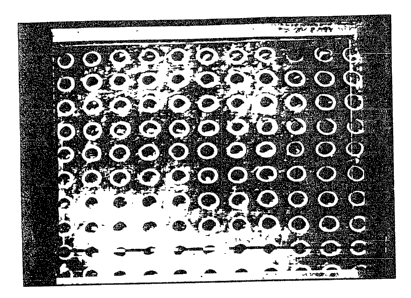
EXAMPLE 1
A membrane of hyaluronic acid benzyl ester with 100x esterifi-
ration (as described in EPA 0216453 filed on 7.7.86) in the form of
CA 02043527 2002-11-12
7
a square of 12 x 12 cm and 25~,m thickness was perforated using a
computerized UV Laser device operating at a frequency of 2'73 um
under the following operating conditions: working frequency 200 Hz,
output energy 250 mJ. Using a suitable screening system, holes
having a diameter of 40~m were obtained at a distance apart of 80
~m,as shown in Figures la and 1b.
The perforated biocompatible membranes according to the present
invention can be used advantageously for the in vitro culture of
epithelial cells, especially keratinocytes.
For this purpose the membranes can be fixed to the base of cell
culture vessels, to metal grids or to any other structure suitable
for cell cultures at the air/culture medium interface, using
sterile vaselin, sterile silicone or other cementing systems which
allow easy removal of the membrane, or by systems involving the use
of biological material such as collagen, fibrin or fibrin glue:
These membranes can be incubated in culture media suitable for the
growth of epithelial cells either alone or in the presence of other
cells, such as irradiated fibroblasts, as described in the cited
literature, without within the time scheduled for growth and hole
colonization causing alteration in mechanical properties which
would compromise their handleability and strength within the
particular application.
Some of the tests carried out are described below to illustrate the
use of the membranes of the present invention.
EXAMPLE 2
The following test was conducted to demonstrate the absence of any
inhibition by hyaluronic acid derivative membranes on the in vitro
growth of human keratinocyte cell cultures.
Membranes denominated HYAFF 11 cut sterilely into 2 x 2 cm squares
and consisting of hyaluronic acid benzyl ester with 100% esterifi
ration (as described in EP 0216453 filed on 7.7.86) were applied to
the base of the culture vessels by means of sterile silicone.
2 x 10⁵ human keratinocytes were seeded onto these in a volume of
0.5 ml, in the presence of 4 x 10⁵ lethally irradiated 3T3
fibroblasts at the second passage.
The capsules were incubated at 37~C for 2 hours in a 5% C02
atmosphere to allow the cells to attach to the matrix. After this
period 5 ml of CEC culture medium (Green H. et al., J. Proc.
Nation. Acad. Sri., 76, 5665-5668, 1979) were- added and the
capsules again incubated. The culture medium was changed every 2
days. The cells were treated with trypsin 9 days after seeding and
counted. All experiments were conducted in duplicate.
RESULTS
No, of human keratinocytes % inhibition
per plate (x 10 5)
Control 27 0%
2p HYAFF 11 membrane 27
These results show that the biomaterial used has no inhibiting
effect on keratinocyte cultures.
EXAMPLE 3
Growth of human keratinocytes using the perforated biocompatible
membranes of the invention, obtained by the method described in
Example 1
HYAFF 11 membranes consisting of hyalurontc acid benzyl ester with
9
100x esterification (as described in EPA 0216453 filed on 7.7.86)
in the form of 3 x 3 cm squares were cemented to the base of 6 cm
diameter Petri capsules using sterile vaselin. Lethally irradiated
3T3 fibroblasts were seeded on the membranes to a concentration of
700,000 cells per plate, under the conditions described in Example
2. After adhesion of the 3T3 cells, ie after about 24 hours, a
cell suspension of human keratinocytes originating from secondary
cultures was added at a concentration of 38,000 cells per cm2. The
culture conditions were analogous to those described in Example 2.
The development of the keratinocyte culture was followed daily
using a phase contrast microscope. The development of inoculated
epithelial cells was observed on the membrane, these having reached
confluence 8-10 days after seeding.
Of particular importance is the fact that even on the second day
after seeding, numerous holes contain keratinocytes, their growth
being more active within the holes than on the surface, to totally
fill them around the 6th day (Figures 2, 3 and 4).
A further fact of great importance is that when analyzed by
histological techniques the cells within the holes demonstrate a
basaloid appearance documented by the findings of Figures showing
frequent mitosis (Figures 5 and 6), denoting high reproductive
vitality. These Findings were confirmed by immunohistochemical
methods using specific antibodies (Mab).
The epithelial cells grown within the holes can therefore be
considered overall to be in the active proliferation stage and thus
effectively usable on transplantation areas.
The artificial skin according to the present invention, obtained by
CA 02043527 2002-11-12
1U
the aforesaid procedures, therefore consists of a biocompatible and
preferably bioreabsorbable support membrane consisting of materials
of natural, synthetic or semisynthetic origin, and having a
thickness of between 10 and 500 ~m,and preferably between 20 and 40
Vim, characterised by comprising an ordered series of holes of a
defined and constant size between 10 and 1000 ~.m, separated from
each other by a constant distance of between 50 and 1000 Vim,
together with autologous or heterologous keratinocyte microcolonies
in the active proliferation stage present within the holes.
This artificial skin can be easily shaped by the operator on the
basis of the areas to be treated, and has a mechanical strength
which enables it to be handled without difficulty and be sutured.
Once implan~;.ed on the lesion area, the keratinocyte microcolonies
create growth nuclei of rapid-growing epithelial tissue, which in a
short time completely re-epithelialize the area on which the
transplantation has been carried out.
It is used by withdrawing it from the culture vessel, removing
all traces of culture medium by a sterile physiological solution
and applying it to the area to be treated without needing to pay
Particular attention to the direction of application, as it is
equally effective if applied on either of its two sides, in
contrast to traditional keratinocyte cultures.
The artificial skin according to the present invention can be used
to cover even extensive lesions of the body surface of traumatic
origin such as burns, of surgical origin such as withdrawal areas
in plastic surgery, or pathological origin such as stasis ulcers or
bedsores.