Note: Descriptions are shown in the official language in which they were submitted.
3~
THROMBECTOMY METHOD AND DEVICB
CROSS REFERENCE TO CO-PENDING APPLICATIONS
None.
~hCKGROUND OF THE INVENTION
1. Field of the Invention - The present
invention relates generally to medical devices and
procedures, and more particularly, relates to medical
devices and procedures for removing thrombus deposits
from the cardiovascular system.
2. Description of the Prior Art - Procedures and
apparatus have been developed for ease in removing
tissue and various deposits. U.S. Patent No. 4,790,813
issued to Kensey and U.S. Patent No. 4,842,579 issued
to Shiber describe techniques for the removal of plaque
deposited in arteries by mechanical ablation using
rotating cutting surfaces. These relatively traumatic
approaches are directed to the treatment and removal of
very hard substances.
In current medical procedures, thrombus deposits
are often removed using a catheter such as is described
in U.S. Patent No. 4,328,811 issued to Fogarty. In
this system-, a surgical cutdown is performed to access
the vessel -and allow catheter entry and advancement to
a point beyond the deposit. The balloon is inflated
and the catheter is withdrawn pulling the deposit along
with it.
Pressurized fluids have also been used in the past
to flush undesirable substances from body cavities.
U.S. Patent No. 1,902,418 describes such a system for
domesticated animals. The more modern approaches tend
~ ~ ~ 3 ~
to use vacuum rather than gravity as the primary means
for removal of the deposits or tissue and relatively
low fluid pressures to cut into and fragment the
substances to be ablated.
U.S. Patent No. 3,930,505 issued to Wallach
describes a surgical apparatus for the removal of
tissue from the eye of a patient. As with similar
systems, Wallach uses a relatively low pressure jet of
water (i.e. 15 to 3500 psi) to disintegrate the tissue,
and a suction pump to perform the actual removal.
A similar approach applied to the cardiovascular
system is discussed in U.S. Patent No. 4,690,672 issued
to Veltrup. Veltrup also provides a much lower
pressure jet of water (i.e. less than 450 psi) to
fragment deposits. As with Wallach, Veltrup uses a
vacuum pump for evacuation of the fragments. The
distal end of the Veltrup catheter is readily
repositionable to permit manual entrapment of the
deposits to be fragmented.
08/06/90 2
SUMMARY OF THE INVENTION
The present invention overcomes the disadvantages
of the prior art systems by performing the entire
procedure at positive pressures. This eliminates the
need for a vacuum pump and provides the added safety
feature of an intravascular environment which is always
positively pressurized as during normal functioning of
the cardiovascular system. This tends to prevent
collapse of the vessel. ~he system also controls the
exposure of the vessel to over pressurization and
prevent distension.
According to the present invention, the only
energy added to the system is via an extremely high
pressure stream of saline solution. This stream serves
to dislodge thrombus deposits, position them, and then
emulsify them. Thrombus particles are attracted to the
jet due to the localized high velocity and low
pressure. Recirculation patterns and fluid entrainment
bring the thrombus continually into close proximity of
the jet. once emulsified by the jet, the particles are
removed by flow through the evacuation lumen generated
as a result of stagnation pressure which is induced at
the mouth of the evacuation lumen by the action of at
least one fluid jet directed at and impinging on the
lumen mouth.
The procedure is practiced by percutaneously or
intraoperatively entering the vascular system of the
patient at a convenient location with a cannula. The
catheter is inserted either directly or over a
previously positioned guide wire and advanced under
08/06/90 3
~ ~J1~8 1~
fluoroscopy to the site of the vascular occlusion or
obstruction which generally contains an aggregation of
blood factors and cells or thrombus deposit, which is
normally identified by angiography. One or more
balloons may be inflated to stabilize the distal end of
the catheter and provide a degree of isolation of the
area to be treated.
Sterile saline is pressurized by a disposable pump
and directed through a flexible metallic tube within
the catheter. One or more jets at the distal end of
the catheter direct the pressurized stream generally in
the direction of the mouth of the evacuation lumen at
the distal end of the catheter with a component
directed toward the vessel wall. One function of the
jet(s) alone or in combination with a distal balloon,
is to dislodge thrombus deposits from attachment to the
vessel wall. Other functions of the jet(s) are to
attract and emulsify the thrombus deposits and create
the stagnation pressure which evacuates the emulsion.
A metering device is utilized at the proximal end
of the evacuation lumen to regulate the flow rate of
the emulsified thrombus out of the catheter. Because
the entire system operates at a positive pressure, the
output must be metered`to prevent excess evacuation.
Safety monitors turn the system off if one of the
lumens or jets becomes clogged. An optional monitor at
the distal end of the catheter can monitor power
delivery and degree of blockage. An alternative
embodiment of the invention provides an extra lumen for
monitoring of temperature and/or pressure at the site
~8/06/30 4
2~'~3~
of the thro~bectomy. The evacuation lumen permits the
passage of an angioplasty dilatation catheter or
angioscope for intravascular viewing.
08/06/90 5
C~ 2 ~
BRIEF DESCRIPTION OF THE DRAWING8
Other objects of the present invention and many of
the attendant advantages of the present invention will
be readily appreciated as the same becomes better
understood by reference to the following detailed
description when considered in connection with the
accompanying drawings, in which like reference numerals
designate like parts throughout the figures thereof and
wherein:
FIG. 1 is a schematic diagram of the overall
system employing the present invention;
FIG. 2a is a mechanical view of disposable pump;
FIG. 2b is a cross-sectional view of the
disposable pump;
FIG. 2c is a conceptual view of the safety
monitor;
FIG. 2d is a cross-sectional view of an
alternative source of pressurized fluid;
FIG. 3 is a cross-sectional view of the manifold;
FIG. 4 is a conceptual view of the operation of
the manifold;
FIG. 5a is a close up view of the distal end of
the cat~eter system of the present invention;
FIG. 5b is a longitudinal sectioned view of the
distal end of the catheter system;
FIG. 5c is a view from the distal end of the
catheter system;
FIG. 6 is a cross-sectional view from immediately
proximal of the balloon;
08/06/90 6
~$ ~7~
FIG. 7 is a cross-sectional view across the
balloon inflation port;
FIG. 8 is a cross sectional view taken distal of
the balloon;
FIG. 9 is a cross-sectional view taken near the
distal tip of the catheter system;
FIG. 10 is a longitudinal sectioned view of the
distal end of a catheter system employing an
alternative embodiment of the present invention;
FIG. 11 is a cross-sectional view taken proximal
to the proximal balloon of the alternative embodiment;
FIG. 12 is a cross-sectional view of the
alternative embodiment from the i~flation port of the
proximal balloon;
FIG. 13 is a cross-sectional view of the
alternative embodiment taken distal of the proximal
balloon;
FIG. 14 is a cross-sectional view of the
alternative embodiment taken distal of the mouth of the
evacuation lumen;
FIG. 15 is a cross-sectional view of the
alternative embodiment taken proximal of the distal
balloon;
FIG. 16a is a sectioned view of the effluent
2~ safety switch; and,
FIG. 16b is a cross-sectional view of the effluent
safety switch.
08/06/90 7
~3 ~J~I~q j~ 3 ~
DETAILED DESCRIPTION OF THE PREFERRED EM~ODIMENT~
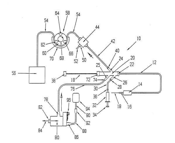
FIG. 1 is a schematic view of the preferred
embodiment of catheter system 10 employing the present
invention. The details supplied herein should be taken
as representative and not lim~ting of the many
embodiments which may be efficaciously employed within
the scope of the present invention.
Catheter system 10 has a standard two lumen
catheter 12, which is extruded of a flexible material,
such as polyolefin, PTFE, PVC, polyurethane, or other
suitable material in the normal fashion. Near the
distal end of catheter 12 is located inflatable balloon
14, which is preferably an elastic balloon having no
predefined outside diameter size limitation upon
inflation. In this manner, balloon 14 can conform to
the exact dimensions of the vessel to hold distal end
16 of catheter 12 in a fixed position. Alternatively,
inflatable balloon 14 can be an inelastic balloon with
a predefined shape and size to permit it to be also
used for dilatation as in translumenal angioplasty.
Distal end 16 of catheter 12 is described in more
detail below.
Guide wire 18, as manipulated by knob 38, is
optionally available for positioning catheter 12 as an
over-the-wire system. Guide wire 18 passes through the
larger of the two lumens of catheter 12 as described in
more detail below.
Manifold 20 is molded of a rigid plastic. The
main branch couples to the larger of the lumens of
08/06/90 8
2 ~3
catheter 12 and has a standard seal assembly 72 applied
to the proximal end to sealingly engage guide wire 18.
Secondary branch 24 is also coupled to the larger
lumen to provide for evacuation of the emulsified
thrombus deposits. Secondary branch 24 sealingly
engages distal end 42 of effluent tubing 54 via seal
assembly 40. The operation of safety monitor 44,
monitor switch 50, and cable 52 are explained in
further detail below.
Flexible effluent tubing 54, including distal end
42, is coupled to safety monitor 44 as described in
more detail below. The flow of effluent through
flexible effluent tubing 54 is metered by rollers 62,
64, and 66 as rotated by rotor 60 in the direction of
arrows 68. It must be emphasized that the effluent in
flexible effluent tubing 54 is under pressure and,
therefore, need not be pumped by peristaltic pump
assembly 58, which merely restricts and meters the
flow. This metering could equally well be accomplished
with a timed mechanical valve (not shown) which
controls the outflow rate. After metering, the
effluent from flexible effluent tubing 54 is deposited
in disposal bag 56.
Secondary branch 26 of manifold 20 is sealingly
coupled to inflation tubing 30 by seal assembly 28.
Inflation and deflation of inflatable balloon 14 is
controlled by plunger 34 of syringe 32 in the customary
manner~ Syringe 32 is sealingly coupled to inflation
tubing 30 by coupling assembly 36.
08/06/9~ 9
The saline solution used to emulsify the thrombus
deposit is derived from standard sterile saline bag 94,
which may be commercially available. The saline
solution is transferred to disposable pump 80 via
hypodermic needle 90 and tubing 88 and couplings 92 and
86. This is a low pressure fluid path.
Disposable pump 80 is a positive displacement
piston pump. It is made to be completely disposable
for sanitary reasons. Disposable pump 80 is driven
reciprocally as shown by arrows 84 by a motor driven
cam (not shown) against cam bearing surface 82. As a
convenient means to correlate infused volume of saline
solution with volume of evacuated effluent, a single
electric motor can be used to drive both disposable
pump 80 and rotor 60. Control of these volumes is
important to prevent rupture or collapse of the vessel
wall. Closer tolerance control can be achieved at
greater complexity usinq pressure and/or flow meters.
The high pressure output of disposable pump 80 is
coupled to tubing 76 by high pressure coupling assembly
78. Tubing 76 has a flexible metallic inner tube
inside of a flexible plastic or rubber outer tube as
shown in more detail below. Tubing 76 is sealingly
coupled to secondary branch 25 of manifold 20 by seal
assembly 74. Safety monitor 96 operates as explained
below to turn off the drive motor if the tubing or jets
become clogged.
oa~o6/so 10
~7, $ ~ u
FIG. 2a is a partially sectioned view of
disposable pump 80. As explained above, disposable
pump 80 is designed to be discarded after a single use
for sanitary reasons. It is a positive displacement
piston pump. All referenced components are as
previously described.
High pressure coupling assembly 78 is shown in
greater detail to highlight that metallic tube 75 is
brazed at point 77 to produce the required high
pressure joint. Outer tubing 71 is a low pressure
connection which may be attached with adhesive. The
entire high pressure coupling assembly 78 is attached
to disposable pump 80 with threads 73 and compressing a
high pressure seal.
Safety monitor 96 comprises two safety features.
Pressure plug 100 is attached to disposable pump 80 by
threads 98. Pressure plug 100 is designed to release
and vent the system to the atmosphere at pressures
above 30,000 - 40,000 psi. The second safety feature
serves to electrically disconnect the drive motor
whenever the pressure is too high. Increased pump
pressure forces contact 104 toward electrical contact
with contact 106 as a result of pushing out of pressure
plug 100 as attached at point 102 (shown in detail
below), thereby closing the electrical circuit to a
relay and turning off the drive motor. Insulators 95
and 97 maintain contacts 104 and 106 open under normal
pressure conditions.
The saline input to the disposable pump includes a
hypodermic needle 90 which penetrates a puncture port
08/06/90 ll
1~3 2 ~
on a bag of saline. The saline is delivered through
coupling 92 t~ tube 88 and through coupling 86 into the
inlet of the disposable piston pump 80.
08~06/90 12
FIG. 2b is a cross-sectional view of disposable
pump 80. As a matter of convenience the disposable
pump 80 is oriented slightly different from FIG. 1.
All referenced components are as previously described.
Cam 310 is rotated by a drive motor (not shown) as
discussed above. The action of cam 310 imparts a
reciprocal motion to cam bearing surface 82 causing
connecting rod 302 to move horizontally. This moves
piston 300 in the direction of arrows 311. Movement to
the left eniarges the effective volume of chamber 305
creating a relatively low pressure. This permits entry
of sterile saline fluid from fluid entry port 312 (see
also Fig. 1) through ball valve 306 under tension of
spring 307.
Movement of piston 300 to the right decreases the
effective volume of chamber 305 forcing sterile saline
solution to exit via ball valve 309 under sufficient
pressure to overcome the tension of spring 308. Note
that ball valve 306 will be forced closed as piston 300
is moved to the right. The saline solution is expelled
through high pressure tube 75.
Seals 301 and 303 and springs 307 and 308 are
s~lected consistent with the fluid pressures to be
developed. Bellows 304 provides an additional seal for
the system. Cam 310 may be designed to provide a
relatively smooth flow of sterile saline, or it may be
implemented as a Geneva or similar cam to enhance the
pulsatile delivery of the sterile saline to change the
emulsification action at the distal tip of catheter 12.
08/06/90 13
V ~ }i~3~
Pressure plug 100 can be adjusted so that if the
pressure reaches an upper limit, such as 30,000-40,ooo
psi, the pressure will be released and the safety
monitor 96 will turn the motor off.
08/06/90 14
r ;- ~ ~
; 2~ 12~3
FIG. 2c is a schematic view of safety monitor 44~
The emulsified thrombus i5 evacuated in line 314. If
the entrance to the evacuation port becomes blocked,
the pressure in line 314 will drop and cause membrane
322 to retract around line 314 which has an opening
port 324 which has passage to the membrane. As the
membrane retracts due to a blockage in the evacuation
tube, the contacts 340 and 336 are opened and thereby
trigger a relay 329 which will turn off the drive motor
328.
FIG. 2d is a cross-sectional view of an
alternative source of pressurized fluid. This approach
replaces the function of disposal piston pump 80.
Using this technique, the high pressure tubing 118,
plugs 53 and 48, tapered ring 119, and saline bag 95
are inserted into the conformal housing 46 and
tightened down using threads 45. Chamber 43 is
pressurized by supplying pressurized non-sterile water
or other fluid through inlet 51 forcing sterile saline
to exit from port 49 of tubing 118. A seal 81 is made
between the bag 95 and the high pressure tubing 76
which delivers the high pressure saline. The high
pressure tubing 118 i5 brazed into a tapered sealing
ring 119. A seal is made between the bag 95 and the
ring 119 and also between the bag 95 and end plug 48 by
tightening down plug 53. The outer plastic tubing 76
is adhesively bonded to plug 53. Bottom plugs 61 and
69 are held in place by threads 63, 65, and 67 and
sealed by seal 83 as plug 69 is tightened down.
Whenever employing this alternative embodiment,
care must be exercised not to rupture sterile saline
bag 95 under the extreme pressures reguired by the
present invention. High pressure fluld is supplied to
tubing 85 from a positive displacement pump (not
shown).
08/05/90 16
FIG. 3 ls a cross sectional view of manifold 20.
Because this component is molded as two halves, which
are solvent-bonded together, the view also happens to
show one of the two halves. As explained above,
catheter lZ is a two lumen catheter. In the preferred
mode, each of the two lumens has two distinct
functions. Therefore, manifold 20 serves to provide
passage for a high pressure tubing and balloon
inflation through one lumen and passage of a guide wire
and evacuation through the other lumen.
The larger lumen of catheter 12 is lumen 110. It
is used for passage of guide wire 18 (not shown in this
view) and for evacuation of effluent and possible
passage of an angioplasty dilatation catheter or
angioscopic probe. Lumen 110 terminates inside the
manifold 20 at the proximal end of the flexible tubular
member and provides passage of a guide wire or other
diagnostic or therapeutic device. Guide wire 18 is
sealed by compressible circular seal 136 which is
compressed by surface 140 as threaded knob 72 is
tightened on threads 138. It is important -to seal
guide wire 18 in this way as guide wire 18 must be
movable with respect to catheter 12 to properly
manipulate distal tip 16 of catheter 12 into position.
Lumen 110 is also terminated at secondary branch
24. This is accomplished by removing a portion of the
outer wall of lumen 110 at point 120. This provides
fluid coupling between lumen 110 and lumen 134 of
secondary branch 24.
~8/06/90 17
~3~
The smaller lumen of catheter 12 is lumen 112.
One of its functions is as a fluid passageway for the
inflation of balloon 14. This function is accomplished
by removing a portion of the outer wall of lumen 112 at
point 114 to fluid couple lumen 112 to lumen 116 of
secondary branch 25.
The remaining purpose of lumen 112 is to provide
for passage of metallic tubing 118. Because of the
extremely high pressures involved, the saline solution
is conveyed in a metallic tubing 118, which is
preferably stainless steel hypo tubing. To handle the
pressures involved, the hypo tubing is run as a
continuous length along catheter 12. The proximal end
of metallic tubing 118 passes through the outer wall of
lumen 112 and into secondary branch 25. A larger
diameter hypo tube is brazed onto hypo tube 118 at
point 123. This larger tubing is covered by protective
plastic tubing 71. Manifold 20 is solvent-bonded
together prior to assembly of the catheter, and points
124, 126, 128, 130 and 133 are used to introduce an
adhesive which serves as a seal to separate each path
and each lumen. Point 132 shows the bonding of the
outer plastic tube which surrounds the high pressure
supply tube to the manifold.
08/06/90 18
2 ~ L~ 2 ~
FIG. 4 is a schematic view of manifold 20 wherein
all referenced elements are as previously described.
This figure is purposely not drawn to scale to better
illustrate the operation of manifold 20.
08/~6/90 19
FIG. 5a is a close up view of balloon 14 and
distal tip 16 of catheter 12. Attachment between
catheter 12 and balloon 14 occurs at overlap points 144
and 146. These overlap points are sealingly attached
with adhesive or heat sealing.
Catheter 12 is a dual lumen catheter extruded from
rubber or a polymer as described above. Cap 150 is
fixedly attached at the distal tip of catheter 12 as
shown. Preferably cap 150 is made of a radiopaque
metal such as platinum, tantalum or stainless steel to
provide ease of location under fluoroscopy.
Extending beyond cap 150 is metallic tubing 118.
This is necessary to permit the jet or jets which
dispense the saline solution to be directed at cap 150
(i.e~ the distal tip of lumen 110). Because metallic
tubing 118 is so flexible, it must be backed by metal
plate 156 to provide the necessary rigidity. Metallic
tubing 118 is bent as explained below. To conform,
metal plate 156 is angled to form rounded distal
surface 160. This annular shaped tip would allow
passage of a guide wire, angioscope, or angioplasty
dilatation catheter.
08/06/90 20
~3 .~ ~ q ., i ~,
FIG. 5b is a longitudinal sectioned view of the
structure of FIG. 5a, wherein referenced elements are
as previously described. Also shown in this view is
balloon inflation port 148 which provides fluid
communication between lumen 112 and balloon 14. Septum
142 separates lumen 110 from lumen 112. Lumen 112 is
sealed distal to the balloon using an adhesive seal 113
attaching the high pressure tube 118 and filling lumen
112.
Metallic tubing 118 is bent into a circular shape
perpendicular to the axis of the catheter beginning at
bend 152. Metal plate 156 bends at point 158 to
provide rigidity at that point. Jet 164 is a small
diameter orifice on the order of .0005 to .003 of an
inch. It directs a stream of saline solution at cap
150 (i.e. mouth of lumen 110) at 5,000 to 30,000 psi.
This pressure is sufficient to emulsify thrombus
deposits located between jet 164 and cap 150. This
stream of saline solution also creates a stagnation
pressure about cap 150 sufficient to propel the
emulsion into and through lumen 110 (see also FIG. 1).
This stream of saline solution is of high velocity
which creates a localized area of low pressure around
the stream which attracts thrombus deposits for
emulsification and removal.
08/06/90 21
`J ~ '3
FIG~ 5c is a view from the distal end of catheter
system 10 wherein referenced elements are as described
above. The central opening would allow passage of a
guide wire, angioscope or angioplasty dilatation
catheter.
08t06/90 22
s '~ 3 ~ ~ ~
FIG. 6 is a cross-sectional view from the proximal
end of catheter 12 to balloon 14, wherein referenced
elements are as previously described. Metallic tubing
118 is shown within lumen 112. The cross-sectional
area of lumen 112 which is in excess of that needed for
metallic tubing llB provides the fluid passageway for
inflation of balloon 140 Lumen 162 of metallic tubing
118 has a diameter of about .003-.OlO inch. It conveys
saline solution at 1,000 to 30,000 psi through the main
body of the catheter.
08/06/90 ~3
2 ~
FIG. 7 is a cross-sectional view taken through
balloon 14 and balloon inflation port 148. The
remaining elements are as previously described.
0~/0~/90 24
~ 2 ~ 2 ~
FIG. 8 is a cross-sectional view taken distal to
balloon 14. The distal end of the balloon inflation
lumen is plugged with adhesive 113 to provide an
enclosed space for balloon inflation.
08/06/90 25
d ~
FIG. 9 is a cross-sectional view taken just
proximal of the saline solution jets. Shown in
addition to jet 164 are jets 154a, 154b, 154c, and
154d, which are similar in size and range to jet 164.
Jet 164 is directed generally back toward the
evacuation channel to generate a stagnation pressure,
create a localized area of low pressure to attract
thrombus deposits, emulsify any thrombus which is
brought into its path and keep the opening to the
evacuation lumen clean and open.
Jets 154a, 154b, 154c, and 154d can number from
zero to eight with a preferred number of three to six
~ets, although not limiting and are directed with some
radial component toward the vessel wall as drawn and
may also have some axial direction towards the
evacuation opening. These jets remove thrombus which
is attached to the vessel wall and establish a
recirculation pattern which entrains thrombotic
material and brings it into contact with jet 164 for
further emulsification and removal.
08/06/9~ 26
~ '~f~
FIG. lO is a close up view of the distal end of an
alternative embodiment of the present invention
including balloon 204 which is located distal to the
active components. Balloon 204, along with balloon
202, can be used to isolate a portion of the vessel
during the procedure. Fluid recirculation between the
balloons brings the thrombus into contact with the jet
for emulsification and removal. In this em~odiment,
lumens 206 and 208 function as lumens llU and 112,
respectively. Cap 220 is similar to cap 150. A
thermistor (not shown) can be used with either the
preferred or alternative embodlment. The thermistor
concept should only be added as a possibility which
will help in monitoring the degree of occlusion and/or
power delivery. Metal tube 212 has the same function
as metal plate 155 in the preferred embodiment.
Metallic tubing 210, bend 214 and jet 216 directly
correspond to similar components in the preferred
embodiment. An adhesive 221 and 223 is applied in the
lumen of distal tubing 225 to provide an enclosed space
and allow balloon 204 to be inflated through the distal
balloon inflation port 219.
38/06/9~ 27
FIG. 11 is a cross-sectional view of the
alternative embodiment from proximal to balloon 202.
In this embodiment, a three lumen catheter is used.
Lumen 206 is the largest lumen, which is used for
passage of the guide wire and evacuation of the
effluent. Annular space 222 is used for inflation of
balloon 204 and for passage of metallic tubing 210.
Lumen 226 provides for inflation of balloon 204.
Annular space 224 could be used to permit an external
device (not shown) to measure the pressure and/or
temperature within the treatment area to determine when
thrombus deposits are completely emulsified.
28
FIG. 12 is a cross-sectional view of the
alternative embodiment as viewed through balloon 202.
Shown is balloon inflation port 218.
08/06/90 29
2 ~
FIG. 13 is a cross-sectional view of the-
alternative e~bodiment as viewed distal of balloon 202.
Guide wire 228 is shown located within lumen 206.
08/06/90 30
FIG. 14 is a cross-sectional view of the
alternative embodiment as viewed distal to cap 220.
08/06/90 31
r~ J ~
FIG. 15 is a cross-sectional view of the
alternative embodiment as viewed proximal to balloon
204. Shown is a jet 216 directed back towards the
evacuation lumen and a plurality of jets numbered 217a,
217b, 217c and 217d, which are directed with some
radial component toward the vessel wall. These
outwardly diracted ]ets may not be necessary since the
distal balloon can be used to dislodge the thrombus off
of the wall. Metal tubing 230 extends to the distal
balloon for inflation.
08/06/90 32
R ` r~
FIG. 16a is a longitudinal sectioned view of
safety monitor 44. It is placed over flexible membrane
330 of distal end 42 and flexible effluent tubing 54.
Fluid communication is supplied by port 332.
08/0~/90 33
- ' 2 ~3~ ?, ~
FIG. 16b i5 a cross sectioned view of safety
monitor 44. It funct:ions much as a safety monitor with
contacts 336 and 340 being closed whenever pressures
are reduced due to blockage of the evacuation tube.
Fluid communication to the membrane is supplied by port
332.
08/06/90 34
Various modif ications can be made to the present
invention without departing from the scope thereof.
I CLAIM:
08/06/90 35