Note: Descriptions are shown in the official language in which they were submitted.
~0~9~5~
CELL CAPSULE EXTRUSION SYSTEMS
5 Background of the Invention
The technical field of this invention
concerns the encapsulation of living cells for the
production of biologically active factors.
There is considerable interest at present in
the biologically active products of living cells,
including, for example, neurotransmitters, hormones,
cytokines, nerve growth factors, angiogenesis
15 factors, blood coagulation factors, lymphokines,
enzymes and other therapeutic agents. There is also
substantial interest in developing new methods and
systems for producing such biological factors as well
as in delivering these factors to subjects for
20 therapeutic purposes.
For example, Parkinson's disease is
characterized by the degeneration of the dopaminergic
nigrostriatal system. Striatal implantation of
25 polymer rods which release sustained amounts of a
neurotransmitter, dopamine, has been reported to
alleviate experimental Parkinsonism in rodents,
indicating that the release of dopamine alone in the
proper target structure may be able to correct this
30 functional deficiency.
-2- 20~90~6
In contrast to the limited capacity of a
polymeric matrix drug release system, encapsulated
dopamine-releasing cells have been proposed as a
means to provide a continuous supply of
5 neurotransmitters. The encapsulation of
neurotransmitter-secreting cells by a permselective
membrane which permits diffusion of the biological
factor may not only prohibit the escape of
mitotically active cells, but also prevent host
10 rejection in the case of cross-species
transplantation.
A number of researchers have proposed the
use of microcapsules -- tiny spheres which
15 encapsulate a microscopic droplet of a cell solution
-- for both therapeutic implantation purposes and
large scale production of biological products.
However, there are a number of shortcomings to the
microencapsulation approach: the microcapsules can be
20 extremely difficult to handle (and retrieve, after
implantation); their volume is limited; and the types
of encapsulating materials which can be used are
constrained (by the formation process) to polymers
which can dissolve in biocompatible solvents.
_3_ 20~9056
An alternative approach has been
macroencapsulation, which typically involves loading
cells into hollow fibers and then closing the
extremities at both ends with a polymer glue. In
5 contrast to microcapsules, macrocapsules offer the
advantage of easy retrievability, an important
feature in therapeutic (especially, neural)
implants. However, the construction of macrocapsules
in the past has often been tedious and labor
10 intensive. Moreover, due to unreliable closure,
conventional methods of macroencapsulation have
provided inconsistent results.
There exists a need for better techniques
15 for macroencapsulation of cells for both therapeutic
implantation and industrial production purposes.
Encapsulation techniques which can be practiced in a
an automated fashion, and which permit the usage of a
wider range of materials and/or provide more reliable
20 closure would satisfy a long felt need in the art.
-4- 20~90~
Summary of the Invention
Methods and systems are disclosed for
encapsulating viable cells which produce
5 biologically-active factors. The cells are
encapsulated within a semipermeable, polymeric
membrane by co-extruding an aqueous cell suspension
and a polymeric solution through a common port to
form a tubular extrudate having a polymeric outer
10 coating which encapsulates the cell suspension.
In one aspect of the invention, methods are
disclosed in which the cell suspension and the
polymeric solution are extruded through a common
15 extrusion port having at least two concentric bores,
such that the cell suspension is extruded through the
inner bore and the polymeric solution is extruded
through the outer bore. The polymeric solution
coagulates to form an outer coating. As the outer
20 coating is formed, the ends of the tubular extrudate
can be sealed to form a cell capsule. In one
illustrated embodiment, the tubular extrudate is
sealed at intervals to define separate cell
compartments connected by polymeric links.
Strings of cell capsules formed in this
manner have a number of advantages over conventional,
cell-encapsulating products. The multi-compartment
form ensures that-breaks in the tubular membrane can
30 be contained to individual cell capsules. Moreover,
the design is particularly advantageous in preparing
implantable cell cultures for delivery of
-5~ 9 ~5 ~
biologically-active factors to a subject for
therapeutic purposes. The string of cell capsules
can be coiled, twisted or otherwise deposited in
various shapes to provide a dense and compact
5 structure for implantation. Because the cell
capsules are connected to each other, they can also
be readily retrieved, if necessary, following
implantation. The string-like nature of these
products is particularly preferable over individual
10 spherical microcapsules which typically are retrieved
by aspiration (often resulting in a high percentage
of unretrievable capsules and, consequently,
inflammation in the subject).
Multi-compartment cell capsule strings can
be formed from the tubular estrudate of the present
invention by sealing the estrudate at intervals using
various techniques. For esample, the estrudate can
be sealed by compressing it at intervals using
20 mechanical or pneumatic force. Alternatively, the
pressure under which the cell suspension or the
polymeric solution is estruded can be modified to
collapse the tubular estrudate at intervals and
define separate cell compartments. In yet another
25 technique, the flow of the cell suspension can be
interrupted or otherwise impeded at intervals to
likewise collapse the tubular estrudate and define
cell compartments.
CA 020490~6 1998-08-0~
The products of the present invention are
particularly well-suited for use in therapeutic implant
devices, such as those disclosed in U.S. Patent
4,892,538, "In Vivo Delivery of Neurotransmitters by
Implanted, Encapsulated Cells" by Aebischer et al. issued
January 9, 1990. In U.S. Patent 4,892,538, techniques
are disclosed for implanting encapsulated
neurotransmitter-secreting cells into a target region
within a subject's brain, such that the encapsulated
cells secrete a neurotransmitter and thereby permit
constitutive delivery of a therapeutic agent to treat a
neurological deficiency, such as Parkinson's disease.
Alternatively, artificial organs capable of secreting
other biological factors, such as hormones (e.g.,
1~ insulin, thymic factors and the like) can also be
constructed using the tubular extrudates and multi-
compartment cell capsule strings of the present
invention.
The cell capsules are also well-suited for use in
bioreactors and other ln vitro culturing systems, for the
production of drugs and other useful biological
materials. In such applications, cells which produce
such materials, either naturally, by mutation or by
recombinant design, are encapsulated and allowed to
2~ synthesize the materials which can be collected following
secretion into a circulating culture medium.
Alternatively, the biological materials can be
accumulated within the cell capsules (e.g., by
appropriate control of the porosity) and then harvested
by removing the strands from the culture medium, lyzing
the polymeric membranes and recovering the synthesized
materials in concentrated form.
20~90S6
--7--
The polymeric coating is preferably a
semipermeable membrane, that is to say, a porous
structure capable of protecting transplanted cells
from autoimmune or viral assault, as well as from
5 other detrimental agents in the external environment,
while allowing essential nutrients, cellular waste
products and cell secretions to diffuse
therethrough. As used herein, the term ~selectively
permeable~ or ~semipermeable~ is used to describe
10 biocompatible membranes which allow diffusion
therethrough of solutes having a molecular weight up
to about lS0,000 (Mr).
The permeability of the polymeric coating
15 can be varied by controlling the viscosity of the
polymeric solution, such that upon coagulation, the
coating will form with a network of microchannels to
provide diffusion pathways. In one embodiment, this
can be achieved by employing a water-miscible solvent
20 as a component of the polymeric solution and
maintaining a pressure differential between the
aqueous cell suspension and the polymeric solution
during extrusion. As the tubular extrudate forms,
water from the aqueous cell suspension infiltrates
25 into the coagulating polymer to replace the solvent
as the solvent is driven outward by the pressure
difference. Upon coagulation, the water which has
infiltrated into the polymeric coating provides a
network of pores. The optimal pressure and viscosity
30 will, of course, vary with the solvent and polymer
employed but can be readily ascertained for any
particular polymer/solvent combination by those
skilled in the art without undue experimentation.
-8- 20490S~
In another aspect of the invention, systems
are disclosed for encapsulating cells to produce the
tubular extrudate and multi-compartment cell capsule
products described above. This system can include an
5 extrusion head assembly (e.g., a spinneret or the
like) having a first inner bore and a second,
concentric, outer bore, as well as a cell suspension
supply means for supplying the aqueous cell
suspension to the inner bore of the extrusion head
10 assembly, and a polymeric solution supply means for
supplying the polymeric solution to the outer pore of
the extrusion head assembly. As the cell suspension
and polymeric solution are co-extruded, they form a
tubular extrudate having a polymeric outer coating
15 which encapsulate the cell suspension.
The tubular extrudate can be sealed at
intervals by any one of a number of mechanisms. In
one illustrated embodiment, two wheels with occluding
20 elements on their periphery cooperate in rotation to
periodically pinch the tubular extrudate and thereby
seal it. This mechanical compression system can be
replaced by a variety of other mechanical or
pneumatic compression systems to seal the tubular
25 extrudate at intervals.
20~9~6
Alternatively, the system can include a flow
control means for varying the pressure differential
between the aqueous cell suspension and the polymeric
solution during co-extrusion. For example, each of
5 the components supply means can include an infusion
pump which is operated by a computer or other control
element. In the normal operation, the infusion pumps
are controlled to maintain a pressure differential
between the aqueous cell suspension and the polymeric
10 solution, such that the polymer solvent is driven
outward during coagulation. By periodically varying
the pressure, the tubular extrudate can be collapsed
at intervals to define individual cell compartments.
This can be accomplished, for example, by reducing
15 the aqueous solution pressure. In some instances, it
may be preferable to terminate the flow of the
aqueous solution entirely and create a vacuum to
ensure a complete seal between compartments.
Various other techniques can likewise be
employed to interrupt the flow of the aqueous
solution at intervals and thereby cause the tubular
extrudate to collapse and form multiple
compartments. For example, a retraction mechanism
25 can be incorporated into the extrusion head assembly
for moving the inner bore relative to the outer bore,
such that the flow of the aqueous solution is
interrupted to define separate cell compartments at
intervals.
~490~6
--10--
The systems disclosed herein can further
include a quenchent bath for coagulating the
polymeric solution following extrusion, and various
mechanisms for drying the tubular extrudate as it
5 emerges from the extrusion head, including blowers,
or evacuation chambers. The extrusion head assembly
can incorporate additional bores to provide multiple
coatings or to deliver a quenchent fluid about the
tubular estrudate. The system can also include a
10 sedimentation chamber for the cell suspension, or an
equivalent cell packing mechanism, to increase the
cell density within the aqueous cell suspension.
The invention will next be described in
15 connection with certain illustrated embodiments;
however, it should be clear that various additions,
subtractions or modifications can be made by those
skilled in the art without departing from the spirit
or scope of the invention.
2 0 1 9 0 5 ~e
--11--
Brief Description of the Drawinqs
FIG. 1 is an overall schematic diagram of a
system for encapsulating viable cells according to
5 the invention;
FIG. 2 is a more detailed schematic diagram
of an extrusion head assembly for use in the system
of FIG. l;
FIG. 3 is a schematic diagram of an
alternative extrusion head assembly for use in the
system of FIG. l;
FIG. 4 is a schematic diagram of a mechanism
for periodically sealing a tubular extrudate
according to the invention to form a
multi-compartment cell culturing vehicle;
FIG. 5 is a schematic diagram of a mechanism
for forming tethered cell capsules;
FIG. 6 is a graph showing dopamine release
versus time for capsules containing dopamine
25 secreting cells produced according to the present
invention with three different solvent systems;
FIG. 7 is graph showing dopamine release by
PC12 cells under normal and potassium-stimulated
30 conditions at various times following encapsulation
according to the invention;
2049~6
-12-
FIG. 8A is a graph showing the release of
catecholamines from encapsulated PC12 cells; and
FIG. 8B is a graph showing the release of
5 catecholamines from encapsulated chromaffin cells.
2049~56
-13-
Detailed Description
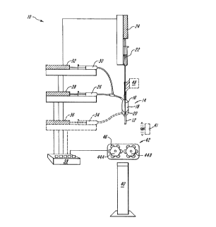
In FIG. 1, a system 10 is shown for
producing a tubular extrudate 12 according to the
5 present invention, including an extrusion head 14
- having a first (innermost) bore 16, a second outer
bore 18 and, optionally, a third (outermost) bore
20. The system 10 further includes a cell suspension
supply 22 and an associated pump 24, a polymer
10 solution supply 26 and an associated pump 28 and,
optionally, a flush solution supply 30 with a pump
32. Additionally, the system can also, optionally,
include a outer flowing quenchent supply 34 with an
associated pump 36. All of the pump elements can be
15 controlled manually or, preferably, by an automated
controller (e.g., a microprocessor) 38. The system
10 can also include a quenchent bath 40, which would
normally be disposed directly below the extrusion
head 14 during operation. Alternatively, the system
20 can include a blower 41 or the system can be employed
within an evacuated or other reduced pressure chamber
to aid in solvent removal.
When the system 10 is employed to shape the
25 tubular extrudate into a multi-compartment cell
capsule string, a sealing means can be employed. One
such sealing element 42 is shown in FIG. 1, including
two motorized wheels 44A and 44B which have a series
of protuberances 46 which cooperate during rotation
30 to periodically pinch and seal the tubular extrudate
as it passes between the wheels 44A and 44B.
- 2~49~56
-14-
Alternatively, a retraction means 48 can be employed
to periodically retract the inner bore so as to
interrupt the flow of the cell suspension. The
effect of these retractions is to periodically seal
5 the tubular extrudate and again form multiple
compartments. In yet another alternative approach,
the controller 38 can vary the pressure applied by
pump 24 (and/or pump 28) to create periodic
interruptions in the flow of the cell suspension.
In FIG. 2, the extrusion head 14 is shown in
more detail, including an inner bore 16 for delivery
of a cell suspension and an outer bore 18 for
delivery of a polymeric solution. As the cell
15 suspension and the polymeric solution are extruded
through the common extrusion pore 19, the polymeric
solution coagulates to form an outer coating about
the cell suspension.
In FIG. 3, an alternative extrusion head 14A
is shown in more detail comprising an inner bore 16
for the delivery of the cell suspension, a second
bore 18 (surrounding the inner bore) for delivery of
the polymeric solution, and an outer most bore 20 for
25 delivery of a flowing quenchent fluid, such as
saline. In this embodiment, a smooth coating can be
obtained by simultaneously extruding the cell
suspension and polymeric solution through common pore
19 while applying a flowing quenchent fluid during
30 the extrusion (e.g., from the outer most bore 20 in
the extrusion head assembly 14A.)
-15- a~ 5~
In FIG. 4, the sealinq element 42 of FIG. 1
is shown in more detail. Motorized wheels 44A and
44B are mounted on opposite sides of the tubular
extrudate 12, such that upon rotation protuberances
5 46 on the wheels periodically come in contact with
the estrudate 12 to pinch and seal the estrudate 12
as it esits the estrusion head 14. The wheels 44A
and 44B can be mechanically linked and operated by a
conventional motor under the control of a controller,
10 such as shown in FIG. 1. The result of the periodic
sealing of the estrudate 12 is a multi-compartment
macrocapsule strand 50 having a polymeric membrane 52
surrounding an encapsulated cell solution 54 with
individual cells 56 disposed therein. The individual
15 cell capsules are joined to each other by
connective filaments 58 where the protuberances 46 of
the sealing means 42 have pinched the extrudate 12.
Various polymers can be used to form the
20 membrane coatings of the present invention, including
polymers derived from solutions which would otherwise
be incompatible with the propagation of living
cells. Because of the unique estrusion process
disclosed in the present invention, solvents which
25 would otherwise be tosic are quickly driven away from
the aqueous cell suspension during the membrane
formation process, thereby permitting the use of many
new and potentially useful polymeric materials. For
esample, polymeric membranes can be formed from
30 polyacrylates (including acrylic copolymers),
polyvinylidenes, polyurethanes, polystyrenes,
polyamides, cellulose acetates, cellulose nitrates,
polysulfones, polyacrylonitriles, as well as
derivatives, copolymers, and mistures thereof.
-
C5~
-16-
The solvent for the polymer solution will
depend upon'the particular polymer chosen for the
membrane material. Suitable solvents include a wide
variety of organic solvents, such as alcohols and
5 ketones, generally, as well as dimethylsulfo~ide
(DMSO), dimethylacetamide (DMA) and dimethylformimide
(DMF), in particular. In general, water-miscible
organic solvents are preferred.
The polymeric solution or ~dope~ can also
include various additives, including surfactants to
enhance the formation of porous channels, as well as
antio~idants to se~uester o~ides that are formed
during the coagulation process. Moreover, when the
15 cell capsules of the present invention are designed
for implantation, materials, such as
anti-inflammatory agents and cell growth factors, can
also be incorporated into the polymeric membrane to
reduce immune response or stimulate the cell culture,
20 respectively. Alternatively, these materials can be
added to the multi-compartment cell capsule strands
after formation by a post-coating or spraying
process. For e~ample, the strands can be immersed in
a solution which contains an anti-inflammatory agent,
25 such as a corticoid, an angiogenic factor, or a
growth factor following e~trusion to post-coat the
cell capsules.
~,~
,f
2~49~
-17-
Post coating procedures can also be used to
provide a protective barrier against immunogens and
the like. For esample, after formation, the cell
capsule strands can be coated (e.g., by immersion,
5 spraying or applying a flowing fluid during
extrusion) with a surface protecting material, such
as polyethylene oxide or polypropylene oxide (e.g.,
having a molecular weight of about 10,000 Daltons or
greater), to inhibit protein interactions with the
10 capsules.
Various techniques can also be employed to
control the smoothness or roughness of the outer
surface of the polymeric coating. In some instances,
15 a very smooth outer coating can be preferable to
reduce scar tissue attachment and other
immunoreactions during implantation. Such a smooth
coating can be obtained by simultaneously immersing
the tubular extrudate in a quenchent, such as a bath
20 of physiological saline, or by applying a flowing,
quenchent fluid during the extrusion (e.g., from a
third, concentric, outermost bore in an extrusion
head assembly). Alternatively, in some applications
a rough outer surface with larger pores may be
25 desired, for example, in instances where capillary
ingrowth during implantation is desired, and such a
rougher outer surface can be obtained by coagulation
in air.
-18~
Various cell lines can be encapsulated
according to the present invention. As noted above,
the multi-compartment cell culture strings are
particularly useful for the constitutive delivery of
5 neurotransmitters, such as dopamine, which is
secreted by cells of the adrenal medulla, embryonic
ventral mesencephalic tissue and neuroblastic cell
lines. PC12 cells (an immortalized cell line derived
from a rat pheocromocytoma) are particularly
10 preferred in some applications because of their
ability to secrete large amounts of dopamine over
long periods of time. Other neurotransmitters
include gamma aminobutyric acid (GABA), serotonin,
acetylcholine, noradrenaline and other compounds
15 necessary for normal nerve functions. A number of
cell lines are known or can be isolated which secrete
these neurotransmitters. Cells can also be employed
which synthesize and secrete agonists, analogs,
derivatives or fragments of neurotransmitters which
20 are active, including, for esample, cells which
secrete bromocriptine, a dopamine agonist, and cells
which secrete L-dopa, a dopamine precursor.
In other embodiments of the invention, the
25 encapsulated cells can be chosen for their secretion
of hormones, cytokines, nerve growth factors,
angiogenesis factors, antibodies, blood coagulation
factors, lymphokines, enzymes, and other therapeutic
agents.
-19- 2~905~
The aqueous cell suspensions can further
include various additives to protect the cells during
the extrusion process or to stimulate their growth
subsequently. Such additives can include, for
5 example a nutrient medium or growth factors which are
incorporated into the aqueous suspension, as well as
various physiologically-compatible substrate
materials to enhance cell growth. The substrate
material can be a dispersive material which prevents
10 the encapsulated cells from clumping together or an
anchorage material which provides additional sites
for cell attachment. Examples of dispersive
materials include negatively-charged polysaccharides
and hydrogels, such as polyalginates, polyfuracellans
15 and polycarrageens, while the anchorage substrate
material can be a proteinaceous material, such as
collagen, laminin, or positively-charged
polysaccharides or polyamino acids, such as
polyglucosamines, poly(n-acetyl)gulcosamines or
20 polygalactosamines. Alternatively, the cell
suspension or the polymeric solution (or both) can
include a foaming agent or a blowing agent which can
distort the inner surface of the polymeric coating to
increase the anchorage surface area of the tubular
25 interior.
The products of the present invention can
take various forms, including simple tubular
extrudates as well as multi-compartment cell capsule
30 strings. The shape of the multi-compartment strings
can be tubular, resembling sausages, or nearly
spherical, resembling strings of pearls. The maximum
-20- ~Q~Q5~
outer diameter of the strand will typically range
from about 0.1 to about l.0 millimeters. The
membrane wall thic~ness will typically range from
about lO to about lO0 micrometers. The strand length
5 of the strands will vary depending upon the
particular application.
The products can also take the form of
~tethered~ cell capsules, that is, one or more
lO individual cell compartments are connected to a long
polymeric tube or string. In FIG. 5, such a tethered
cell capsule 51 is shown havinq a polymeric membrane
52 surrounding an encapsulated cell solution 54 with
individual cells 56 disposed therein. The cell
15 capsule 51 further includes a long polymeric filament
59 which can be formed by the same apparatus as
described above in connection with FIG. 4 by
interrupting the flow of the cell solution and
constraining the polymeric solution to form a solid
20 tether. The tether also can be post coated with a
material (e.g., a polyurethane or the like) which
imparts additional strength to the filament. Such
tether cell capsules can find a variety of
applications, particularly when implanted in a
25 subject for constitutive delivery of active factors.
In use, the cell capsule can be located as close to
the target region (e.g., in the brain, peritoneal
cavity or elsewhere) as desired while the other end
of the tether can be fi~ed at a convenient anchor
30 point or disposal in a readily accessible location
for retrieval.
~ 04G~ oS(~
-21-
The invention will nest be described in
connection with certain illustrative, non-limiting
e~amples:
EXAMPLES
An estrusion system similar to that illustrated
in FIG. 1 was used, consisting of three
electronically controlled programmable infusion
10 pumps, a jet spinneret, two motor-controlled, coasial
wheel systems on the perimeter of which occluding
polyt~etrafluoroethylene tubes were mounted, and an
uptake system.
The macrocapsules were formed by injection
of a polymeric solution into the outer tube of the
spinneret. A coagulant, typically the encapsulated
cells in their culture medium, was simultaneously
injected in the spinneret inner tube. The
20 encapsulating membrane was formed by a dry-jet, wet
spinning process, i.e., the fast stabilization of the
polymer solution emerging from the spinneret nozzle
by the internal quench medium coupled with further
stabilization in a quench bath. The closure of the
25 fiber was performed by mechanically squeezing the
forming hollow fiber with the coasial wheel system
prior to immersion in the quench bath. Near the
spinneret head, the solvent concentration was
sufficiently high to allow proper fusion of the fiber
30 wall. Following each round of encapsulation, pure
solvent was flushed automatically through the lumen
of the spinneret to avoid clogging of the nozzle.
-22-
PC12 cells, an immortalized cell line
derived from a rat pheocromocytoma which secretes
large amounts of dopamine, were cultivated on
collagen-coated tissue culture dishes in RPMI 1640
5 medium supplemented with 10% heat inactivated horse
serum and 5% fetal calf serum. Dissociated bovine
adrenal medullary cells, a non-dividing cell type
which secretes dopamine, were maintained in DMEM
medium supplemented with 5% fetal calf serum. Prior
10 to encapsulation, the cells were harvested and loaded
at a concentration of 1 X 10⁵ cells/ml in a 3 ml
syringe. A 15 percent vinylchloride-acrylonitrile
copolymer solution in either dimethylsulfoside
(DMSO), dimethylformamide ~DMF), or dimethylacetamide
15 (DMA) was loaded into a 5 ml glass syringe. Both
solutions were then coestruded through the spinneret,
and the capsules were collected in a physiologic
saline solution. The capsules were rinsed and placed
in individual wells containing the appropriate
20 culture media.
Basal and potassium-evoked release of
catecholamines was quantified under static incubation
conditions by ion-pair reverse-phase high performance
2~ liquid chromatography (HPLC) equipped with
electrochemical detection at 2 and 4 weeks.
Morphological analysis, including light, scanning,
and transmission electron microscopy, was performed
on representative samples for each time period.
-23~
All cell-loaded capsules released dopamine
into the medium under basal conditions at all time
periods. High potassium treatment increased dopamine
release from both PC12 and adrenal medullary cells.
5 Dopamine output by PC12 cells, but not adrenal
medullary cells, increased with time. The increase
in dopamine release by the PC12 cell-loaded capsules
over time is believed to be related to cell
proliferation within the polymer capsule. No
10 significant difference in dopamine release could be
observed from PC12-loaded capsules estruded with the
three different solvent systems (DMSO, DMF, DMA),
which suggests that the encapsulation technique of
the present invention may prevent cell damaqe
15 inflicted by solvents tFIG. 6). Due to the higher
pressure of the inner bore system, the solvent was
quickly driven toward the outside of the polymer
~, ,
capsule which prevented estended cell-solvent contact.
Morphological analysis revealed the presence
of small clusters of PC12 cells randomly dispersed
throughout the lumen of the capsule. At the electron
microscope level, well-preserved PC12 cells, with
their typical electron-dense secretory granules,
25 could be observed. Cell division within the capsule
space was suggested by the presence of numerous
mitotic figures. Although initially coestruded as a
cell suspension, adrenal chromaffin cells formed
packed aggregates one week after encapsulation.
-24- ~ 0~ Q~
FIG. 7 shows the results cf an ~ vitro
assay in which PC12 cells were encapsulated accordinq
to the present invention and monitored for release
dopamine at two and four weeks following
5 encapsulation. Dopamine levels were measured under
both normal (controlled) conditions and also under a
high potassium stimulation, which is known to induce
depolarization of the cells and, consequently, to
increase the secretion of dopamine in viable cells.
10 As can be seen from the graph, there was little
activity at two weeks; however, at four weeks the
encapsulated cells e~hibited dopamine secretions not
only under normal conditions but also eshibited a
strong response to the potassium stimulation,
15 indicating that the cells were indeed viable in their
encapsulated state.
FIG. 8A and 8B shows the results of further
in vitro assays in which the secretions of PC12 cells
20 and chromaffin cells, respectively, were monitored
four weeks after encapsulation according to the
present invention. Again, the cells were stimulated
by high potassium concentrations and the medium while
the PC12 cells released only dopamine, the chromaffin
25 cells released a variety of catecholamines. The
graph shows the levels of noradrenaline (NE),
epinephrine (EPI), and dopamine (DA).
.~
-25- 20~9QS~
Due to their fluid dynamics, the
macrocapsules extruded in accordance with the present
invention will allow the use of a wider range of
polymer/solvent systems and can constitute a more
5 efficient encapsulation technique. The results show
that immortalized and differentiated
dopamine-secreting cells will survive in
macroencapsulation. The ability of these capsules to
spontaneously release dopamine over time suggests
10 that polymer encapsulation can provide an alternative
to the transplantation of non-encapsulated or
microencapsulated dopamine-secreting cells in the
treatment of Parkinson's disease.