Note: Descriptions are shown in the official language in which they were submitted.
2~505~1
APPARaTUS AND NETHOD FOR PRECISELY CONTROLLING THE EXCISION
OF OBSTRUCTIVE ~ISSUE IN A HUMAN BLOOD VESSEL
BACXGROUND OF THE INVENTION
Field of the Invention:
This invention constitutes a means and method for the
excision of tissue from within the lumen of a human blood
vessel by the use of a Precision Atherectomy Catheter (PAC)
system. Although much of the description herein concerns
atherectomy of plaque from within an artery, this invention
is more generally applicable to the excision of any tissue
from any blood vessel of a human body.
Description of the Prior Art:
There are numerous treatments to remove tissue from
lumens within the vessels in a human body including surgical
interventions such as endarterectomy and by-pass surgery
using veins or artificial graft materials. Balloon
angioplasty is becoming increasingly popular for the
dilation of arterial stenoses without the excision of the
plaque. More recantly atherectomy, the excision from an
artery of atheromatous plaque, has been successfully used to
open arterial stenoses.
In UK Patent Application GB-A 2,044,103 by D.N. Ross
dated October 15, 1980 there is described a device for
removing plaque within an artery by drawing together two
cutting edges that are initially placed on either side of an
arterial stenosis. One significant disadvantage of the Ross
invention is that it cannot cut a passageway through the
plaque that is larger than the outside diameter of the
catheter.
U.S. Patent 4,765,332, issued August 23, 1988 to Robert
E. and Tim A. Fischell, entitled "Pullback Atherectomy
Catheter System," teaches a retrograde cutting catheter
that can be advanced over a guide wire with a single cutting
edge that can be rotated or mechanically vibrated, but does
not teach a means for forming a passageway through the
plaque that is larger than the outside diameter of the
catheter.
U.S. Patent Application No. 447,187 filed on December
7, 1989 by Robert E, Fischell and Tim A. Fischell (which is
included herein by reference) describes an atherectomy
catheter which also is incapable of forming a passageway
though a blood vessel that is larger than the outside
diameter of that catheter.
The European Patent Application No.EP-A 0 163,502 filed
on May ~, 1985 by J.B. Simpson describes an atherectomy
catheter which can form a passageway through an arterial
stenosis that is larger than the outside diameter of the
lcatheter. This is accomplished by inflating a balloon
opposite a window in a housing at the catheter's distal end
and pushing a cutter through the plaque and then pushing the
plaque forward into a plaque collection chamber. The window
is then rotated to a new position in the plaque and the
process is repeated several times until a passageway is
formed that is a larger diameter than the catheter's outer
diameter when the balloon is not in~lated. Although the
Simpson invention does solve the problem of opening a
passageway in the artery that is larger than the outside
diameter of the catheter when the balloon in not inflated,
there is no control over the thickness of plaque that is
removed with each pass of the cutter. Thus at a low
pressure in the balloon very little plaque would enter the
window and therefore very little plaque would be removed.
At a very high balloon pressure, much more plaque would be
pushed into the window and therefore there would be a much
deeper cut into the plaque and considerably more plaque
would be removed (as compared to low pressure) and in fact
the arterial wall could be (and in practice has been)
perforated. Furthermore, for the same balloon pressures,
harder plaque would not enter the window as much as softer
3 ~50~1~
plaque, and in that case, less plaque would be removed by
the cutter. In summary, with the Simpson atherectomy
catheter, the amount of plaque removed on each forward
thrust of the cutter is indeterminate and arterial
perforation does occur.
SUMMARY OF THE INYENTION
The Precision Endoluminal Tissue Excision Catheter
(PAC) system as described herein is designed to overcome
many of the shortcomings of the prior art devices. The PAC
system as described herein is applicable only to blood
vessels in parts of the human body which can be compressed;
e.g., the arteries in the arms, legs, neck or abdomen.
PAC uses a pressure cuff surrounding the portion of the
body at the site of the tissue obstruction to compress the
tissue as the atherectomy catheter moves through and cuts
that tissue. After the atherectomy is completed, the
pressure cuff is returned to ambient pressure and the
catheter is removed. The pressure of blood in the artery or
vein then moves the wall of the blood vessel outward so that
the passageway formed is in fact a larger diameter than the
catheter's outside diameter.
An additional characteristic of the PAC design is that
the obstructive tissue (typically p]aque) is always cut off
the vessel wall by an exact amount. Since modern
angiography provides an indicat~on of plaque thickness on
the wall, by limiting the plaque thickness that can be
removed on a single pass of PAC through that tissue, the PAC
system can provide assurance that the vessel wall will not
be perforated.
Since there are now ultrasonic imaging catheters that
provide precise measurement of plaque thickness on an
arterial wall, this ultrasonic imaging can now be used in
conjunction with the PAC system to provide further safety by
4 ~50~11
preventing perforation of the arterial wall.
Thus one objective of the PAC system is to make a
passageway through obstructive tissue in a human blood
vessel that is larger than the outside diameter of the
catheter.
Another objective of the PAC system is to measure the
thickness of the obstructive tissue on the blood vessel wall
and to use compression combined with a specific cutter
configuration to excise not more than that known thickness
of obstructive material from the wall of the blood vessel.
Still another objective of the PAC system is to easily
remove the plaque collected in the plaque collection
chamber.
BRIFF DESCRIPTION OF T~E DRAWINGS
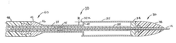
FIG. 1 is a longitudinal cross section of the distal
end of the catheter subsystem of the PAC system shown in
its closed position.
FIG. 2 is a longitudinal cross section of the distal
end of the catheter subsystem of the PAC system shown in its
open position.
FIG. 3 illustrates the catheter subsystem and the
pressure cuff subsystem of the PAC system with the catheter
subsystem shown lying in its closed position within a
stenosis in an artery of a leg and with the pressure cuff
uninflated.
FIG. 4 illustrates the PAC system with the catheter
subsystem shown in the open position within a leg artery
and with the pressure cuff uninflated.
FIG. 5 illustrates the PAC system with the distal end
of the catheter subsystem shown in its open position and
with the pressure cuff inflated so as to compress the
plaque against the closing catheter's distal cylinder.
FIG. 6 shows the PAC system with the catheter subsystem
pulled back and closed and the pressure cuff deflated.
20~5ll
FIG. 7 shows the PAC system in conjunction with an
endoluminal ultrasonic imaging system being used for
atherectomy in a human leg.
FIG. 8 is a cross-sectional view showing an ultrasonic
transducer placed near the distal end of the closing
catheter of PAC's catheter subsystem.
DETAILED DESCRIPTION OF THE DRAWINGS
The PAC system consists of two major subsystems: the
catheter subsystem and the pressure cuff subsystem. FIG. 1
is a longitudinal cross-sectional view showing the distal
end of the catheter subsystem 10 in the closed position.
The catheter subsystem 10 consists of 3 principal parts:
the guide wire 14, the cut/collect catheter 30 and the
closing catheter 40. From FIG. 1 we see that the
cut/collect catheter 30 has a cutting cylinder 32 which has
a sharpened edge 32a at its proximal end and at its distal
end it is joined (typically welded) to the central support
34. At the distal end of the central support 34 is a
distal projection 36 which is designed to hold onto an
elastomer, flexible tip 38. When the flexible tip 38 is
molded onto the distal projection 36, a releasing agent is
first applied to the distal projection 36 so that after the
molding process, the flexible tip 38 is free to rotate about
the distal projection 36 of the central support 34. In the
proximal direction, the central support 34 has a central
cylinder 35 which surrounds the guide wire 14.
The closing catheter 40 consists of a distal cylinder
45 which has a flared end 47 at is distal end and at its
proximal end is connected to a cone 46 which is connected to
the straight section 42 of the metal distal portion of the
closing catheter 40. The section 42 also has a proximal
projection 44 designed to securely hold onto a plastic
cylinder 48. At its proximal end (wh~ch is shown in FIG~
~05U51 .~
7), the plastic cylinder 48 of the closing catheter 40
extends outside the patient's body.
Located between the outer surface of the distal
cylinder 45 and inner surface of the cutting cylinder 32 is
the plaque collection chamber 12. When the atherectomy
procedure is completed, the catheter subsystem 30 will be
in the closed condition as shown in FIG. 1, and the plaque
that is to be cut and collected will lie within the plaque
collection chamber 12.
FIG. 2 shows a longitudinal cross-sectional view of the
distal end of the catheter subsystem 10 of the PAC system
with the catheter subsystem 10 shown in the open position.
The open position is achieved by pulling the closing
catheter 40 backwards (i.e., in a retrograde direction)
relative to the cut/collect catheter 30 whose proximal end
also extends outside the body. FIG. 2 shows a plastic
cylinder 37 attached to the central cylinder 35 of the
central support 34 of the cut/collect catheter 30. It is
this plastic cylinder 37 of the cut/collect catheter that
extends proximally outside the patient's body as shown in
FIG. 7. Only when starting from its open position, as
shown in FIG. 2, is the catheter subsystem 10 capable of
excising plaque from the walls of a human blood vessel.
One thing to note in FIG. 2 is the radial distance
between the outer cylindrical surface of the distal cylinder
45 and the cutting edge 32a of the cutting cylinder 32. It
is this radial offset distance R which determines the
precise thickness of obstructive tissue that is cut off as
the cut/collect catheter 30 is pulled back in a retrograde
direction over the tissue while the closing catheter 40
remains stationary.
At its proximal end, lying outside the patient's body,
the plastic cylinder 37 is typically connected to a rotating
device such as is described in U.S. Patent Application
Serial No. 447,187 by Robert E. and Tim A. Fischell. This
rotating device is used to spin the cutting edge 32a as it
7 ~5~51`
is pulled back through the plaque to be excised thus
enhancing the cutting action.
The cutting cylinder 32 of the cut/collect catheter 30
is typically fabricated from a hardenable, 400 series
stainless steel. All other metal parts would typically be
made from 304 stainless steel or a metal with equival~nt
characteristics. All plastic parts would typically be made
from elastomer materials such as polyethylene, polyurethane,
Nylon or equivalent plastic materials. The outer diameter
of the catheter subsystem would typically lie between 1.0
and 5.0 mm.
FIG. 3 shows the entire PAC system which consists of
the catheter subsystem 10 and pressure cuff subsystem 20.
It should be remembered that the catheter subsystem 10
consists of a guide wire 14, a cut/collect catheter 30 and a
closing catheter 40. The pressure cuff subsystem 20
consists of an inflatable cuff 22, a pumping means 24 with a
pressure relief valve 25 and a pressure gauge 26 all of
which are illustrated in FIG. 3.
To achieve the condition shown in FIG. 3, it is
typical to have an insertion sheath inserted at the groin
of a patient. After the insertion sheath is in place, a
guide wire is advanced through the sheath and through the
stenosis consisting of plaque located somewhere in an artery
or a vein. FIG. 3 illustrates plaque forming a stenosis of
an artery in the leg. After the guide wire is placed through
the stenosis, the catheter subsystem lO having a tapered
distal end flexible tip 38 is advanced over the guide wire
14 and through the stenosis until the cutting edge 32a of
the cutting cylinder 32 lies just distal to the stenosis. To
accomplish this positioning within the stenosis, the
catheter subsystem 10 is advanced in the closed position;
i.e., with the cone 46 of the closing catheter 40 pushed
against the proximal end of the cutting cylinder 32.
8 2 V ~
The next step in this procedure (as shown in FIG. 4)
is to pull back on the plastic cylinder 48 that lies outside
the body which causes the closing catheter 40 to be pulled
back while keeping the cut/collect catheter 30 stationary.
Pulling the closing catheter 40 back exposes the cylinder
45 at the distal end of the closing catheter 40 and the
cutting edge 32a of the cutting cylinder 32. Also, as can
be seen in FIG. 4, the inside diameter of the lumen in the
plaque would typically remain at the same diameter as the
outside diameter of the catheter subsystem 10. This is
because plaque is a reasonably plastic material and will
remain at essentially that same diameter (or slightly less)
to which it was dilated by the insertion of the catheter
subsystem 10 as shown in FIG. 3. One can also see in FIG. 4
that the pressure cuff subsystem 20 remains in the
uninflated condition.
FIG. 5 shows the next step in this procedure wherein
the inflatable cuff 22 of the pressure cuff subsystem 20
is inflated to a higher pressure. This inflation can be
accomplished by pumping on a pressure bulb 24 as typically
accomplished when measuring blood pressure. The exact level
of the pressure attained will be indicated by the pressure
gauge 26. When the inflatable cuff 22 is inflated as
indicated by the pressure gauge 26, the effect is to
collapse the arterial wall around the outer diameter of the
catheter subsystem 10, and more importantly the plaque is
pushed onto the outer cylindrical surface of the distal
cylinder 45 of the closing catheter 40. In this position a
precise thickness of plaque is ready to be excised.
Specifically, the precise plaque thickness to be excised is
the equal to the radial offset R as shown in FIG. 2.
With the inflatable cuff 22 inflated to a reasonably
high pressure, the cut/collect catheter 30 is pulled
backwards so that the cutting edge 32a of the cutting
cylinder 32, typically while rotating, is pulled back
through the plaque thus cutting a precise thickness of
9 2 () 5 1) ~ 1 ~
plaque away from the remaining plaque which adheres to the
arterial wall. The pullback of the cut/collect catheter 30
continues until the proximal end of the cutting cylinder 32
is in contact with the cone 46 of the closing catheter 40.
After this cutting has been completed, all the excised
plaque will be situated in the plaque collection chamber 12
(see FIG. l).
FIG. 6 illustrates the next step in using the PETEC
system. Specifically FIG. 6 shows that the pressure cuff
subsystem 20 has been deflated. This can be accomplished
by opening a valve 25 on the pumping means 24 the pressure
being indicated by the pressure gauge 26. Such valves are
typically found on pressure cuffs used to measure blood
pressure. We also see in FIG. 6 that the proximal end of
the cutting cylinder 32 is placed tightly against the cone
46 of the closing catheter 40. In FIG. 6, the entire
catheter subsystem 10 (except the guide wire 14) has been
pulled back beyond the residual plaque of the stenosis.
Because the artery can expand after the inflatable cuff 22
is deflated, the luminal diameter inside the residual plaque
will be a larger diameter than the outside diameter of the
catheter subsystem 10. This is a most important objective
in the field of atherectomy, in that, the ideal atherectomy
system will remove plaque from the arterial wall to a larger
luminal diameter than the outside diameter of the catheter
used to perform the procedure. By this method,
comparatively small catheters can be percutaneously placed
through the patient's skin and yet the catheter can
accomplish the function of removing plaque from the blood
vessel to a comparatively large diameter. The method
described herein can therefore be used to leave only a small
residual of obstructive tissue on the wall of the blood
vessel.
After the cutting has been achieved and the amount of
plaque adhering to the wall significantly reduced, the
entire catheter subsystem 10 is removed from the body.
2~505~:L
When the catheter subsystem 10 is outside the body, the
closing catheter 40 is pulled back from the cut/collect
catheter 30. The process of opening the distal end of the
catheter subsystem 10 results in the flared end 47 on the
distal cylinder 45 sweeping out all the plaque contained
in the plaque collection chamber 12 (see FIGS. 1 and 2).
Because the outer edge of the flared end 47 fits closely
within the inside surface of the cutting cylinder 32, all
the plaque captured in the plaque collection chamber 12 will
be automatically pulled out of the plaque collection chamber
12 when the closing catheter 40 is pulled back. The plaque
thus removed can undergo pathologic examination to determine
the nature of the excised tissue.
Although the procedure described herein uses a
pressurized cuff set at a comparatively high pressure when
the cut/collect catheter 30 is pulled back through the
plaque, alternative methods of using such a pressure cuff
subsystem are available. For example, it may be desirable
to pressurize the inflatable cuff 22 to a considerably
higher pressure when the distal end of the catheter
subsystem 10 has been opened. However, in
contradistinction to the aforementio~ed method, the
inflatable cuff 20 could be deflated just prior to pulling
back the cutting edge 32a of the cutting cylinder 32 through
the plaque. ~his method could be effective because the
plaque is comparatively plastic and the mere application of
pressure on the arterial wall can push the plaque against
the cylinder 45 so that, even when the pressure is relieved
there will be a considerable amount of plaque in contact
with the outer cylindrical surface of the distal cylinder
45. The plaque would be excised when the cutting cylinder
32 is pulled back until its proximal end is in contact with
the cone 46 at the distal end of the closing catheter 40.
One reason why this method might be preferred is that it
would tend to avoid perforation of the normal arterial wall.
This method would provide cutting of only that obstructive
11 20~0~1~
tissue which extends inwardly from the wall of the blood
vessel and not the blood vessel wall itself.
Typical pressures that would be used for the procedure
illustrated in FIGS. 3, 4, 5 and 6 would be just above the
patient's systolic pressure. For example, if the patient's
systolic pressure in his leg was measured to be 150 mm of
Hg, it would be useful to use pressures that are 0 to 50 mm
of Hg above this pressure. However pressures between 50 and
250 mm Hg may be successfully used for this procedure. When
the technique of pressurizing the cuff and deflating the
cuff just prior to pulling back the cutting cylinder 32 of
the cut/collect catheter 30 is used, one might go to
pressures as high as 300 mm of Hg so that one can be sure
that the plaque has been plastically deformed onto the outer
surface of the distal cylinder 4S of the closing catheter
40.
Although one might conceive of a simple belt being used
in place of the pressure cuff subsystem 20, without an
accurate method for measuring the compressional force on the
delicate blood vessel, damage to the vessel including
perforation may be the result. Thus, a rather precise
indication of compressional pressure on the blood vessel is
essential in order to achieve a consistently safe
atherectomy procedure.
It should be understood that throughout the description
of the P~TEC system given above, the thickness of the
obstructive tissue on the vessel wall is measured by
angiography prior to tissue excision. Since this tissue
thickness can be determined before cutting and since the
precise thickness of tissue removed (as given by the radial
offset R) is known, the risk of vessel wall perforation is
minimized.
It is conceived that catheter subsystems with varying
offsets may be used depending on the measured thickness of
obstructive tissue to be excised from the vessel wall.
Typical dimensions for the radial offset R would be between
12 ~'~5~51~
0.1 and 1.0 mm. For example, if a tissue thickness on the
vessel wall is measured by angiography to be 0.7 mm, then a
catheter subsystem with R = 0.5 mm might be used to excise
a plaque thickness of 0.5 mm. This would open a vessel to a
sufficiently large diameter while avoiding the risk of
perforating the vessel wall.
Another method for measuring tissue thickness on the
wall is by ultrasonic imaging. Specifically U.S. Patent
No. 4,917,097 entitied "Apparatus and Method for Imaging
Small Cavities," which is incorporated herein by reference,
describes a system which is capable of measuring the
thickness of tissue on the vessel wall to 0.1 mm. A
separate catheter with this measurement capability could be
used to measure obstructive tissue wall thickness prior to
using the PETEC system for excising tissue. It is further
conceived that the closing catheter 40 could be used as a
sonography catheter to measure the thickness of obstructive
tissue. Specifically, FIG. 7 shows the pressure cuff
subsystem 20 wrapped around a human leg into which the
catheter subsystem 10 has been percutaneously advanced over
a guide wire 14. The guide wire 14, the plastic cylinder 48
of the closing catheter 40 (see FIGS. 1 and 2) and the
plastic cylinder 37 of the cut/collect catheter 30 (see FIG.
2) are all shown extending proximally outside of the
patient's body. An endoluminal sonography system 50 is also
shown in FIG. 7. This system 50 consists of electronic
equipment 52 which is used to generate ultrasonic signals
and to measure the reflected signal from within the blood
vess~l. The equipment 52 typically includes a CRT display
54. Electrical wires 56 and 58 are shown connecting into
the plastic cylinder 48 of the closing catheter 40
Although only two wires are shown, additional wires may be
required to obtain detailed ultrasonic imaging of the vessel
wall.
FIG. 8 shows the distal end of the wires 56 and 58 as
they are connected into an ultrasonic transducer 59 that is
13 2050~
placed at the distal end of the plastic tube 4~. The
technique for obtaining endoluminal images of blood vessels
is well known in the art and will not be described in any
more detail herein.
Using the system illustrated in FIGS. 7 and 8, it is
possible to precisely measure the thickness of obstructive
tissue on the blood vessel wall. This measurement can be
accomplished with a separate catheter or with the addition
of sensing means at the distal end of the closing catheter
as shown in FIG. 8. When combined with the PETEC's
capability to excise a precise thickness of obstructive
tissue, the ultrasonic imaging system provides an excellent
combined system for precisely excising such tissue and
obtaining the maximum luminal opening with the least risk
of vessel wall perforation.
Although the discussion herein is principally concerned
with a catheter that cuts in the retrograde direction, the
invention that is taught herein is equally applicable to
atherectomy catheters that cut in the anterograde direction
such as that described in the Fischell et al U.S. Patent
No. 4,898,575 or the Simpson European Patent Application
No. EP-A 0 1~3,502. Furthermore, various blade
configurations such as those described in the prior art
could be used in conjunction with the technique for
compressing plaque as described herein. Furthermore,
ablation techniques such as using grinding, laser
vaporization or high energy ultrasonic vibrators could be
used successfully with the tissue compression methodology
described herein.
Various other modifications, adaptations, and
alternative designs are, of course possible in light of the
above teachings. Therefore, it should be understood at this
time that within the scope of the appended claims, the
invention may be practiced otherwise than as specifically
described herein.