Note: Descriptions are shown in the official language in which they were submitted.
_ 2050601
PRODUCTION IN BACTERIA AND YEAST
OF HEMOGLOBIN AND ANALOGUES THEREOF
Field of the Invention
The present invention relates to the intracellular
assembly of a hemoglobin-like protein in biologically
functional, substantially soluble form through co-expression
of alpha- and beta-globin-like polypeptides in bacterial or
yeast cells.
It further relates to the genetic fusing of the two
alpha subunits of hemoglobin to form a novel polypeptide, di-
alpha globin, which may be considered a
a
WO 90/13645 2 0 5 0 6 01 P~/US90/02654
- 2 -
partially assembled intermediate leading to a hemoglobin-like
protein, and the use of this compound in the production of
synthetic hemoglobins having an increased intravascular half-
life as compared to stroma-free hemoglobins. It also relates
to the analogous polypeptide di-beta globin.
Information Disclosure Statement
It is not always practical to transfuse a patient
with donated blood. In these situations, use of a red blood
cell substitute is desirable. The product must effectively
transport 02, just as do red blood cells. ("Plasma expanders",
such as dextran and albumin, do not transport oxygen.) The two
types of substitutes that have been studied most extensively
are hemoglobin solutions and fluorocarbon emulsions.
A. Structure and Function of Hemoglobin
Hemoglobin (Hgb) is the oxygen-carrying component of
blood. Hemoglobin circulates through the bloodstream inside
small enucleate cells called erythrocytes (red blood cells).
Hemoglobin is a protein constructed from four associated
polypeptide chains, and bearing prosthetic groups known as
hemes. The erythrocyte helps maintain hemoglobin in its
reduced, functional form. The heme iron atom is susceptible to
oxidation, but may be reduced again by one of two enzyme
systems within the erythrocyte, the cytochrome b5 and
glutathione reduction systems.
About 920 of the normal adult human hemolysate is Hgb
A (designated alpha2 beta2, because it comprises two alpha and
two beta chains). The alpha chain consists of 141 amino acids.
The iron atom of the heme (ferroprotoporphyrin IX) group is
bound covalently to the imidazole of His 87 (the "proximal
histidine"). The beta chain is 146 residues long and heme is
bound to it at His 92. Apohemoglobin is the heme-free analogue
of hemoglobin; it exists predominantly as the ap-globin dimer.
2050601
- 3 -
Separated, hems-free, alpha and beta globins have
been prepared from the hems-containing alpha and beta subunits
of hemoglobin. The separated hems-free globin chains are
folded very differently, even though the hems-containing
subunits are highly similar in secondary structure and basic
folding features. This shows that the binding of the
prosthetic hems group to globin subunits has quite different
effects on alpha and beta globin. Yip, et al., J. Biol. Chem.,
247: 7237-44 (1972).
Native human hemoglobin has been fully reconstituted
from separated hems-free alpha and beta globin and from hemin.
Preferably, hems is first added to the alpha globin subunit.
The hems-bound alpha globin is then complexed to the hems-free
beta subunit. Finally, hems is added to the half-filled globin
dimer, and tetrameric hemoglobin is obtained. Yip, et al.,
PNAS (USA), 74: 64-68 (1977).
In cell-free systems prepared from unfractionated
rabbit reticulocyte hemolysates, globin is actively synthesized
for approximately five minutes, and then protein synthesis
abruptly ceases. Prior addition of hemin prevents or delays
the cessation of synthetic activity, as a result of the effect
of hemin on an inhibitory protein known as "hemin-regulated
inhibitor" (HRI). Hemin deficiency has a more severe effect on
alpha chain synthesis than on beta chain synthesis as alpha
~globin mRNA is less efficient than beta-globin mRNA in
initiating polypeptide chain synthesis. It has been speculated
that alpha chains are released from their site of synthesis
only in the presence of free beta chains, which immediately
complex the released alpha chains to form aQ-globin dimers.
These then combine with hems to form tetrameric hemoglobin.
Winterhalter and Huehns, J..Biol. Chem., 239: 3699 (1964). It
is certainly known that the addition of hems to a~3-globin
dimers (apohemoglobin) leads to the rapid formation of
hemoglobin.
w..
2050601
4
The human alpha and beta globin genes reside on
chromosomes 16 and 11, respectively. Bunn and Forget,
Hemoglobin: Molecular, Genetic and Clinical Aspects, W.B.
Sannders Co, Philadelphia, PA, 1986 p. 172. Both genes have
been cloned and sequenced, Liebhaber, et al., PNAS 77: 7054-
58 (1980) (alpha-globin genomic DNA); Marotta, et al., J.
Biol. Chem., 252: 5040-53 (1977) (beta globin cDNA); Lawn, et
al., Cell, 21:647 (1980) (beta globin genomic DNA).
Hemoglobin exhibits cooperative binding of oxygen by
the four subunits of the hemoglobin molecule (two alpha-globins
and two beta-globins in the case' of Hgb A), and this
cooperativity greatly facilitates efficient oxygen transport.
Cooperativity, achieved by the so-called heme-heme interaction,
allows hemoglobin to vary its affinity for oxygen. Hemoglobin
reversibly binds up to four moles of oxygen per mole of Hgb.
Oxygen-carrying compounds are frequently compared by
means of a device known as an oxygen dissociation curve. This
curve is obtained when, for a given oxygen carrier, oxygen
saturation or content is graphed against the partial pressure
of oxygen. For Hgb, the percentage of saturation increases
with partial pressure according to a sigmoid relationship. The
PSO is the partial pressure at which the oxygen-carrying
solution is half saturated with oxygen. It is thus a measure
of oxygen-binding affinity; the higher the PSO, the more
loosely the oxygen is held.
When the oxygen dissociation curve of an oxygen-
carrying solution is such that the Pso is less than that for
whole blood, it is said to be "left-shifted."
The oxygen affinity of hemoglobin is lowered by the
presence of 2,3-diphosphoglycerate (2,3-DPG), chloride ions and
hydrogen ions. Respiring tissue releases carbon dioxide into
r~
WO 90/13645 PCT/US90/02654
2050601
- 5 -
the blood and lowers its pH (i.e. increases the hydrogen ion
concentration), thereby causing oxygen to dissociate from
hemoglobin and allowing it to diffuse into individual cells.
The ability of hemoglobin to alter its oxygen
affinity, increasing the efficiency of oxygen transport around
the body, is dependent on the presence of the metabolite 2,3-
DPG. Inside the erythrocyte 2,3-DPG is present at a
concentration nearly as great as that of hemoglobin itself. In
the absence of 2,3-DPG "conventional" hemoglobin binds oxygen
very tightly and would release little oxygen to respiring
tissue.
Aging erythrocytes release small amounts of free
hemoglobin into the blood plasma where it is rapidly bound by
the scavenging protein haptoglobin. The hemoglobin-haptoglobin
complex is removed from the blood and degraded by the spleen
and liver.
B. Blood Substitutes, Generally
It is clear from the above considerations that free
native hemoglobin A, injected directly into the bloodstream,
would not support efficient oxygen transport about the body.
The essential allosteric regulator 2,3-DPG is not present in
sufficient concentration in the plasma to allow hemoglobin to
release much oxygen at venous oxygen tension.
Nonetheless, solutions of conventional hemoglobin
have been used as RBC substitutes. The classic method of
preparing hemoglobin solutions employs outdated blood. The red
cells are lysed and cellular debris is removed, leaving what is
hopefully "stromal-free hemoglobin" (SFH).
Several basic problems have been observed with this
approach. The solution must be freed of any toxic components
of the red cell membrane without resorting to cumbersome and
2050601
- 6 -
tedious procedures which would discourage large-scale
production. DeVenuto, "Appraisal of Hemoglobin Solution as a
Blood Substitute", Surqg~y. Gvnecolocry and Obstetrics, 149:
417-436 (1979) .
Second, as expected, such solutions are "left-
shifted" (lower PSO) as compared to whole blood. Could, et
al., "The Development of Polymerized Pyridoxylated Hemoglobin
Solution as a Red Cell Substitute", inn. Emerg. Med. 15: 1416-
1419 (Dec. 3, 1986) .
Third, SFH has a half-life in the circulatory system
of only about 2-4 hours. This is because oxy Hgb partially
dissociates into a dimer (ap) that is small enough to be
filtered by the kidney.
Finally, SFH has a high colloid osmotic pressure
(COP). Thus, administration of SFH in a dose that would have
the same oxygen-carrying capacity as a unit of packed red blood
cells is inadvisable, since the high osmotic pressure
would cause a massive influx of water from the cells into the
bloodstream, thus dehydrating the patient's tissues. This
consideration limits the dose of SFH to that which provide a
final concentration of about 6-8 gm Hgb/dl.
In~an effort to restore the desired PSO, researchers
added 2,3-DPG to the hemoglobin solution. Unfortunately, 2,3-
DPG was rapidly eliminated from the circulation. Scientists
then turned to other organic phosphates, particularly pyridoxal
phosphate. Like 2,3-DPG, these compounds stabilized the "T
state" of the Hgb by forming a salt bridge between the N-
termini of the two beta chains. The pyridoxylated hemoglobin
had a Pso of 20-22 torr, as compared to 10 torn for SFH and 28
torr for whole blood. While this is an improvement over SFH,
the pyridoxylated Hgb remains "high affinity" relative to whole
blood.
~s
2050601
7
C. Chemical Crosslinking of Hemoglobin Subunits
The properties of hemoglobin have been altered by
specifically chemically crosslinking the alpha chains between
the Lys99 of alphal and the Lys99 of alpha2. Walder, U.S.
4,600,531 (1986) and 4,598,064 (1986); Snyder, et al., PNAS
(USA) 84: 7280-84 (1987); Chaterjee, et al., J. Biol. Chem.,
261: 9927-37 (1986). The P50 was 29 mm Hg, and renal
excretion was abrogated by the crosslinking, but the plasma
half-life was increased just 2-3 fold.
This chemical crosslinking was accomplished by
reacting bis(3,5-dibromosalicyl) fumarate with deoxyhemoglobin
A in the presence of inositol hexaphosphate. This reaction has
a low yield (10-20%). Moreover, purification is required to
eliminate derivatives modified at other sites (there are 42
other lysine residues and the amino terminal amino groups of
the four chains at which competing reactions could occur).
A further problem with the use of a "diaspirin"
crosslinking agent is that it can participate in a side
reaction yielding a carcinogenic halophenol.
In the hemoglobin analogue of the present invention,
the N-terminal., valine and C-terminal arginine of the alpha
globins axe connected by means of an amino acid or peptide
linker, without resort of special coupling agents.
The beta chains have also been chemically
crosslinked. Kavanaugh, et al.~, Biochemistry, 27: 1804-8(
1988). Kavanaugh notes that the beta N-termini are 16 A apart
in the T state and 20 A apart in the R state. Not
surprisingly, the introduction of a DIDS bridge between the N-
termini of T state hemoglobin hindered the shift to the R
_f,
WO 90/13645 2 0 5 0 b 01 P~/US90/02654
_ g _
state, thereby decreasing the OZ affinity of the molecule.
While the Kavanaugh analogue has desirable oxygen binding and
renal clearance characteristics, it too is obtained in low
yield.
D. Gene Expression, Generally
Gene expression embraces the transcription of DNA
into messenger RNA, and the translation of messenger RNA into
protein. The process of transcription begins when an enzyme,
DNA-directed RNA polymerase, binds to DNA. The binding site
for this enzyme is often called the "promoter," and the binding
of the enzyme to the promoter may be controlled by various
repressors or inducers of transcription. The RNA polymerase
slides along the DNA molecule, manufacturing a messenger RNA
transcript. When it encounters a second regulatory element,
the "terminator," the enzyme falls off, and the mRNA transcript
is formed.
Messenger RNA is used by the ribosomes, the protein
factories of the cell, as a template for the construction of
the corresponding protein. The ribosomal binding site
comprises the so-called Shine Delgarno (SD) sequence and a
properly spaced initiation (start) codon. Beginning at a
special RNA triplet known as the initiation codon, transfer
RNAs bind to corresponding codons of the messenger. Each
transfer RNA is two-handed; it binds to the messenger codon by
means of a complementary anti-codon, while holding the
corresponding amino acid in position to be linked into the
growing polypeptide chain. The chain falls off when the
ribosome encounters one of three special triplets known as
"stop" codons. That part of the original gene which
corresponds to the messenger sequence from the initiator codon
to the last codon before the stop codon is known as the coding
sequence. There is also a 5'-flanking sequence, which begins
with the promoter, and a 3'-flanking sequence which ends with
the terminator.
CA 02050601 2000-05-31
77481-17
9
E. Polycistronic Expression
It is possible for a single messenger RNA transcript
to have one promoter, but two or more pairs of start and stop
codons that define distinctly translatable sequences. Each
such sequence is known as a "cistron," and the polypeptide
corresponding to the cistrons are thus co-expressed under the
control of the single promoter.
The majority of bacterial operons are polycistronic,
that is, several different genes are transcribed as a single
message from their operons. Examples include the lactose
operon with three linked genes (lacZ, lacy and lacA) and the
tryptophan operon with five associated genes (tr~~E, trpD, trpC,
t~r.~B, and tr~A). In these operons, the synthesis of messenger
RNA is initiated at the promoter and, within the transcript,
coding regions are separated by intercistronic regions of
various lengths. (An operon is a cluster of genes that is
controlled as a single transcriptional genetic unit).
Translational efficiency varies from cistron to cistron.
Kastelein, et al., Gene, 23: 245-54 (1983).
When intercistronic regions are longer than the span
of the ribosome (about 35 bases), dissociation at the stop
codon of one cistron is followed by independent initiation at
the next cistron. With shorter intercistronic regions, or with
overlapping cistrons, the 30S subunit of a terminating ribosome
may fail to dissociate from the polycistronic mRNA, being
instantly attracted to the next translational initiation site.
Lewin, Gene Expression, John Wiley and Sons, New York, N.Y.:
1977, 143-148.
Unlike bacterial mRNAs, eukaroyotic mRNAs are
generally monocistronic in nature. Lewin, Gene Expression,
157.
2050601
- 10 -
Synthetic polycistronic operons have been constructed
and expressed in both prokaryotes and eukaryotes.
Lee, et al., Nucleic Acids Res., 12: 6797 (1984)
describe a special case of a synthetic polycistronic operon in
which all of the cistrons express the same polypeptide. This
homopolycistronic structure was constructed to maximize the
gene dosage effect.
Schoner, et al., PNAS, 83: 8506-10 (1986) translated
a synthetic two-cistron mRNA in ~ coli. The first cistron was
a short, arbitrary AU-rich sequence, while the second cistron
was a mammalian gene (bGH). It was found that "read through"
translation occurred if the stop codon of the first cistron
followed the SD element of the second cistron and lay close to
the start codon of the second cistron. Schoner's purpose was
to overcome his failure to express Met-bGH with its native
codons at high levels, possibly as a result of inhibition of
translation by local secondary structures. The first cistron
was engineered to favor ribosome binding (by placing the SD
sequence and the AUG initiation codon in an AU-rich region free
of local secondary structure). See also Schoner, et al., Meth.
Enzymol., 153: 401-416 (.1987), which reveals that bGH
overproduction by this technique was associated with the
formation of protein granules.
Saito, et al., J. Hiochem., 101: 1281-88 (1987)
expressed a synthetic somatomedin C gene in ~ coli using a two
cistron system. They theorized that the instability of
somatomedin C, a basic polypeptide, might be overcome by
complexing it with an acidic polypeptide. Thus, they
constructed a two-cistron system which could express both
polypeptides. The termination codon of the first cistron
overlapped the initiation codon of the second cistron. The
transformants accumulated somatomedin C at high levels.
However, the somatomedin C was recovered in the form of
insoluble pellets (see page 1282).
i~
2050601
- 11 -
The ribosomes of mammalian cells are likewise capable
of reinitiating translation at an initiation codon downstream
from a termination codon. Thus, Boel, et al., FENS Lett.,
219:181 (1987) expressed a dicistronic transcription unit in
mammalian (CHO) cells. This unit directed synthesis of both
the precursor of human pancreatic polypeptide and of a
selectable genetic marker (mouse DHFR).
Lodan, W088/05486 (1988) describes the production of~
dicistronic 'mRNA which encodes both a protein of interest
(e. g., tissue plasminogen activator)~and a selectable phenotype
(e. g., neomycinresistance). The common promoter was, in each
of the examples a derivative of the Harvey murine sarcoma
virus, and the dicistronic mRNA was translated in suitable
eukaryotic cells.
GENETECH, EP Appl 117,058 (1984) discloses the expression
in vertebrate host cells of a dicistronic expression vector
wherein one cistron codes for the desired protein (e. g., HbsAg)
and a second codes for a second protein (e. g., DNFR) whose
synthesis is subject to environmental control (e. g., with
methotrexate).
F. Fused Genes and Proteins, Generally
Genes may be fused together by removing the stop
codon of the first gene, and joining it in phase to the second
gene. Parts of., genes may also be fused, and spacer DNAs which
maintain phase may be interposed between the fused sequences.
The product of a fused gene is a single polypeptide, not a
plurality of polypeptides as is expressed by a polycistronic
operon. Different genes have been fused together for a variety
of purposes. Thus, Gilbert, U.S. 4,338,397 (1982) inserted a
rat preproinsulin gene behind a fragment of the E. coli
penicillinase gene. His purpose was to direct E. coli
transformants to secrete the expression product of the fused
Z-a
2050601
- 12 -
gene. Fused genes have also been prepared so that a non-
antigenic polypeptide may be.expressed already conjugated to an
immunogenic carrier protein. The present invention, however,
contemplates the joining of two copies of the same gene.
The use of linker DNA sequences to join two different
DNA sequences is known. These linkers are used to provide
restriction sites for DNA cleavage, or to encode peptides
having a unique character that facilitates purification of the
encoded fusion protein or a fragment thereof. See, e.g.,
Rutter, U.S. 4,769,326 (1988).
The lectin of Pisum sativum seeds is synthesized as a
single 275-amino acid preproprotein consisting of a signal
sequence followed first by the beta chain and then by the alpha
chain. The signal sequence is removed in the endoplasmic
reticulum, and in the protein bodies the resulting "prolectin"
is cleaved into a 187-AA beta chain and a 58-AA alpha chain.
(Further processing results in truncation at the carboxyl
termini). While the pea seed isolate is thus a heterodimer, it
was discovered that the uncleaved naturally-occurring
"prolectin" also binds carbohydrates, and that this "prolectin"
could be expressed in ~ coli and recovered in functional form.
Stubbs, et al., J. Biol. Chem., 261: 6141-44 (1986).
Toth, U.S. 4,774,180 (1988) teaches the expression of
polyprotein. This polyprotein .was made from a fused DNA
sequence encoding both a first polypeptide which catalyzes the
reaction of glycine with ATP to form glycyl-adenylate and a
second polypeptide which reacts glycyl adenylate with tRNA~~Y
to obtain the glycine-charged tRNA. These two polypeptides are
the alpha and beta subunits~ of glycine tRNA synthetase which
has an aZpz quaternary structure. The two subunits, in the
cola genome, are encoded by a single dicistronic gene. Toth
2050601
- 13 -
linked the coding region of the alpha chain to the coding
region of the beta chain by means of a linker encoding six
amino acids. See also Toth and Schimmel, J. Biol. Chem. , 261:
6643-46 (May 1986).
Ladner, U.S. 4,704,692 (1987) describes an expert system for
finding linkers which may be used to convert two naturally
aggregated but chemica~.ly separated polypeptide chains into a
single polypeptide chain with a similar conformation after
folding. This system relies on a database containing amino
acid sequences for which 3-D structures are known. The
database is examined for candidate amino acid sequences with a
span similar in length to the interchain gap to be bridged.
The direction and orientation of~the candidate peptides in then
checked. The algorithm assumes that these peptides will
maintain the same length and orientation regardless of the
flanking sequences.
Ladner, W088/06601 (1988) presents a hypothetical approach
to the preparation of "pseudodimeric" analogues of dimeric
repressor proteins. In essence, an amino acid linker is
introduced to convert the dimeric molecule into a single chain.
According to Ladner, this linker may be designed directly by
the method of the '692 patent; alternatively, the linker-
encoding DNA is a random oligonucleotide and in v_ivo selection
is used to find a pseudodimer whose linker permits the molecule
to fold correctly and bind sequence-specifically to DNA.
Hallewell, et al., J. Biol. Chem., 264: 5260-68
(1989) prepared an analogue of CuZn superoxide dismutase. Each
dismutase molecule is a dimer of two identical subunits; a
copper ion and a zinc ion are liganded to the subunit. The
dimer interaction in CuZn superoxide dismutase is so strong
that the subunits have not been separated without inactivating
the enzyme. The enzyme has considerable conformational
similarity to immunoglobulins; Nallewell, et al., joined two
human superoxide dismutase genes, either directly or With DNA
w .
2050601
- 14 -
encoding a 19-residue human immunologlobulin IgAl hinge region
and .expressed the fused genes in a transformed host. In
attempting to express the directly joined genes, recombination
occurred to eliminate one of the tandem genes in some plasmid
molecules. Hallewell, at al., postulated that the direct
connection distorted the dimer, causing the exposure of
hydrophobic areas which then had a toxic effect. This would
have provided selection pressure favoring gene deletion. No
recombination was detected with the IgAl linker construction.
Unfortunately, it cannot be assumed that a
pseudodimeric fusion protein containing a peptide linker will
fold properly so to be a functional equivalent of its parental
heterodimer.
G. Expression of Soluble Proteins
Efforts to produce heterologous proteins in
transformed cells sometimes result in the precipitation of some
or all of the protein as insoluble inclusion bodies, also known
as refractile bodies. See, e.g., Paul, et al., Eur. J. Cell
Biol., 31:171-174 (1983) '(human proinsulin/E. co ' trpE fusion
protein); Denefle, et al., Gene, 56:61-70 (1987) (angiogenin);
Langley, et al., Eur. J. Biochem., 163:.313-321 (1987) (bovine
growth hormone): Petrov, et al., Biology of the Cell, 61:1-4
(1987) (calcitonin): Richardson, et al., Biochim. Biophys.
Acta, 950:385-94 (1988) (ricin B chain): Davis, et al.,
Biochemistry, 26:1322-26 (1987) (tumor necrosis factor): Lee,
et al., Biochim. Biophys. Res. Commun., 151:598-607 (1988)
(gamma interferon): Meng, et al., J. Chromatogr., 443:1.83-92
(1988) (somatomedin C). Tsuji, et al., Biochemistry, 26:3129-34
(1987) (interleukin-2). The term "refractile" refers to the
ability to observe these bodies by phase contrast microscopy.
Frequently, this insoluble protein retains only a fraction of
the expected biological activity, possibly due to incorrect
folding. It has been suggested that inclusibn bodies are
formed when molecules of partially folded proteins interact
2050601
with each other faster than they can fold into their native,
active conformation. Kruger, et al., Biopharm, 40 (March
1989); Haase-Pettingell and King, J. Biol. Chem., 263:4977-83
(1988). "Factors contributing to the formation of inclusion
bodies in recombinant bacteria remain obscure and it is not
easy to predict the physical state of the product of a newly
expressed eukaryotic gene in E. coli." Petrov, supra.
While the formation of these inclusion bodies
results in enrichment of the recombinant protein, and is
therefore sometimes desirable, it also necessitates
solubilization of the aggregates and regeneration of the
protein's biological activity. Petrov, supra at 4, comments,
"sometimes these obstacles seem to be the most critical point
of the recombinant technology."
Attempts have been made to solubilize and renature
these proteins. Wetzel, U.S. 4,599,197 (1986); Builder, U.S.
4,620,948 (1986); Olson, U.S. 4,511,503 (1985); Jones, U.S.
4,512,922 (1985). However, such efforts can be laborious and
uncertain of success. See Giantini and Shatkin, Gene,
56:153-160 (1987). As stated by Weir and Sparks, Biochem.
J., 245: 85-91 (1987), "proteins vary considerably in their
optimal conditions for renaturation; various factors such as
pH, salt concentration and type, rate of removal of
denaturant, concentration of the target protein and of
contaminants may strongly affect the recovery of authentic
protein." These complications are avoided if the protein of
interest is expressed in soluble form.
Gatenby, et al., Eur. J. Biochem., 168: 227-31
(1987) has discussed difficulties in the preparation of the
2050601
15a
higher plant enzyme ribulose-bisphosphate carboxylase. This
enzyme has the subunit structure LgSg, where L is a large
subunit and S is a small subunit. In nature, a binding
protein apparently
~,
2050601
- 16 -
maintains L in soluble form prior to assembly with S. Attempts
to assemble an active higher plant RuBPCase in ~ coli have
been frustrated by the formation of an insoluble, inactive
aggregate of L. .
H. Bacterial Expression of Human Alpha and Beta Globins
Nagai and 'Thorgerson (Nature, ~0, 810-812, 1984)
expressed in E. coli a hybrid protein consisting of the 31
amino-terminal residues of the lambda cII protein, an Ile-Glu-
Gly-Arg linker, and the complete human beta globin chain.
They cleaved the hybrid immediately after the linker with blood
coagulation factor Xa, thus liberating the beta globin chain..
Later, Nagai, et al., P.N.A.S. (U.S.A.), 8:7252-55 (1985) took
the recombinant DNA-derived human beta globin, naturally
derived human alpha globin, and a source of heme and succeeded
in producing active human hemoglobin. Because the alpha globin
was derived from erythrocytes, the final product may contain
undesirable erythrocyte membrane constituents.
More recently, an efficient bacterial expression
system for human alpha globin was reported (Hoffman and Nagai;
WO 88/09179 (1988).
T'h3s led to the production of wholly synthetic human
hemoglobin by separate expression of the insoluble globin
subunits in~ separate bacterial cell lines, and ~n situ
refolding of the chains in the presence of oxidized heme
cofactor to obtain tetrameric hemoglobin. This procedure is
laborious and low in yield. It requires the use of denaturing
solvents (urea and guanidine), and chemical reduction of ferric
ion to the ferrous state (see example). One object of the
present invention is to overcome these disadvantages.
While human alpha and beta globins may be expressed
separately in E, cola, Walder, Proceedings, Biotech USA 1988
(San Franciso, Nov. 14-16, 1988) warns at page 360, "isolated
alpha and beta [globin) chains are unstable and tend to
2050601
- 17 -
precipitate." If human alpha and beta globin are not produced
in soluble form, they must be solubilized with denaturing
agents and then refolded to restore activity. Moreover, when a
wild-type alpha globin gene is expressed in ~ co ', alpha
globin accumulates only slowly. It is not certain whether this
is due to inefficient translation or to the action of host
proteases, but WO 88/09179 teaches that this problem may be
overcome by fusing a short section of the beta globin gene to
the alpha globin gene, so that a hybrid protein is produced.
This hybrid protein must then be cleaved, e.g., with a
protease, to release the 'globin. If the protease is not
completely selective (perhaps because of contamination by other
proteases), the desired cleavage product may not be the only
one obtained. In any event, that product must be separated
from other ~, ~ polypeptides, and any contaminants
associated with the protease.
Sperm whale myoglobin has been expressed in ~ col',
demonstrating that bacteria can incorporate prosthetic heme
groups into a protein expressed from a cloned eukaryotic gene.
Springer and Sligar, PNAS (USA) 84: 8961-65 (1987). Walder
says, "it remains to be seen if hemoglobin can be similarly
made if both the alpha and beta chains are expressed within the
same cell." While there is a high degree of tertiary structure
similarity between myoglobin (a single chain protein) and the
individual alpha and beta ~globin subunits, hemoglobin is a
heterotetrameric protein, the primary globin sequences have no
more than a 27% homology and myoglobin is now known to enjoy
significantly higher 'stability than either alpha or beta
globin. Thus, ~t could not be predicted that co-expression of
alpha and beta globin in the same cell would result in
intracellular assembly of a functional hemoglobin, which
requires proper folding of the alpha and beta chains and
incorporation of heme.
q:ffl .
WO 90/13645 2 0 5 0 6 01 P~/US90/02654
- 18 -
I. Human Gene Expression in Yeast, Generally
A number of human proteins have been expressed in
transformed yeast cells, especially Saccharomyces cerevisiae,
either cytoplasmically or by secretion into the culture medium.
King, et al., Biochem. Soc. Transac., 16:1083-1086 (1988).
But success is not guaranteed. Thim, et al., FEBS Lett.,
212:307-312 (1987) experienced difficulty in obtaining properly
crosslinked insulin from yeast cells in which the intact
proinsulin-encoding gene had been inserted. They overcame this
problem by constructing a modified proinsulin gene which
encoded the B and A chains linked by a hexapeptide spacer. The
product of this gene was cleaved and the two chains were
properly folded and crosslinked by the cells.
Richardson, et al., Biochim. Biophys. Acta, 950:385-
94 (1988) expressed the B chain of the heterodimeric protein
ricin in E. coli. They reported that it was hard to obtain
high levels of secretion of a yeast alpha factor leader/ricin B
chain fusion protein. No attempt was made to co-express and
assemble the ricin A and B chains.
Murakami, et al., DNA, 6:189-97 (1987) reported
production of a heme-containing fused enzyme in transformed
yeast cells.
Horwitz, et al., PNAS (USA), 85:8678-82 (Nov. 1988)
described the construction of yeast strains which secrete
functional mouse variable region/human IgG1 constant region
chimeric antibodies into the culture medium. They characterize
their paper the first report of the secretion of a foreign
multimeric or heterodimeric protein in yeast. But see also
Carlson, Mol. Cell. Biol., 8:2638-46 (June 1988), showing
transcription and translation of heavy and light-chain cDNAs
into polypeptides which associate and bind antigen.
2050601
19 -
Heggs, et al., Nature, 283:835 (1980) attempted to
express a chromosomal rabbit beta globin gene in ~ cerevisiae.
However, these yeast cells were unable to properly splice the
intron-containing globin mRNA transcript.
No admission is made that any reference cited herein
is prior art. The description of the work and the citation of
publication date are based solely on the published information
and' the applicants reserve the right to question the accuracy
of that information.
SUI~tARY OF THE INVENTION
It is the object of this invention to overcome the
aforementioned deficiencies of the prior art. For example,
Applicants have achieved the first complete expression an
~ssembly of tetrameric hemoglobin in cells which do not produce
hemoglobin in nature. Prior work has related to the separate
expression of alpha and beta globin and their extrace,~ular
combination with heme to form hemoglobin.
A central feature of the present invention is the
intracellular assembly of alpha and beta globin-like
polypeptides and intracellular incorporation of heme to form a
biologically functional hemoglobin-like protein. This
intracellular assembly is achieved by expressing the alpha and
beta globin-like polypeptides in the same cell so that fold
together and incorporate heme. An important characteristic of
this invention is a substantial reduction the formation of
insoluble globin aggregates, in particular of beta globin, as
compared to what is observed when globins are separately
expressed in ~, coli or S. cerevisiae. Co-expression may be
achieved from genes on two separate but compatible plasmids in
the same cell, or from two different operons on the same
plasmid, or from a single polycistronic operon.
2050601
In one embodiment, the alpha and beta globin-like
polypeptides are co-expressed in bacterial cells. The
corresponding genes may be included in the same synthetic
operon (i.e., driven by one promoter), or placed in separate
operons with separate promoters (which may be the same or
different). Preferably, expression of the alpha and beta
globin is enhanced by placing a "ribosomal loader" sequence
as hereafter described before each globin gene. This is
particularly advantageous in the case of alpha globin which
is more difficult to produce in quantity.
In another embodiment, the alpha and beta globin-
like polypeptides are co-expressed in yeast cells.
Improvements in both the yield of the alpha globin and the
solubility of beta globin are obtained.
A further aspect of the invention is the production of novel
intermediates, di-alpha globin-like polypeptide and di-beta
globin-like polypeptide (and mutants thereof), which can be
expressed in a cell and assembled with each other or with
beta or alpha globin-like polypeptides, respectively, into a
hemoglobin-like protein. While intracellular assembly is not
strictly required, di-alpha and di-beta globin may be
considered specially adapted to intracellular assembly of a
functional hemoglobin since expression of, e.g., a di-alpha
globin is analogous in some respects to intracellular
assembly of two alpha globin subunits, differing from
assembly as previously discussed in that the association is
accomplished by expression of a covalent peptide linker
rather than by noncovalent interaction of the subunits. Di-
alpha and di-beta-globin-like polypeptides may be expressed
in, preferably,
CA 02050601 2000-03-08
77481-17
20a
bacterial cells or in yeast cells.
More specifically, the present invention provides a
di-alpha globin-like polypeptide consisting essentially of
first and second alpha globin-like polypeptide sequences
connected by one or more peptide bonds, directly or indirectly,
between the normal C-terminus of the first alpha globin-like
polypeptide and the normal N-terminus of the second alpha
globin-like polypeptide into a single polypeptide chain, said
chain being capable of associating with beta globin and
incorporating heme to form a hemoglobin-like protein with
reversible oxygen-binding activity.
The present invention also provides a di-beta globin-
like polypeptide consisting essentially of first and second
beta globin-like polypeptide sequences connected by one or more
peptide bonds, directly or indirectly, between the normal C-
terminus of the first beta globin-like polypeptide and the
normal N-terminus of the second beta globin-like polypeptide
into a single polypeptide chain, said chain being capable of
associating with alpha-globin and incorporating heme to form a
hemoglobin-like protein with reversible oxygen-binding
activity.
The present invention also provides a hemoglobin-like
protein having reversible oxygen binding activity, said protein
selected from the group consisting of multimeric proteins
composed of: (a) a di-alpha globin-like polypeptide and two
beta globin-like polypeptides, said di-alpha globin-like
polypeptide associating with said beta globin polypeptides and
incorporating heme to form a human hemoglobin-like protein; (b)
a di-beta globin-like polypeptide and two alpha globin-like
polypeptide, said di-beta globin-like polypeptide associating
with said alpha globin polypeptides and incorporating heme to
form a hemoglobin-like protein; and (c) a di-alpha globin-like
CA 02050601 2000-03-08
' ~ 77481-17
20b
polypeptide and a di-beta globin-like polypeptide, said di-
alpha globin-like polypeptide and said di-beta globin
polypeptides associating with each other and incorporating heme
to form a hemoglobin-like protein.
The present invention also provides a recombinant DNA
molecule comprising expressible first and second DNA sequences
encoding first and second alpha globin-like polypeptide
sequences fused directly or through a linker DNA sequence
encoding a linker amino acid sequence, said first and second
polypeptide sequences and if included said linker amino acid
sequence being expressed as a single polypeptide chain, said
chain being capable of associating with beta globin and
incorporating heme to form a hemoglobin-like protein with
reversible oxygen-binding activity.
The present invention also provides a recombinant DNA
molecule comprising expressible first and second DNA sequences
encoding first and second beta globin-like polypeptide
sequences fused directly or through a linker DNA sequence
encoding a linker amino acid sequence, said first and second
polypeptide sequences and said linker amino acid sequences
being expressed as a single polypeptide chain, said chain being
capable of associating with alpha globin and incorporating heme
to form a hemoglobin-like protein with reversible oxygen-
binding activity.
The present invention also provides a method of
producing a hemoglobin-like protein with reversible oxygen
binding activity wherein the two alpha subunits of native
hemoglobin are replaced by a single di-alpha globin-like
polypeptide, which comprises providing a host transformed with
a recombinant DNA molecule according to the invention,
cultivating said host under conditions whereunder it expresses
said di-alpha globin-like polypeptide, and combining said
CA 02050601 2000-03-08
77481-17
20c
polypeptide with beta globin and heme to obtain a hemoglobin
like protein.
The present invention also provides a method of
producing a hemoglobin-like protein with reversible oxygen
binding activity wherein the two beta subunits of native
hemoglobin are replaced by a single di-beta globin-like
polypeptide, which comprises providing a host transformed with
a recombinant DNA molecule according to the invention,
cultivating said host under conditions whereunder it expresses
said di-beta globin-like polypeptide, and combining said
polypeptide with alpha globin and heme to obtain hemoglobin-
like protein.
The present invention also provides a method of
determining a functional linker for a di-alpha globin-like or
di-beta globin-like polypeptide, suitable for use in the
production of a hemoglobin-like protein, which comprises (a)
providing a family of recombinant DNA vectors, each vector
encoding a di-alpha globin-like polypeptide or a di-beta
globin-like polypeptide characterized by a polypeptide linker
of one or more amino acids, said family collectively encoding a
plurality of different polypeptides differing in the linker
amino acid sequence, (b) transforming cells with said family of
vectors, (c) producing a di-alpha or di-beta hemoglobin-like
protein in said cells, (d) screening said cells for the
production of a hemoglobin-like protein by determining which
cells react with carbon monoxide in a manner indicating the
presence of a hemoglobin-like protein, and (e) determining the
amino acid sequence of the linker of the di-alpha hemoglobin or
di-beta hemoglobin produced by the cells which screened
positively in step (d) above.
The present invention also provides a method for the
production of a hemoglobin-like protein wherein an alpha
CA 02050601 2000-05-31
77481-17
20d
globin-like polypeptide and a beta globin-like polypeptide are
each expressed in transformed, non-erythrocyte cells, the
improvement comprising expressing the alpha globin and beta
globin-like polypeptides in the same cell in such manner that
the alpha and beta globin-like polypeptides are assembled and
combined with heme so as to intracellularly produce a
biologically functional hemoglobin-like protein.
The present invention also provides a pharmaceutical
composition comprising a hemoglobin-like protein disclosed
herein, together with a pharmaceutically acceptable diluent or
carrier, for supplementing human blood.
These facets of the invention are now discussed in
greater detail.
WO 90/13645 PCT/US90/02654
2050601
- 21 -
Yeast Expression of Hemoctlobin-Like Proteins
Applicant have discovered that it is possible to
produce human hemoglobin (or mutants thereof) in yeast,
especially Saccharomyces cerevisiae. The use of a yeast
expression system obviates the need to separate the hemoglobin
from bacterial endotoxins. We have also found that alpha and
beta globins with the correct N-terminal amino acid may be
obtained without first expressing the globin as a part of
selectively cleavable fusion protein. We believe that this is
because the yeast enzyme methionyl aminopeptidase is capable of
removing the N-terminal methionine from Met-alpha-globin and
Met-beta-globin to expose the desired N-terminal amino acid
(Valine). Production of altered oxygen affinity mutants as
discussed in W088/09179 is of special interest. Such mutants
may be produced by site-specific mutagenesis of globin genes
followed by cloning and expression in yeast.
In a preferred embodiment, expression is controlled
by a "gal-gap49" hybrid promoter as hereafter defined.
Co-Expression of Alpha and Beta Globin Genes in Yeast Cells
In a preferred embodiment, the alpha and beta globin
genes are both expressed within the same yeast cell.
Expression of the beta globin gene alone results in the
production of beta globin as a largely insoluble, difficult-to-
extract protein comprising less than 20 of the total cell
protein. Expression of the alpha globin gene alone results in
production of alpha globin at very low levels (under 0.50 of
the total cell protein). In neither case is heme incorporated.
When, however, the alpha and beta globin genes are co-
expressed, the transformed yeast cells fold the alpha and beta
globin chains together and incorporate heme groups to obtain
functional recombinant human hemoglobin in soluble form,
accumulating to about 10% of the total cell protein, without
any change in the promoters operably linked to the genes.
2050601
- 22 -
The alpha and beta globin genes may, in turn, be
carried on different plasmids or on the same plasmid within the
host cell.
o s o o- a
in Bacterial Cells.
Applicants have translationally coupled alpha and
beta globin genes to a small "ribosomal loader" gene encoding a
small consumable peptide that will lead the ribosome directly
into the ATG of the desired alpha and beta globin message and
thus enhance translational efficiency. The have also placed
the alpha and beta globin genes in the same operon so they are
transcribed into a single polycistronic mRNA transcript. The
globins are then translated as separate polypeptide chains
which subsequently are folded together and joined with
intracellular heme by transformed cells to form the hemoglobin
tetramer. Applicant's method overcomes the problem associated
with separate purification of precipitated alpha and beta
globins. .
The polycistronic expression and assembly of a
heterooligomeric human protein in soluble, active form in a
heterologous host has not been previously reported. It is
especially noteworthy that this was a mammalian protein
expressed in a prokaryotic (bacterial) host. It should further
be considered that this protein incorporates prosthetic groups,
which add a further complication to the goal of proper post-
translational processing.
In one embodiment, Met-FX-alpha globin and Met-FX-
beta globin are co-expressed, where FX denotes a leader peptide
which a recognition site fob Factor Xa cleavage activity. FX-
alpha globin and FX-beta globin assemble to form a mutant
- 23 -
hemoglobin with reversible oxygen binding activity, albeit
higher in affinity for oxygen than native hemoglobin.
Alternatively, the FX leader, or other fused leader, may be
cleaved to obtain a duplicate of native Hgb.
In another embodiment, Met-alpha globin and Met-beta
globin are co-expressed. This eliminates the need for a
cleavage step.
In a third embodiment, des-val-alpha globin and des-
val beta globin are co-expressed. Native alpha and beta globin
both begin with valine. The valine.may, however, be replaced
with methionine, which is of similar hydrophobicity.
In further embodiments, one or more codons of the
native genes are altered so that an alpha and/or beta globin-
related protein characterized by one or more amino acid
differences (insertions, deletions or substitutions) from the
native species is produced.
g~-Alpha and Di-Heta Globins
A new protein, di-alpha globin, has been prepared,
which consists of two alpha globin amino acid sequences
. covalently connected by peptide bonds, preferably through an
intermediate linker of .one or more amino acids. Surprisingly,
these "genetically fused" alpha globin chains were
capable of appropriately folding and combining with beta globin
and heme to produce functional hemoglobin analogue. The term
"genetically fused" refers to the method of production.
Two copies of the globin gene are fused together, preferably
with a spacer DNA encoding the amino acid linker, so the
construct directly encodes the desired di-alpha globin. The
term "analogue" is used because in native hemoglobin, the
alphal and alpha2 subunits are noncovalently bound. The
analogous preparation of di-beta globin is also envisioned.
WO 90/13645 ~ ~ ~ ~ ~ PCT/US90/02654
- 24 -
The preparation of "genetically crosslinked"
hemoglobins avoids the disadvantages of chemical crosslinking.
The latter is inefficient and often requires deoxygenation of
the hemoglobin solution and the presence of another molecule
(e. g., inositol hexaphosphate or 2,3-DPG) to prevent competing
reactions.
In a preferred embodiment, the di-alpha globin and/or
the beta globin contain mutations which reduce the oxygen-
binding affinity of the hemoglobin analogue in solution so as
to approach the oxygen-binding characteristics of whole blood.
The di-alpha hemoglobin advantageously exhibits a
substantially longer half-life in the circulatory system than
does conventional (des-val) recombinant hemoglobin.
Preferably, in humans, the half-life exceeds 9 hours at a dose
of at least 1 gm/kgm body weight. This would be expected to
correspond to a half-life of about 3 hours in rats given a
comparable dose.
The di-alpha and di-beta globins can be expressed in
both bacteria and yeast.
BRIEF DESCRIPTION OF THE DRAWINGS
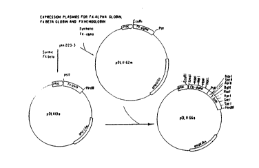
Figure 1: Flowchart for construction of plasmids for
expression of FX-alpha globin (pDL II-62m), FX-
beta globin (pDL II-l0a), and FX-hemoglobin (pDL
II-66a) are schematized.
Fiaure 2: Flowcharts for construction of plasmid pDL III-
la (2a) bearing dicistronic Des-Val-Alpha globin
gene under control of Tac promoter, and
polycistronic di-alpha/beta co-expression
plasmid pDL III-47a (2b).
CA 02050601 2000-05-31
' ' 77481-17
Figure 3: Flow chart for construction of plasmid for
co-expression of Met-alpha and Met-beta
globins, pDL III-13e.
Figure 4: Oligonucleotides for construction of
5 synthetic FX-alpha and FX-beta globin
genes. The top strand is shown 5' to 3'
and the bottom strand as 3' to 5'. Areas
of overlap between complementary synthetic
oligonucleotides are shown as areas where
10 both strands are shown in the same case
letters. The PstI site that joins FX-alpha
and FX-beta occurs at the overlap of SJH I-
35a and SJH I-36b.
Figure 5: Synthetic gene for expression of Met-FX-
15 alpha and Met-FX-beta globin. Region A
contains the alpha globin gene and region B
the beta globin gene. The location of the
Factor X sequence and the two
Shine-Delgarno sequences (SD#1 and SD#2) in
20 both regions is indicated. Selected
restriction sites are also found. The
translated amino acid sequences for the
ribosomal loader and Met-FX-alpha/and beta-
globin are given.
25 Figure 6: Elution profile and absorbance spectrum for
FX-hemoglobin.
Figure 7: Oligonucleotides for construction of mutant
hemoglobins (differing in amino acid
sequence from conventional hemoglobin).
CA 02050601 2000-05-31
77481-17
25a
Figure 8: Oligonucleotides for construction of
plasmids which do not encode the Factor Xa
substrate recognition site.
Figure 9: Plasmid pDL III-13E.
2050601
- 26 -
F3,gwre 10: Oxygen Binding of Des-Fx Hgb
Figure 11: Plasmids pDL III-14c (lla) and pDL III-38b (llb).
Figure 12 Shows the sequence of a preferred synthetic gene
for expression of (des-Val)-alpha-(GlyGly)-alpha
and des-Val beta globin. A_ shows the region
(EcoRI to PstI) containing Shine-Delgarno
ribosomal binding sites (SD#1 and SD#2), the
sequence expressing the octapeptide (Met...Glu)
which serves as a cotranslational coupler, and
the sequence encoding the two nearly identical
alpha globin-like polypeptides and the
interposed Gly-Gly linker. The first alpha
,globin sequence begins "Met-Leu", that is, it
contains an artifactual methionine, omits the
valine which' is the normal first residue of
mature alpha globin, and continues with the
second residue, leucine. The second alpha
globin sequence begins "Val-Leu", immediately
after the underlined "Gly-Gly" linker. Start
and stop codons are underlined. _B shows the
analogous region (PstI to HindIII) containing
the coding sequence for des-Val beta globin. ~1_
and ~ are connected at the PstI site to form a
single polycistronic operon.
~qure 13: Shows the structure of the final expression
vector pDL III-47a. "PTac" is the Tac promoter,
and "ampicillin" is the ampicillin resistance
gene. Figure 13a shows an XbaI-BamHI fragment
of PDL III-47a.
FiQUre 14: Plasmid pSGE0.0E4
Figure 15: Plasmid pSGEl.lE4
2050601
- 27 -
~,iqure 16: Plasmid pDL IV-67a
~iaure 17: Plasmid pJR VI-54a
giaure 18: Plasmid pDL di-alpha/beta/beta
~Laure 19: Flowchart showing the construction of various
expression vectors featuring lambda P~
regulation of various polycistronic globin
operons.
Figure 20: Nucleotide sequence of GAL-GAP promoter, with
restriction sites indicated. The region from
SphI to EcoRV contains a synthetic GAL~_~o
regulatory region (M. Johnston and R. Davis.
1984. Molecular and Cellular Biology, 4:1440-
1448). The UAS is in the region numbered 29-63
on this Figure. The region from EcoRV to the
Xbal site contains the consensus GAP491
transcriptional start region, with the
approximate start of transcription being at 395.
(L. McAlister and M.J. Holland. 1983. J.
Biol. Chem., 260:15019-15027: J. Holland, et
al. 1983. J. Hiol. Chem., ?8:5291-5299.)
~iaure 21: Flowcharts showing construction of beta-globin
expression cassette (21a and 21b).
]Figu a 2 Flowcharts showing construction of beta-globin
expression vector pGS4988 (22).
~iaure 23: Flowcharts (23a and 23b) showing construction of
an alpha-globin expression cassette and of
vectors pGS289 and pGS389 for co-expression of
0
2050601
- 28 -
alpha and beta globin from the same plasmid.
Note that alpha and beta globin are expressed
from separate~promoters.
Figwre 24: Absorption spectra for yeast-produced
recombinant and native human hemoglobin.
~,gure 25: Flowchart (25a) showing construction of di-
alpha/beta hemoglobin yeast expression vector
and map of plasmid pGS3089 (25b).
FicLure 26: Map of plasmid pGS3089RGV desR.
DETAILED DESCRIPTION OF THE PREFERRED EMBODIMENTS
Tie structure of conventional hemoglobin is well
known. For example, see the entire text of
Bunn and Forget, eds., emoct obin~ Molecular Genetic and
Clinical Aspects (W. B. Saunders Co., Philadelphia, PA: 1986)
and of Fermi and Perutz "Hemoglobin and Myoglobin," in Phillips
and Richards, Atlas of Molecular Structures in Biolocty
(Clarendon Press: 1981).
The primary structure of a polypeptide is defined by
its amino acid sequence and by identification of any
modifications of the side chains of the .individual amino acids.
About 92% of the normal adult human hemolysate is Hgb
A (designated alpha2 beta2, because it comprises two alpha and
two beta chains). The alpha chain consists of 141 amino acids
(See Figure 12). The iron atom of the heme
(ferroprotoporp?~yrin IX) group is bound covalently to the
imidazole of mss- 87 (the "proximal histidine"). The beta chain
is 146 residues long (see Figure 12) and heme is bound to it at
his 92.
WO 90/13645 2 0 5 0 6 01 p~/US90/02654
- 29 -
Other recognized hemoglobin species are Hgb A2 (a2 a2
d2 ) , Hgb A~ a , Hgb A~ b , and Hgb A~ ~ , as well as the rare species
Hgb F (a2 gamma2), Hgb Gower-1 (Zeta2 epsilonz), Hgb Gower-2
(alphaz epsilon2), Hgb Portland (Zeta2 gamma2), and Hgb H
(beta4) and Hgb Bart (gamma4). They are distinguished from Hgb
A by a different selection of polypeptide chains.
Segments of polypeptide chains may be stabilized by
folding into one of two common conformations, the alpha helix
and the beta pleated sheet. In its native state, about 75% of
the hemoglobin molecule is alpha-helical. Alpha-helical
segments are separated by segments wherein the chain is less
constrained. It is conventional to identify the alpha-helical
segments of each chain by letters, e.g., the proximal histidine
of the alpha chain is F8 (residue 8 of helix F). The non-
helical segments are identified by letter pairs, indicating
which helical segments they connect. Thus, nonhelical segment
BC lies between helix B and helix C. In comparing two variants
of a particular hemoglobin chain, it may be enlightening to
attempt to align the helical segments when seeking to find
structural homologies. For the amino acid sequence and helical
residue notation for conventional human hemoglobin Ao alpha and
beta chains, see Bunn and Forget, supra, and Table 1 herein.
The tertiary structure of the hemoglobin molecule
refers to the steric relationships of amino acid residues that
are far apart in the linear sequence, while quaternary
structure refers to the way in which the subunits (chains) are
packed together. The tertiary and quaternary structure of the
hemoglobin molecule have been discerned by X-ray diffraction
analysis of hemoglobin crystals, which allows one to calculate
the three-dimensional positions of the very atoms of the
molecule.
In its unoxygenated ("deoxy", or "T" for "tense")
form, the subunits of hemoglobin (alphal, alpha2, betal, and
beta2) form a tetrahedron having a twofold axis of symmetry.
2050601
- 30 -
The axis runs, down a water-filled "central cavity". The
subunits interact with one another by means of Van der Waals
forces, hydrogen bonds and by ionic interactions (or "salt
bridges"). The alphalbetal and alpha2beta2 interfaces remain
relatively fixed during oxygenation. In contrast, there is
considerable flux at the alphalbeta2 '(and alpha2 betal)
interface. In its oxygenated -("oxy", or "R" for "relaxed"
form), the intersubunit distances are increased.
The tertiary and quaternary structures of native
oxyhemoglobin and deoxyhemoglobin are sufficiently well known
that almost all of the nonhydrogen atoms can be positioned with
an accuracy of 0.5 A° or better. For human deoxyhemoglobin,
see Fermi, et al., J. Mol. Biol., 175: 159 (1984), and for
human oxyhemoglobin, see Shaanan, J. Mol. Biol., 171: 31
(1983)_
While analyses. of hemoglobin structure tend to focus
on the alpha-beta interfaces, it is known that the distance
between the amino terminus of one alpha subunit and the
carboxyl terminus of the other is about 5.6 A° in the deoxy
configuration and 3.3 A° in the oxy configuration.
For the purpose of the appended claims, a hemoglobin-
like protein is an oxygen binding protein with a plurality of
heme prosthetic groups and a P5o of at least about 6 torr
and composed
of a plurality of polypeptides each of which is a human alpha
(or di-alpha) globin-like or human beta (or di-beta) globin-
like polypeptide. A human alpha globin-like polypeptide is
native human alpha globin or a mutant thereof differing from
the native sequence by one or more substitutions, deletions or
insertions, while remaining at least 75o homologous with human
alpha globin, and still capable of incorporating heme and
associating with beta globin. A beta globin-like subunit is
analogously defined. Subunits of animal hemoglobins or mutants
thereof which are sufficiently homologous with human alpha or
70484-21
2050601
- 31 -
beta globin are embraced by the term "human alpha or beta
globin-like polypeptide." For example, the subunits of bovine
hemoglobin are within the scope of these terms. The alpha and
beta globin-like polypeptides may be referred to collectively
as "globins".
For the purpose of the appended claims, a di-alpha
globin-like polypeptide is one which consists essentially of
two alpha globin-like polypeptide sequences connected by
peptide bonds between the normal C-terminus of the first alpha
globin-like pollpeptide and the normal N-terminus of the second
alpha globin-like polypeptide. These two sequences may be
directly connected, or connected through a peptide linker of
one or more amino acids. Alpha globin chains crosslinked at
the N- and C-terminals other than by peptide bonds (e.g., by
DIDS) are excluded. The di-alpha globin-like polypeptide must
be capable of folding together with beta globin and
incorporating heme to form functional hemoglobin-like protein.
The di-beta globin-like polypeptide is analogously defined.
The di-alpha and di-beta globin-like polypeptides may
collectively be referred to as "pseudodimeric globin-like
polypeptides".
A "Met FX alpha globin" is an alpha globin-like
polypeptide comprising an N-terminal methionine, a oligopeptide
which acts as a recognition site. for Factor Xa (e. g., Ile-Glu-
Gly-Arg), and an alpha globin-like sequence (e. g., Val-His-Leu-
Thr-Pro...) which may correspond to wild-type alpha globin or
to a mutant thereof as taught herein. The term "Met FX alpha
globin" is sometimes abbreviated as "FX alpha globin". "FX
beta globin" is an analogously defined beta globin-like
polypeptide.
"Met-alpha globin" is an alpha globin-like
polypeptide with an extra N-terminal methionine. The secon
amino acid is valine, which is the first amino acid of mature
wild-type alpha globin. Met-beta globin is analogously
2050601
- 32 -
defined. A "Des-FX alpha globin" gene (or "dFX alpha globin")
is a Met-alpha globin gene obtained by excising the FX codons
from a Met-FX alpha globin gene. Note that "Met-Hgb" is used
to refer to methionyl Hgb formed from methionyl-alpha globin
and methionyl-beta globin.
"Des-Val-alpha globin" (or "dVal alpha globin") is an
alpha globin-like polypeptide wherein methionine is substituted
for the valine which begins the sequence of mature wild-type
alpha globin. Des-Val-beta globin is analogously defined.
Des-Val-alpha/alpha globin (di-Des-Val-alpha globin) is a "di-
alpha globin" in which a "Des-Val-alpha" sequence is linked via
an appropriate peptidyl linker to an alpha globin-like sequence
which begins with Val.
The alpha and beta globin-like chains need not
correspond exactly in sequence to the alpha and beta globins of
"conventional" hemoglobin. Rather, mutations may be introduced
to alter the oxygen affinity or stability of the hemoglobin, or
the ease of expression and assembly of the individual chains.
By way of example and not limitation, several mutant
hemoglobins have been prepared by the method of this invention.
Guidance as to further mutations is provided lay the copending
application of Hoffman and Nagai, WO 88/09179 (1988).
The DNA sequences encoding the individual alpha (or
di-alpha) and beta (or di-beta) globin chains may be of
genomic, cDNA and synthetic origin, or a combination thereof.
Since the genomic globin genes contains introns, genomic DNA
must either be'expressed in a host which can properly splice
the.premessenger RNA or modified by excising the introns. Use
of an at least partially synthetic gene is preferable for
several reasons. First, the codons encoding the desired amino
acids may be selected with a view to providing unique or nearly
unique restriction sites at convenient points in the sequence,
thus facilitating rapid alteration of the sequence by cassette
F)
2050601
33
mutagenesis. Second, the codon selection may be made to
optimize expression in a selected host. For codon
preferences in E. coli, see Konigsberg, et al., PNAS, 80:687-
91 (1983). For codon preferences in yeast, see the next
section. Finally, secondary structures formed by the
messenger RNA transcript may interfere with transcription or
translation. If so, these secondary structures may be
eliminated by altering the codon selections.
Of course, if a linker is used to genetically fuse
subunits, the linker will normally be encoded by a synthetic
DNA. While the di-alpha globin and the beta globin may be
expressed separately and then combined with each other and
heme in vitro, they are preferably placed on one plasmid.
The present invention is not limited to the use of
any particular host cell, vector, or promoter. However, the
preferred host cells are bacterial (especially, E. coli) and
yeast (especially S. cerevisiae) cells. The promoter
selected must be functional in the desired host cells. It
preferably is an inducible promoter which, upon induction,
provides a high rate of transcription. A preferred bacterial
promoter is the Tac promoter, a trp/lac hybrid described
fully in DeBoer, U.S. 4,551,433 (1985) and commercially
available from Pharmacia-LKB. Other promoters which might be
used include the temperature sensitive lambda PL and PR
promoters, as well as the lac, trp, trc, pIN (lipoprotein
promoter and lac operator hybrid), gal and heat shock
promoters. The promoter used need not be identical to any
naturally-occurring promoter. Guidance for the design of
promoters is provided by studies of promoter structure such
as that of Harley and Reynolds, Nucleic Acids Res., 15:2343-
61 (1987) and papers cited therein.
2050b01
33a
The location of the promoter relative to the first structural
gene may be optimized. See Roberts, et al., PNAS (USA),
76:760-4 (1979). The use of a single promoter is favored.
Suitable yeast expression systems are described in detail
elsewhere in this specification.
I~
CA 02050601 2000-05-31
77481-17
34
The vector used must be one having an origin of
replication which is functional in the host cell. It desirably
also has unique restriction sites for insertion of the globin
genes and the desired regulatory elements and a conventional
selectable marker. A vector may be modified to introduce or
eliminate restriction sites to make it more suitable for
further manipulations.
The alpha and beta globin chains may be expressed
either directly or as part of fusion proteins. When expressed
as fusion proteins, the latter may include a site at which they
may be cleaved to release the alpha and beta globin free of
extraneous polypeptide. If so, a site sensitive to the enzyme
Factor Xa may be provided, as taught in Nagai and Thorgenson,
EP Appl 161,937 (1985).
Alternatively, the alpha and beta fusion proteins may
be synthesized, folded and heme incorporated to yield a
hemoglobin analogue.
The direct expression of the alpha and beta globin
subunits is desirable. Factor Xa is a blood derivative.
Preparations of Factor Xa may therefore contain undesirable
blood-associated substances or etiologic agents. In any event,
the hemoglobin must be separated from the Factor Xa.
Nagai and Thorgenson, EP Appl 161,937 teach the
construction of fused genes in which DNA coding for a
polypeptide of interest is immediately preceded by DNA encoding
a cleavage site for Factor Xa, a serine protease. Certain of
the peptide sequences to be cleaved by Factor Xa are quoted
below (wherein the cleavage site is denoted by an "_"):
Ile-Glu-Gly-Arg=Val-His-Leu-Thr CII FxB-globin
CA 02050601 2000-05-31
77481-17
34a
Ile-Glu-Gly-Arg=Thr-Ala-Thr-Ser Hu prothrombin
Ile-Glu-Gly-Arg=Thr-Ser-Glu-Asp Bo prothrombin
2050601
- 35 -
Ile-Asp-Gly-Arg=Ile-Val-Glu-Gly Hu prothrombin
Ile-Glu-Gly-Arg=Ile-Val-Glu-Gly Bo prothrombin
Ala-Glu-Gly-Arg=Asp-Asp-Leu-Tyr Hu antithrombin III
In the above list, "CIIFXp-globin" refers to a hybrid
fusion protein comprising the 31 amino-tern~inal residues of the
lambdaCII protein, the Factor Xa recognition sequence "Ile-Glu-
Gly-Arg," and the complete amino acid sequence of human beta
globin (which begins "Val-His-Leu-Thr-..."). It will be
evident from study of Figure 4 of the present invention that
FX-alpha and FX-beta globins of Example 1 correspond to the
native globin preceded by "Met-Ile-Glu-Gly-Arg."
In bacterial mRNA, the site at which the ribosome
binds to the messenger is a polypurine stretch which lies 4-7
bases upstream of the start (AUG) codon. The consensus
sequence of this stretch is 5'...AGGAGG...3', and is frequently
referred to as the Shine-Dalgarno sequence. Shine and
Dalgarno, Nature, 254: 34 (1975). The exact distance between
the SD sequence and the translational start codon, and the base
sequence of this "spacer" -region, affect the efficiency of
translation and may be optimized empirically. Shepard, et al.,
DNA 1: 125 (1985) ; DeBoer, et al. , DNA 2: 231 (1983) : Hui, et
al., EMBO J., 3: 623 (1984).
In addition, the SD sequence may itself be modified
to alter expression. Hui and DeBoer, PNAS (USA), 84:4762-66
(1987). Comparative studies of ribosomal binding sites, such
as the study of Scherer, et al., Nucleic Acids Res., 8:3895-
3907 (1907), may provide guidance as to suitable base changes.
If the hemoglobin is to be expressed in a host ether than ~,
co i, a ribosomal-binding site preferred by that host should be
provided. Zaghbil and Doi, J. Bacteriol., 168:1033-35 (1986).
A
WO 90/13645 2 0 5 0 6 01 P~'/US90/02654
- 36 -
Any host may be used which recognizes the selected
promoter and ribosomal binding site and which has the
capability of synthesizing and incorporating heme. Bacterial
and yeast hosts are preferred.
The intracellularly assembled hemoglobin may be
recovered from the producing cells and purified by any art-
recognized technique.
Polycistronic Co-Expression of Alpha and Beta Globins and Their
Assembly Into Hemoglobin
In one embodiment, expression of the alpha and beta
globin genes is driven by a single promoter, and the genes are
arranged so that a polycistronic messenger RNA transcript is
transcribed, from which the separate alpha and beta globin
polypeptides are subsequently translated. However, the present
invention includes the co-expression of the alpha and beta
globin genes from separate promoters, i.e., the host
transcribes separate alpha and beta globin mRNAs.
The use of a single promoter is favored on
theoretical grounds. Ideally, alpha and beta globin are
expressed in stoichiometrically equal amounts. While use of a
single promoter does not guarantee equality, it eliminates one
unbalancing influence --differences in transcription owing to
differences in promoter strength and accessibility. If
differences in promoter strength were minimized by use of two
identical promoters on the same plasmid, plasmid stability
would be reduced as there would be a propensity toward
recombination of the homologous regions. We note, however,
that in preliminary experiments we have co-expressed alpha and
beta globins from separate promoters.
Another justification for using a single promoter is
to minimize the number of repressor binding sites.
WO 90/13645 O 6 O 1 PGT/US90/02654
- 37 -
Preferably, the alpha and beta globin genes are
arranged so that the ribosome will translate the alpha globin
cistron first. The rationale is that there is some basis for
believing that alpha globin affects the folding of beta globin.
Nonetheless, the position of the genes may be switched so that
beta globin is synthesized first, as is shown in Example 6.
The stability of the polycistronic mRNA transcript,
the efficacy of its translation into alpha and beta globin, and
the folding of the globin chains into tetrameric hemoglobin may
be modified by varying the length and base sequence of the
intercistronic regions (the region lying between the stop codon
of one cistron and the start codon of the next cistron), the
phasing of a second cistron relative to a first cistron, and
the position and sequence of the ribosomal binding site for the
one cistron relative to the preceding cistron.
In a preferred embodiment, the alpha and beta globin
genes are each preceded by a short "introductory" cistron or
"ribosomal loader" which facilities the subsequent translation
of the globin cistron. In Figure 4, region A contains two
cistrons and a Shine-Delgarno sequence preceeding each cistron.
The first Shine-Delgarno sequence (SD#1) is bound by the
ribosome, which then translates the first cistron, a short
cistron encoding an octapeptide. (This cistron is referred to
as an "introductory cistron or ribosomal loader.) The second
cistron is a globin gene, in this case, an FX alpha-globin
gene. The Shine-Delgarno sequence (SD#2) for facilitating
translation of the second cistron actually lies within the
first cistron. For this reason, the two are said to be
"translationally coupled". Region B is identical in structure,
except that the second cistron encodes FX-beta globin. Between
regions A and B is a 43-base intercistronic region. The
introductory cistrons of regions A and B correspond to the
first cistron of the two-cistron expression system denoted
pCZ144 in Schoner, et al., Meth. Enzymol., 153: 401-16
WO 90/13645 2 0 5 0 6 ~ ~ PCT/US90/02654
- 38 -
(1987). The present invention is not, however, limited to the
particular "starter" cistron taught by Schoner, et al.; other
introductory cistrons that allow for restart of high level
translation of a following cistron may be used.
Guidance as to the design of intercistronic sequences
and as to the location of SD sequences may be obtained by
comparing the translational efficiency of spontaneous or
controlled mutants of the same polycistronic operon, as
exemplified by Schoner, et al., PNAS, 83: 8506-10 (1980). It
is also possible to look for consensus features in the
intercistronic regions of different operons. McCarthy, et al.,
EMBO J., 4: 519-26 (1985) have identified a translation-
enhancing intercistronic sequence in the E. coli atp operon.
The present invention is intended to reduce or avoid
the localization of the hemoglobin or its component
polypeptides into inclusion bodies. Consequently, a further
feature of the invention is that the functional hemoglobin is
substantially found (preferably over 80%) in the soluble
fraction of the cell. It appears that with this invention,
over 90% of the functional hemoglobin can be so directed when
alpha2 betaZ hemoglobin is assembled from alpha- and beta-
globin chains co-expressed from a tetracistronic operon as
described herein. With di-alpha, betaZ hemoglobin, nearly 100%
is soluble when expression is induced at 25°C and less at
higher induction temperatures. These percentages reflect the
percent of all di-alpha and beta chains found in the soluble
fraction of the cell and not actual recovery of protein from
the cell.
Expression in Yeast
In another embodiment the present invention relates
to the production of hemoglobin-like molecules in yeast. Our
preferred host for expression of recombinant human hemoglobin
in yeast is Saccharomyces cerevisiae. However, other fungi or
WO 90/13645 PGT/US90/02654
2050601
- 39 -
yeast may be used for the purpose, such as strains of
Asperaillus or Pichia. For yeast to be a suitable host it must
be capable of being transformed with recombinant vectors,
either replicating or integrating types. This allows the
insertion of the desired DNA sequence for the gene of interest.
It must also be capable of high density cell growth, in
appropriate volume to provide sufficient cell mass to isolate
the desired gene product from the desired reaction vessels,
where ideally the growth would be easily controlled by several
parameters including nutrient formulation, agitation and oxygen
transfer and temperature. It is also desirable to be able to
induce the expression of protein synthesis with the
manipulation of the media, temperature, or by the addition or
consumption of certain chemicals. Finally, to be a suitable
host, the yeast must be capable of producing recombinant
proteins, preferably in excess of l~ of the total cell protein.
This allows more facile isolation of the desired recombinant
protein.
With reference to S. cerevisiae, haploid strains of
potential use include:
BJY3501 Mata pep4::HIS3 prbl-o 1.6R his3 200 ura3-52
canl GAL,
GSY112 As above but Leu2::HISG
GSY112 cir As above but cured of 2~, plasmid
BJY3505 Mata pep4::HIS3 prbl-o 1.6R HIS3 lys2-208
trpl-101 ura3-52 gal2 canl
GSY113 As above but leu2::HISG
RSY330 Mata pep4-3 prbl-1122 hist7 ura3-52 trpl-289
canl gall
BJY2168 Mata prcl-407 prbl-1122 pep4-3 leu2 trpl ura3-
52
BJY1991 Mata prbl-1122 pep4-3 leu2 trpl ura3-52
RSY334 Mata regl-501 pep4-3 prbl-1122 ura3-52 leu2-3,
112 gall
RSY214 Mata pep4-3 prbl ura3-52
To date, strains such as GSY112, GSY113 and RSY334
have been the best hemoglobin producers. Strains such as
RSY334 that carry the regl-501 mutation may be particularly
._ 2050601
- 40 -
important as they uncouple glucose repression from galactose
induction, allowing one to induce with lower levels of
galactose in the presence of glucose. Because this strain
carries the gall mutation it cannot metabolize galactose, so
the galactose concentration remains constant and continues to
function as a inducer.
Diploid strains formed from crosses of any of the
above compatible strains or similar compatible strains may also
be useful as they tend to have faster growth rates than haploid
strains.
For example, the following diploid strains, which co-
express alpha (or di-alpha) and beta globins, are described in
the Examples:
HJY3505 [pGS4988] x RSY330 [pGS4688]
HJY3505 [pGS4988] x HJY 1991 [pGS4688]
Other coatings may likewise be used in practicing the
present invention,.
The use of protease-deficient strains may also be
advantageous.
Yeast expression systems can be divided into two main
categories: (1) Systems designed to secrete protein and (2)
system designed for the cytoplasmic expression of proteins.
The advantages of secretion systems are:
(1) The protein is often easier to purify from
culture media than from total cell extracts.
(2) If the proteins has essential disulfide bonds,
they are more likely to form if the protein
passes through the secretory pathway. This is
2050601
- 41 -
thought to be partly due to the presence of
protein disulfide isomerase in the endoplasmic
reticulum and to a less reducing environment
than in the cytoplasm.
(3) Secretion can also be advantageous if the first
amino acid (after the initiating methionine)
creates a dipeptide sequence that is poorly
processed by methionyl aminopeptidase. The
addiction of a secretory signal sequence may
allow processing by signal peptidase during
secretion resulting in' a protein with the
authentic amino acid at the amino terminus of
the protein.
The disadvantages of a secretion system are:
(1) Generally, the expression levels are much lower
than those that can-be obtained by cytoplasmic
expression. This seems to be, in part, due to a
rate limiting step in secretion.
(2) Not all proteins are secretable.
(3) Often, particularly with S. cerevisiae.
misprocessed forms of the protein accumulate and
these can be difficult ~to purify away from
correctly processed forms.
The most commonly used secretory expression system
used in yeast is the yeast a-factor secretory signal sequences
and the a-factor promoter. See A.J. Brake. Yeast Genetic
Enaineerina Eds. P. Barr, A. Brake, and P. Valenzuela.
Butterworth Publishing, Boston 1989, for a review. The
~.
2050601
- 42 -
invertase signal sequence coupled to a variety of promoters has
also been used to express heterologous proteins in yeast. See
G. Stetler et al., Biotechnology, 7:55-60 (1989), R. Smith et
al., Science, 229:1219-1229 (1985).
The advantages of cytoplasmic expression are:
(1) Expression levels can be quite high with reports
of proteins expressed at >20% of the total cell
protein, usually as a soluble protein.
(2) Some proteins cannot be efficiently secreted.
(3) Proteins that contain glycosylation sites, if
secreted from yeast, will be glycosylated with
the pattern of sugars unique to yeast. Because
these highly hydrophilic, post-translational
modifications lie on the surface of proteins
they are likely to confer antigenicity on the
protein. If the protein is functional without
the carbohydrate side chains it may be
advantageous~to produce the protein without them
rather than with the yeast-specific modification
(see for ex.. Travis et al. 1985. J Biol Chem
,0:4384-4389) .
(4) Some proteins have other specific modifications
that may occur only in the cytoplasm, for
example amino terminal acetylation, modification
with lipids, perhaps heme incorporation and so
forth.
At present, cytoplasmic expression is preferred since
the yeast cells fold together the globin chains and incorporate
heme to produce hemoglobin in yivo. However, it is possible to
separately express and secrete the alpha and beta globin chains
and assemble hemoglobin 'fir vitro.
~,
WO 90/13645 PGT/US90/02654
2050b01
43 -
The globin genes must be placed under the control of
a suitable promoter. .The commonly used yeast promoters
generally fall into two broad categories: regulated and
constitutive. Constitutive promoters that are in wide use
include GAP, PGK (phosphoglycerate kinase) and the a-factor
promoter. Regulated promoters have also been used and these
include the yeast metallothionein promoter (regulated by
copper), the Gall-10 promoter, GAL7 promoter (regulated by
galactose and glucose) the ADHII promoter (regulated by ethanol
and glucose) the PH05 promoter (phosphate regulation) and
several hybrid promoters such as PH05-GAP, GAL-PGK, ADHII-GAP,
and GAL-CYC1.
The use of regulated promoters may be important for
plasmid stability. Often expression of recombinant proteins,
at high levels, inhibits the growth of the host organism. This
disadvantage can result in the accumulation of cells in the
population with few copies of the plasmid present, a decrease
in growth rate and a drop in overall expression levels. By
maintaining the gene in a repressed state and inducing late in
the fermentation, high growth rates can be maintained and
selection against high plasmid copy numbers can be avoided.
It is somewhat difficult to obtain accurate data on
the relative strength of yeast promoters because the only true
measure of this would be based on kinetics of mRNA synthesis.
Most of the data that exists measures only the final protein
concentration that has been obtained. Because there can be
great differences in protein stability, it is necessary to
,compare the same protein and mRNA with multiple promoters on
identical vectors. A study that approaches this was done by
Verbakel et al. (Gene, 61:207-215 (1987)). They compared the
GAPDH, CYC1 GAL7, PH05, and PGK promoters and measured the
WO 90/13645 ~ ~ ~ ~ PCT/US90/02654
- 44 -
expression of a (~-galactosidase/poliovirus VP2 protein fusion.
CYCl resulted in an expression level of 0.14% of the total cell
protein (TCP); GAPDH, 0.22% TCP; PH05, 0.26% TCP; PGK, 0.9%
TCP: and GAL7, 0.96% TCP.
Using our GALGAP hybrid promoter, we have obtained
expression levels of hemoglobin that represents >15% of the
total cell protein.. This most probably represents an increased
promoter strength along with a highly stable protein product
and perhaps mRNA with an extended half life.
The use of a GAL-GAP hybrid promoter is preferred.
Both elements (the GAL AS and the GAP transcriptional
initiation site) are well understood. Studies on the
mechanisms of transcriptional regulation of the GAL regulon
have been fairly extensive. The galactose regulon includes
five genes that encode enzymes required for the utilization of
galactose. Four of these genes (GAL1, GAL7, GAL10, and GAL2)
are expressed only in the presence of galactose. Galactose
induction does not occur in the presence of glucose unless the
yeast strain bears a mutation in the REG1 gene. The GAL1, 7,
and 2 genes are regulated by at least two other genes, GAL80
and GAL4. The GAL4 gene is a transcriptional activator protein
that activates mRNA synthesis from the GAL1, 7, 10 and 2
upstream activator sequences (UAS~a~). Although GAL4 is
constitutively expressed, it is functionally silent in the
absence of galactose. Repression of GAL4 activity, in the
absence of galactose is maintained by the product of the GAL80
gene. The GAL80 protein apparently interacts physically with
GAL4 to prevent transcriptional activation. Presumably
.galactose or a galactose derivative prevents this interaction
to allow GAL4 mediated induction.
Haploid strains of S.cerevisiae have three different
genes encoding the enzyme glyceraldehyde-3-phosphate
dehydrogenase (GAP). These genes have been designated TDH1,
TDH2 and TDH3 and each is present as a single copy per haploid
WO 90/13645 ~ ~ '~ PCT/US90/02654
- 45 -
genome. The TDH3 gene produces approximately 60% of the cell's
GAP enzyme and TDH1 and 2 produce about 12% and 28%,
respectively (McAllister, L and M.J. Holland, 1985. J. Biol
Chem, 260: 15019-15027). Holland's group (Holland et al. 1981.
J. Biol Chem, 256:1385-1395; and Holland et al. 1983. J Biol
Chem 258:5291-5299) has cloned and characterized the three GAP
genes of S.cerevisiae. The clones have been designated pGAPll,
pGAP63, and pGAP491. pGAP491 corresponds to the TDH3 gene and
is therefore, the most highly expressed. This promoter has
been used to express a wide variety of proteins in yeast
including:
Protein REF
a Interferon (1)
Hepatitis B antigen (1)
Thaumatin (2)
Hepatitis B antigen (3)
HIV III reverse (4)
transcriptase
human SOC (5)
al antiprotease (6)
(1) Bitter, G. and
K.M.
Eagan.
1984.
Gene,
32.263-274.
-
(2) Edens, L. et al. .
1984. Cell, 37:629-633
(3) Kitano, K. et al. 1987. Biotechnology 5:281-283.
(4) Hallewell, R.A. et al. 1987. Biotechnology 5:363-
366.
(5) Barr, P.J. et al. 1987. Biotechnology 5:486-489.
(6) Travis, J. et al. 1985. J Biol Chem 2600:4384-4389.
This promoter is commonly used as a 600-850bp
fragment and is essentially un-regulated. In its long form
this is a very powerful promoter. The form we are using
consists of only -200bp 5' of the translational initiation
site. This form, with no added enhancer sequences is
substantially less active than the longer form of the promoter
(Edens, L. et al. Cell, 37:629 (1984)). Our addition of the
GAL enhancer region confers both regulation and high levels of
expression. With only the GAP491 promoter, alpha and beta
globin were produced at a level of less than 0.2% total cell
protein; with the GAL-GAP491 hybrid promoter, expression jumped
to 7-10% total cell protein.
WO 90/13645 2 0 5 0 6 ~ ~ P~T/US90/02654
- 46 -
Several other hybrid promoters are of particular
interest:
GAL-SIGMA
A strong galactose-regulated promoter with
the sigma transcriptional start site.
SIGMA-GAP
A strong peptide hormone-regulated promoter
with the GAP491 transcriptional start
site.
GAL-EF III
A strong galactose-regulated promoter with
the elongation factor III transcriptional
start site.
SIGMA-EF III
A strong peptide hormone-regulated promoter
with the elongation factor III
transcriptional start site.
One could easily conceive of other promoter systems
that would also work. This would include, but not be limited
to, a variety of constitutive promoters. For example, the
yeast mating factors (MFa) promoter or the mating factor a
promoter MF(a), the phosphoglycerate kinase promoter (PGK),
hexokinasel, hexokinase2, glucokinase, pyruvate kinase, triose
phosphate isomerase, phosphoglycerate isomerase,
phosphoglycerate mutase, phosphofructose kinase or aldolase
promoters may all be used. In short, any well expressed yeast
promoter may work for expression of hemoglobin in yeast. A
wide variety of naturally occurring, regulated promoters could
also be used, for example: GAL1-10, GAL7, PH05, ADHII have all
been used to produce heterologous proteins in yeast. A variety
of synthetic or semi-synthetic yeast promoters could also be
T _
2050b01
47
employed such as GAL-PGK, GAL-MFa-1, GAL-Mfal, GAL-SIGMA.
ADHII regulatory sequences could also be coupled to strong
transcriptional initiation sites derived from a variety of
promoters. The PH05 regulatory sequence or the sigma element
regulatory sequences could also be used to construct powerful
hybrid promoters. In addition to yeast promoters, it is
conceivable that one could use a powerful prokaryotic
promoter like the T7 promoter. In this case, one could place
the T7 polymerase under the control of a tightly regulated
yeast promoter. Induction of the phage polymerase in yeast
cells bearing hemoglobin genes under T7 promoter regulation
would allow transcription of the genes by this very efficient
phage polymerase.
Because most of the yeast regulatory sequences
described above serve as targets for proteins that are
positive regulators of transcription, it is conceivable that
these proteins may limit transcription in situations where
the target sequence is present in many copies. Such a
situation may be obtained with vectors such as pClB, pCIT,
pClU or pClN which may be present in excess of 200 copies per
cell. Over-expression of the positive regulator (for example
GAL4) may result in enhanced expression. It is possible to
construct a strain in which the GAL4 gene is altered to
remove its promoter and the promoter replaced with the GAL7
or GAL1-10 promoters, both of which are transcribed more
efficiently than the GAL4 promoter. In this situation, the
positive transcriptional activator protein GAL4 would be
expressed at elevated level at the time hemoglobin expression
was induced.
2050bO1
47a
The consensus sequence for higher eukaryotic
ribosome binding sites has been defined by Kozack (cell,
44:283-292 (1986)) to be G~GCCAUGG. Deviations from this
sequences, particularly at the -3 position (A or G), have a
large effect on translation of a particular mRNA. Virtually
all highly expressed mammalian genes use this sequence.
Highly expressed yeast mRNAs, on the other hand, differ from
this sequence and
n
2050601
- 48 -
instead use the sequence-.A.AAAAUGU (Cigan and Donahue, Gene,
X9:1-18 (1987)). The ribosome binding site that we use for
expression of the a and ~-globins corresponds to the higher
eukaryotic ribosome binding site. It is within the
contemplation of this invention to systematically alter this
RBS to test the effects of changes that make it more closely
resemble the RBS of yeast. It should be pointed out, however,
that alterations at the -2,~ -1 and +3 positions, in general,
have been found to only slightly affect translational
efficiency in yeast and in mammals.
Intracellular expression of genes in S. cerevisiae is
primarily affected by the strength of the promoter associated
with the gene, the plasmid copy number (for plasmid-borne
genes), the transcription terminator, the host strain, and the
codon preference pattern of the gene. When secretion of the
gene product is desired, the secretion leader sequence becomes
significant. It should be noted that with multicopy plasmids,
secretion efficiency may be . reduced by strong promoter
constructions. Ernst, DNA 5:483-491 (1986).
A variety of extrachromosomally replicating vectors
(plasmids) are available for transforming yeast cells. The
most useful multicopy extrachromosomal yeast vectors are
shuttle vectors that use a full length 2~c-circle combined with
an ~. coli plasmid. These vectors carry genes that allow one
to maintain the plasmid in appropriate yeast mutants and
antibiotic resistance markers that allow selection in ~. co '.
Use of the full-length 2~C-circle, in contrast to vectors
containing only a partial 2~ sequence, generally results in
much higher plasmid stability, particularly in yeast strains
that have been cured of endogenous 2u plasmid. The pC series
of vectors described herein are vectors of this type.
Strains could also be constructed in such a way that
the GALGAP hemoglobin expression cassettes were integrated into
chromosomes by using yeast integrating vectors. Although the
za
WO 90/13645 2 0 5 0 6 01 PGT/US90/02654
- 49 -
copy number of the hemoglobin genes would be lower than for
plasmid vectors, they would be quite stable and perhaps not
require selection to be maintained in the host cell. Yeast
integrating vectors include Yip5 (Struhl, et al, PNAS, 76:1035-
39, 1989), Yip1 (Id.), and pGT6 (Tchumper and Carbon, Gene,
10:157-166, 1980). For information on these and other yeast
vectors, see Pouwels, et al., Cloning Vector, VI-I, et sea
(Elsevier, 1985).
The alpha and beta globin genes may be introduced by
separate plasmids, or both upon the same plasmid. The
advantage of a single plasmid system over a double plasmid
system is theoretical. It is generally thought that there is
an upper limit to the total number of plasmid copies per cell.
If it is 1000, for example, the two plasmid system could have
only 500 copies of a-chain plasmid and 500 of the R-chain
plasmid. A single plasmid of 1000 copies per cell would bear
1000 copies of each a- and R-chain gene. The number of copies
may be irrelevant, however, if other factors are limiting. In
fact, several groups favor using strains that contain genes
integrated into various chromosomal loci. Such strains very
stably maintain the foreign gene and do not require special
media to maintain selection for the plasmid.
Highly expressed yeast genes show a very high codon
bias. The genes encoding glyceraldehyde-3-phosphate
dehydrogenase and ADH-I, for example, show a 90% bias for a set
of 25 codons. Highly expressed yeast genes (>1% of the total
mRNA) have yeast codon bias indices of >.90. Moderately
expressed genes (0.1-.050 of the total mRNA) have bias indices
of 0.6-0.8, and genes expressed at low levels (>0.05% of the
total cell protein) have a codon bias of 0.10-0.50 (Bennetzen
and Hall, J. Biol. Chem., 257:3026-3031 (1982)). The
calculated value for the codons of the human a-globin cDNA is
0.23. A similar value can be calculated for the a-globin cDNA.
Because there is a very high correlation between the most
commonly used codons, it is possible that hemoglobin expression
WO 90/13645 2 ~ 5 0 6 ~ ~ PGT/US90/02654
- 50 -
from the human cDNA in yeast may be limited by the availability
of the appropriate tRNA molecules. If this is so, a complete
synthesis of the gene using the most highly favored yeast
codons could improve the expression levels. It is quite
possible that the greatest negative effect of adverse codon use
would be if there was an abundance of codons used in the cDNA
that are represented by low abundance tRNAs. In such a case,
high level expression of hemoglobin could completely drain that
pool of tRNA molecules, reducing translation not only of
hemoglobin but of yeast proteins that happen to use that codon
as well. In the case of the a-globin human cDNA, the most
commonly used leucine codon is CTG (14 of 21), this codon is
never used in highly expressed yeast genes (Guthrie and
Abelson, The Molecular Biology of the Yeast Saccharomyces, Eds.
Stratern, Jones and Broach, 1982. Cold Spring Harbor, NY).
The low codon bias index and the presence of rare yeast codons
in the globin cDNAs have been sufficient incentive for us to
synthesize a modified form of the alpha- and beta-globin genes
using the preferred yeast codons.
"Di-Alpha" and Di-Beta" Hemoglobins
The present invention further contemplates in some
embodiments the combination of (a) one molecule of a di-alpha
globin-like polypeptide with two molecules of a beta globin-
like polypeptide to form a "di-alpha" hemoglobin-like protein;
(b) two molecules of an alpha-globin-like polypeptide with one
molecule of a di-beta globin-like polypeptide to form a "di-
beta" hemoglobin-like protein; or (c) one molecule of a di-
alpha globin-like polypeptide with one molecule of a di-beta
globin-like polypeptide to form a "di-alpha/di-beta"
hemoglobin-like protein.
It should further be noted that the delta, gamma and
epsilon chains have considerable homology with the beta chain
and that the zeta chain has considerable homology with the
2050b01
- 51 -
alpha chain. Di-delta, di-gamma, di-epsilon and di-zeta
polypeptides are therefore within the compass of the invention
and may be used in the preparation of novel hemoglobins of
types other than Hgb Ao.
Preferably the di-alpha linker, (if one is used)
consists of 1 to 3 amino acids which may be the same or
different. A Mono-Gly linker is especially preferred. In
designing such a linker, it is important to recognize that it
is desirable to usa one or more amino acids that will flexibly
connect the two subunits, transforming them into domains of a
single di-alpha globin polypeptide.
The preparation of "di-beta" mutants is also
contemplated. The distance between the N-terminus of one beta
subunit and the C-terminus of the other is 18.4 A in the deoxy
configuration and 5.2 A in the oxy form. Preferably, the di-
beta linker consists of 4 to .9 amino acids which may be the
same or different.
The linker preferably should be unlikely to form a
secondary structure, such as an alpha helix or a beta-sheet.
Certain amino acids have a greater tendency to participate in
such structures. See Chou and Fasman, Biochemistry, 13:222-245
(1974). The amino acids are ranked in order of decreasing
participation below. The preferred linker amino acids are
boldfaced. Glycine is the most suitable amino acid for this
purpose. The most preferred di-alpha linkers are Gly or Gly-
Gly.
A~,pha Helix $eta Sheet
Formers Formers
Glu (1.53) Met (1.67)
Ala (1.45) Val (1.65)
Leu (1.34) Ha Ile (1.60)
HI3
His (1.24) Cys (1.30)
Met (1.20) Tyr (1.29)
Gln (1.17) Phe (1.28)
Val (1.14) Gln (1.23)
WO 90/13645 2 0 5 ~ 6 01 P~T/US90/02654
- 52 -
Trp(1.14) Leu (1.22)
Phe(1.12) ha Thr (1.20)
Lvs(1.07) Trp (1.19)ha
Ile(1.00) Ala (0.97)Ia
Asp(0.98) ArQ (0.90)
Thr(0.82) G~ (0.81)
Ara(0.79) Asp (0.80)iQ
Ser(0.79) Lys (0.74)
Cys(0.77) is Ser (0.72)
Asn(0.73) His (0.71)
Tyr(0.61) ba Asn (0.65)
Pro( 0 . 59 ) Pro ( 0 bf~
.
62~
Gly(0.53) Ba Glu (0.26)BQ
(The letter symbols are Ha, strong a former; ha, a
former; Ia; weak a former; ia, a indifferent; ba, a breaker;
and Ba strong a breaker. The ~i symbols are analogous. Trp is
bQ if near the C-terminal of a /3-sheet region.)
The alpha helix of a polypeptide chain comprises an
average of 3.6 residues per turn. In globular proteins, the
average length is about 17A, corresponding to 11 residues or 3
helix turns. In alpha and beta globin, the helices range in
length from 7 to 21 amino acids (A.A.). The beta pleated sheet
comprises 2.3 residues per turn; the average length is about
20A or 6 residues.
Chou and Fasman define an alpha helix nucleus as a
hexapeptide containing four helix forming residues and not more
than one helix breaker, and a beta sheet nucleus as a
pentapeptide containing three beta sheet forming residues and
not more than one sheet breaker.
The amino acid sequence in the vicinity of the di-
alpha linker is as follows:
residue # 138 139 140 141 1 2 3 4
AA Ser Lys Tyr Arg -(XXX)~ Val Leu Ser Pro
Helix Not H21 HC1 HC2 HC3 NA1 NA2 A1 A2
Helix Pot 079 107 061 079 114 134 079 059
Sheet Pot 072 074 129 090 165 122 072 062
WO 90/13645 2 0 5 0 6 01 P~/US90/02654
- 53 -
(Note: Helix- and sheet forming potentials have been
multiplied by 100 for typographical reasons.)
The di-alpha linker is preferably only 1-3 amino
acids. Thus, it can form an alpha helix only in conjunction
with the linker "termini". A one or two residue linker, even
if composed of amino acids with strong secondary structure
propensities, would be unlikely to assume an alpha helix or
beta sheet configuration in view of the disruptive effect of,
e.g., Arg 141 or Ser 3. If the linker is 3 residues long, it
would be preferable that no more than one residue be a strong
alpha helix former, unless the linker also included a strong
alpha helix breaker.
The amino acid sequence in the vicinity of the di-
beta linker may impose more stringent constraints.
143 144 145 146 1 2 3 4
His Lys Tyr His (XXX) - Val His Leu Thr
-
H21 HC1 HC2 HC3 NA1 NA2 NA3 A1
124 107 061 124 114 124 134 082
071 074 129 071 165 071 122 120
The di-beta linker is longer (preferably 4-9 A.A.)
and therefore more susceptible to secondary structure
formation. It is desirable that the amino acid adjacent to
Val-1 be an alpha helix breaker in view of alpha-helix
propensities of Val-His-Leu. More generally, it is desirable
that the linker not contain (or cooperate with the proximately
linked amino acids to form) an alpha helix nucleus or beta
sheet nucleus.
Amino acids with a high propensity toward alpha helix
formation may be used in the linker if accompanied by "helix
breaking" amino acids. Similarly, Beta sheet formation may be
prevented by "sheet disrupting" amino acids.
2050601
- 54 -
Of course, prediction, of secondary structure using
Chou and Fasman's approach. has its limitations and the ultimate
test of the acceptability of a linker is whether or not the di-
alpha or di-beta hemoglobin has the desired affinity for
oxygen. In particular, a poly-alanine linker, despite its
supposed propensity to alpha-helix formation, may well be of
value since the alanine group is compact and therefore the
linker should be quite flexible if secondary structure does not
form.
In an especially preferred embodiment, di-alpha and
beta globin genes are combined into a single polycistronic
operon. The use of a polycistronic operon is not, however,
necessary to practice the present invention, and the alpha (or
di-alpha) and beta (or di-beta) globin genes may be expressed
from separate promoters which may be the same or different.
While the preferred "genetically fused
hemoglobin" of the present invention is. one comprising a di-
alpha and/or di-beta globin, other globin chains may be
genetically fused and used in the production of
hemoglobins of species other than Hgb Ao (aZpz).
By the means suggested in this specification, we have
had the following achievements:
(1) Expression of Met-FX-alpha globin in E. coli
from a dicistronic operon comprising a introductory cistron and
a Met-FX-alpha globin gene, both transcribed from a Tac
promoter. (Example 2)
WO 90/13645 2 0 5 0 6 01 P~/US90/02654
- 55 -
(2) Expression of Met-FX beta globin in E. coli by
similar means. (Example 2)
(3) Co-expression of Met-FX-alpha globin and Met-FX-
beta globin in E. coli from a tetracistronic operon comprising
an introductory cistron, an FX-alpha globin gene, an
intercistronic sequence, a second introductory cistron, and an
FX-beta globin gene, all controlled by a single Tac promoter.
The alpha and beta globins were intracellularly assembled into
functional FX hemoglobin. The FX Hgb was enzymatically
converted to Hgb. (Example 3)
(4) Co-expression as in (3) above of mutants of FX-
hemoglobin in which the beta globin subunits possessed the Beth
Israel, Cheverly, Providence/MSR, Kansas or beta6~ Val -~ Ile
mutations. (Example 4)
(5) Co-expression as in (3) above of Met-alpha
globin and Met-beta globin and intracellular assembly into Met-
Hgb (a.k.a. Des-FX-Hgb). (Example 5)
(6) Co-expression as in (3) above of Des-Val-alpha
globin and Des-Val-beta globin. (Example 6)
(7) Co-expression as in (6) above, but with the Des-
Val-beta globin gene preceeding the Des-Val-alpha globin gene
within the operon. (Example 6)
(8) Co-expression as in (3) above of (Des-Val)-
alpha-(Gly-Gly)-alpha globin and beta-globin and intracellular
assembly into a di-alpha Hgb. (Example 8)
(9) Co-expression as in (8) above of (Des-Val-alpha-
(GlyGly)-alpha globin and a mutant (Nagai, Arg-Nagai, or
Kansas) of beta globin and intracellular assembly into a mutant
di-alpha Hgb. (Example 9)
2050601
- 56 -
(10) Co-expresion ~as in (3) above of Des-Val-alpha
globin and a Des-Val beta-globin with the Presbyterian mutation
and intracellular assembly into a mutant Des-Val Hgb. (Example
11) .
(11) Co-expression as in (8) above of a (Des-Val)-
alpha-(GlyGly)-alpha globin and of a betal globin with the
Presbyterian mutation. (Example 11).
(12) Expression of di-alpha globin and beta-globin
from separate promoters on the same plasmid (Example 12).
(13) Devised hypothetical protocol for co-expression
of di-alpha globin and two copies of beta globin from same
operon (Example 13).
(14) Devised hypothetical protocol for co-production
of Di-beta Hgb (Example 14).
(15) Devised hypothetical protocol for co-expression
of alpha globin and beta-globin under control of separate
promoters on the same plasmid (Example 16).
(16) Devised hypothetical protocol for co-expression
of alpha and beta globin under control of different promoters
on different plasmids (Example 17).
(17) Co-expression of a and p globin in S. cerevisiae
(Example 19).
(18) Co-expression of di-a globin and p globin in
cerevisiae. (Example 23).
(19) Construction of Des-Val-alpha globin and Des-
Val-beta globin in E.E. coli under lambda P~ control.
2050601
- 57 -
(20) Co-expression of di-alpha globin and Des-Val-
beta globin in E. cola under lambda P~ control.
(21) Co-expression of alpha and beta globin from
separate plasmids in diploid strains of S. cerevisiae.
(22) Co-expression of di-alpha globin and beta globin
Presbyterian mutant in S. cerevisiae.
(23) Preparation of vectors for expression of di-
alpha globins with -Gly- or -Pro- linkers.
(24) Evaluation of different strains and induction
temperatures for expression of di-alpha Hgb in E. o
(25) Co-Expression of alpha and mutant beta globin in
. cerevisiae, assembling to form low-affinity Hgb mutants.
An unexpected and surprising change in oxygen binding
characteristics of hemoglobin was observed upon replacement of
the N-terminal valine with methionine. As illustrated in
Example 11, hemoglobin Ao purified from blood has a P5 o value
of 4.03 with N=2.7. DesFX-hgb produced in E. col', a
hemoglobin identical to Ao except for the addition of a
methionine at the N-termini of the alpha and beta chains, has
essentially the same PSO and N values. (Example 7). Thus, the
addition of a methionine, without altering the adjacent valine
residue, has little or no effect on .oxygen binding. On the
other hand, a two-fold higher P5o value, 7.04, was observed for
desVal-hgb produced in ~. coli, a hemoglobin in which the
normal N-terminal valine of each chain was replaced with
methionine. Cooperativity, as measured by N, was the same,
however, for this molecule as for the others. A similar
comparison was made for two hemoglobins~ each containing
fused alpha chains and. each containing the Presbyterian
mutation, one produced in E. coli (Example 11) and one in
yeast. The E. coli hemoglobin was constructed with a Des-Val
- 58 _ 2050601
alpha chain, i.e., the N-terminus had the normal valine
replaced with methionine. Oxygen binding was characterized by
Pso=33 and N=2.4 (Example 11). The corresponding yeast coding
region begins with an additional methionine codon in front of
the normal valine codon. Because this initial methionine is
removed post translationally in vivo, the purified hemoglobin
has a normal N-terminal valine. For this molecule, Pso=23 to
25 and N=2.5. Thus, in two quite. different cases, the
replacement of an N-terminal valine with an N-terminal
methionine increased the .Pso value. Under physiological
conditions, it is expected that the fused Presbyterian
hemoglobin produced in ~ coli will deliver 20-30% more oxygen
than the similar hemoglobin, with its altered N-terminus,
produced in yeast.
A very large number of different plasmids are
referred to in the Examples which follow. In order to
highlight the relationships among these plasmids, a Table of
vectors has been compiled (See Table 100).
Example 1: Construction of FX-alpha Globin (pDLII-62m) and
Beta Globin (pDLII-l0a) Expression Vectors
Materials and Methods
Unless otherwise stated all electroelutions,
phenol/chloroform extractions, ethanol (EtOH) precipitations,
alkaline-SDS pl.asmid purifications, agarose electrophoresis and
DNA manipulations were carried out essentially as described by
Maniatis et ai,. ("Molecular Cloning" Cold Spring Harbor, New
York, 1982).
The following abbreviations and definitions are used:
ethylenediaminetetraacetic acid (EDTA); sodium dodecylsulfate
(SDS)~ polyacrylamide gel electrophoresis (PAGE);
dimethylsulfoxide (DMSO); dithiothreitol (DTT); isopropyl- eta-
2050601
- 59 -
g-thiogalactopyranoside (IPTG); 2xYT medium (16g bacto
tryptone, lOg Bacto yeast extract, 5g NaCl per liter water):
SDS-PAGE loading buffer (0.125I~ Tris-HCl, pH6.8, 20% v/v
glycerol, 2% SDS, 2% 2-mercaptoethanol, 0.01% bromphenol blue);
phosphate buffered TB medium (24g yeast extract, 12g tryptone,
4 mL glycerol, l7mL 1~ KH2 P04 , 72 mL 1~I K2 HP04 per liter
water): Tris-EDTA buffer (TE: lOm~I Tris, pH8.0, lm~l EDTA);
Tris-borate, EDTA buffer (TBE: 0.089 ~i Tris, 0.089 M_ boric
acid, 0.002 ~I EDTA): Tris-acetate EDTA buffer (TAE: 0.04 ~,i
Tris, 0.04 j~ acetic acid, 0.001 ~ EDTA).
Protein electrophoresis was performed by the method
of Laemmli, U.K. (Nature 1970, ~,?7, 680-685) on 15% SDS-
polyacrylamide gels.
co JM109 cells were made competent as follows:
Two hundred milliliters of 2xYT medium were inoculated with 2mL
of an ~.coli JM109 overnight culture. The 200mL culture was
then incubated with shaking at 37'C for 1 hour. Cells were
harvested by centrifugation at 6000 rpm at 4'C for 4 min in a
Beckman JS13.1 swinging bucket rotor (Beckman Instruments,
Inc., Palo Alto, CA). The cells were resuspended in 50mL total
of a buffer containing 45m~I MnCl2, 60mM CaCl2, 40mM KOAc, pH
6.2, 15% sucrose (w/v) 1.3% RbCl (w/v), and 7.5% (v/v)
glycerol. Following centrifugation as above, the cells were
resuspended in 20mL of the same buffer and incubated at 0'C for
30 minutes. Cells were dispensed in one milliliter aliquots
and stored at -80'C until used.
Unless otherwise ~ stated, all restriction enzyme
digests were performed under conditions suggested by the
manufacturer. DNA concentrations were determined by Beer's law
using measured'absorbance at 260 nm (A26o) against a water
reference standard. For synthetic oligonucleotides the
following extinctions were used: EZeo=O.p5mL/ug/cm and for
double stranded DNA E26o=0.02 mL/ug/cm.
2050601
- 60 -
Globin Gene Synthesis
Each globin gene was constructed from 14 separate
oligonucleotides ranging in length from 50-85 base pairs. The
FX-alpha globin gene was synthesized from oligonucleotides SJH
I-33a-f, SJH I-34a-f and SJH I-35a,b; FX-beta globin gene was
synthesized from SJH I-36a-f, SJH I-37a-f and SJH I-38a,b.
Each globin gene was preceeded by a short loader gene as
previously described. Oligonucleotides were synthesized on a
Biosearch 8600 instrument using beta-cyanoethylphosphoramidite
chemistry on 1,000 angstrom CPG columns (0.2 mmole) (Beaucage,
S.L. and Caruthers, M.H. Tet. Lett. 1981, ~Z, 1859-1862). The
sequence of theme oligonucleotides is given in Figure 4.
Unless otherwise stated, the oligonucleotides were
cleaved from the columns using the following protocol:
Approximately 0.5mL of fresh, concentrated NH40H was drawn into
the column with a 1mL syringe. The NH4 OH was allowed to react
for 2omin then expressed into a glass vial (approximately 4mL
capacity). This process was repeated 2 times, the vial was
filled to greater than 75% capacity with NH4oH, and heated at
55'C, overnight. The samples were lyophilized, resuspended in
O.lmL H20 and the concentration estimated by measuring A26o~
Two hundred micrograms of the individual
oligonucleotides were purified by urea polyacrylamide gel
electrophoresis. To do this, an equal volume of 2x loading
buffer (90% formamide (v/v), 0.5xTBE, 0.05% (w/v) bromophenol
blue, 0.05% (w/v) xylene cyanol) was added to the
oligonucleotide. The sample was heated at 95'C for 10 min. and
applied to a 10% acrylamide gel containing 7M urea and iXTBE.
Electrophoresis was at 800 volts for approximately 2 hrs. The
full length oligonucleotide was visualized under ultraviolet
light. That region of the gel was then excised and incubated
in 3mL of 100m~1 Tris, pH 7.8, 500m~1 NaCl, 5mM EDTA buffer at
60'C, overnight.
Trade-mark
2050601
- 61 -
The oligonucleotide solution was further purified by
reverse phase chromatography as follows: A C18 Sep-Pak*
cartridge (Waters Associates) was washed with lOmL 100%
methanol followed by lOmL of H20. The oligonucleotide solution
was applied to the column, washed with 20mL Hz0 and eluted with
3xlmL aliquots of 50n~ triethylammonium acetate, pH
7.3/methanol (l:l). The purified oligonucleotide was
lyophilized, 'washed with 100% ethanol, dried, and resuspended
in O.lmL H20. The concentration was determined by AZSO~
The synthetic FX-beta gene sequence (included in
Figure 5) was constructed as follows: l0opmole of the
following ol.igonucleoti:des were kinased in 3 separate
reactions. Reaction 1 contained oligonucleotides SJH I-36b, c,
d, e, and f. Reaction 2 contained SJH I-37a, b, c, and e.
Reaction 3 contained SJH I-37d, f, and SJH I-38a. After
combining the appropriate oligonucleotides, the solutions were
lyophilized to dryness and resuspended in l6uL of HZ 0. Two uL
of lOx kinase buffer (0.5~ Tris-HC1, pIi7.4, 0.1~ MgCl=), 0.5 uL
of 100 DTT, and luL of l.On~1 ATP were then added. The
reaction was initiated by addition of luL (2U) of T4
polynucleotide kinase (IBI, Inc., New Haven, CT). After
incubation at 37'C for 1 hour, the reactions were heated to
95'C for 10 minutes to inactivate the kinase. The three
reactions were combined and 100 pmoles of oligonucleotides SJH
I-36a and SJH I-38b were added. After addition of lOuL of
100m~1 Tris, pH 7. 8, 100 nil MgClz , the oligonucleotides were
allowed to anneal by incubating at 65'C for 30 min, 37'C for
30min, and 15'C for 1 hour. Annealed oligonucleotides were
ligated by addition of ATP ( l~, f final ) and DTT ( 10 n~M f final )
and ~4uL (20U) T4 DNA ligase (IBI, Inc., New Haven, CT) and
incubation at 15'C for 1 hour. Aliquots of this ligation
mixture were then cloned directly into M13mp19 (see below).
Oligonucleotides for the construction of FX-alpha
globin were similarly purified, kinased, annealed, and ligated.
Before ligation into Ml3mpl9, the full length FX-alpha globin
Trade-mark
2050b01
- 62 -
gene was purified by electrophoresis through 0.8% agarose in
1XTAE buffer and electroeluted into dialysis tubing using 0.5X
TBE ,as the electroelution buffer by the method of Maniatis gt
~,7,. Eluted DNA was phenol extracted, EtOH precipitated, and
resuspended in 20uL TE buffer. Aliquots were used for cloning
into M13mp19.
Phaqe Vectors
For cloning, a 2, 5 or 10 .fold molar excess of the
individual FX-alpha and FX-beta globin gene sequences were
combined with 200ng of double cut, gel purified Ml3mpl9-RF (New
England BioLabs~; Inc., Beverly, MD) and ligated overnight at
15'C in 50uL ligation buffer (IBI, Inc., New Haven, CT)
containing 2U of T4 lipase. FX-Alpha globin was cloned into
the ~1/~s,~I sites of M13mp19. FX-Beta globin was cloned into
the ~stI/~"ndIII sites of Ml3mpl9.
col' JM109 was transformed with the Ml3mpl9
ligation mixture containing the FX-alpha or FX-beta globin gene
sequences using the following transformation protocol: Nine
microliters of DMSO were added to 0.25mL of competent .coli
JM109 and the cells ware incubated on ice for l0min. Aliquots
of the FX-alpha or FX-beta globin ligation reactions were added
and incubated on ice for 40 minutes and at 42'C for 3 min. One
hundred microliters of a JM109 overnight culture was added to
each transformation mix followed by 60~L of a solution
containing 50~cL of 2% (w/v) 5-bromo-4-chloro-3-indolyl
galactopyranoside in dimethylformamide and lO~sL of 100
isopropylthiogalactoside. Molten B-top agar (lOg Bacto*
tryptone, 8g NaCl, 6g agar per liter), 2.5mL was added and the
mixture poured onto a B-bottom agar plate (lOg Bacto tryptone,
8g NaCl, 12g of agar per liter). Following incubation
overnight at 37'C colorless plaques (i.e., clones containing
inserted DNA) were removed from the plates using sterile
transfer pipettes and inoculated into 1mL of a JM109 overnight
culture diluted 1:100 in 2xYT media.
*Trade-mark
WO 90/13645 2 0 5 0 6 01 PCT/US90/02654
- 63 -
Bacteriophage clones were grown at 37°C for 6 to 8
hrs. and centrifuged in a microcentrifuge for 5 minutes. The
cell pellets were processed for M13mp19 RF using the alkaline-
SDS method and resuspended in 20~,L of TE buffer containing
20~g/mL DNAase free RNAase (Sigma, St. Louis, MO.). Aliquots
(1-3~,L) of the RF preparations were digested with 10-20 units
each of the appropriate restriction enzymes (see above) and
analyzed on 0.8% agarose electrophoresis gels (see above).
M13mp19 clones containing the correct size inserts
were grown in larger quantities to obtain single stranded phage
DNA for sequencing. Thirty-five milliliters of 2xYT medium was
inoculated with 0.3mL of a JM109 overnight culture. After
growth for 1 hour at 37°C, the 35mL culture was inoculated with
200~,L of the appropriate phage-containing supernatant. The
culture was incubated for 6 hours at 37°C and the culture
supernatant collected by centrifugation at 9000 rpm for l0 min
in a JS13.1 rotor. The phage were precipitated from 3lmL of
supernatant by addition of 5mL 4M NaCl and 4mL 40% (w/w)
polyethylene glycol 6000 and recentrifuged as above. The phage
pellet was resuspended in 0.4mL of TE, extracted with
phenol/chloroform/isoamyl alcohol (50:49:1 (v/v)) three times,
chloroform/isoamyl alcohol (49:1 (v/v)) one time, and ethanol
precipitated. The DNA pellet was resuspended in 20~,L TE and
quantitated spectrophotometrically by Az6o. One microgram of
phage DNA was used per set of sequencing reactions.
Sequencing was by the dideoxy method of Sanger
(Sanger, F.S. et al. Proc. Nat. Acad. Sci. USA 1977, 74,
5463-5467) with M13 -20 and M13 -40 universal primers (New
England BioLabs, Beverly, MA) and gene specific primers (for
FX-alpha globin the following primers were used: alpha-1 5'-
CGTATGTTCCTGTCTTT-3'; alpha-2 5'-ACAAACTGCGTGTTGAT-3'; for FX-
beta globin the following primers were used: beta-1 5'-
GCTGGTTGTTTACCCGT-3'; beta-2 5'-ACCCGGAAAACTTCCGTC-3').
WO 90/13645 ~ ~ ~ ~ PCT/LJS90/02654
- 64 -
Expression Vectors pDL II-62m and ~DL II-l0a
The appropriate sequences were excised from the
M13mp19 vector and cloned into the pKK-223-3 (Pharmacia/LKB,
Piscataway, NJ)) expression vector under control of the Tac
promoter (Brosius, J. and Holy, A. Proc. Nat. Acad. Sci. USA
1984, 81, 6929-6933). A DNA sequence encoding alpha globin was
removed by cutting with EcoRI and PstI. The globin containing
fragment was gel purified and ligated into EcoRI and PstI
double cut, gel purified pKK-223-3 using methods described
above. E.coli JM109 cells were transformed with the ligation
reaction containing the desired FX-alpha globin sequence (pDL
II-62m) (Figure 1) and selected for by growth on 2xYT media
containing ampicillin (100 ~,g/mL). Individual clones were
screened for the presence of the desired insert (yielding pDL
II-62m) by alkaline-SDS purification of plasmids and
restriction analysis with the enzymes EcoRI and PstI using the
methods described above. Cloning of the FX-beta globin
sequence into the pKK-223-3 expression vector, yielding pDL II-
l0a (Figure 1) was done analogously except that restriction
analysis and cloning was done with PstI and HindIII.
Example 2: Separate Expression of Synthetic FX-Alpha and
FX-Beta Globin
To assess the expression of the individual FX-globin
gene products, E.coli JM109 clones transformed with either pDL
II-62m or pDL II-l0a were inoculated into 2mL of TB media
containing ampicillin (100~,g/mL). The inoculum was grown at
37°C for 3-4 hours, then divided into two 1mL aliquots. One of
the aliquots was induced by the addition of IPTG (lmM, final)
and grown for an additional 3-4 hours. The cells were
collected by centrifugation, resuspended in 0.5mL SDS-PAGE
loading buffer and heated at 85°C for 10 minutes. The total
cell protein mixture was electrophoresed on 15% SDS-
polyacrylamide gels using authentic hemoglobin as a molecular
weight standard.
m...~.
WO 90/13645 O 5 O 6 O ~ PCT/LJS90/02654
- 65
Example 3: Polvcistronic Coexpression of FX-alpha and FX-
beta Globin Gene Products from the same Operon
~PDLII-66a)i And Conversion of FX-Hemoglobin to
Hemoglobin
To achieve coexpression of FX-alpha and FX-beta
globins from a single polycistronic operon, the FX-beta globin
sequence from pDL II-10a was excised with HindIII and PstI, gel
purified, and ligated into PstI/HindIII cut and gel purified
pDL II-62m. Ligation and transformation conditions were
identical to those described above. Note that each globin
cistron was preceeded by an "introduction" cistron as
previously described, so that the entire Tac promoter driven
operon had four cistrons. Clones were individually examined
for the presence of both FX-alpha and FX-beta globin genes by
digestion of plasmids with EcoRI and separation of fragments by
electrophoresis through 0.8% agarose. Plasmids containing both
genes (pDL II-66a, Figure 1) produced a fragment of
approximately l.Okb following EcoRI digestion.
Clones containing pDL II-66a were grown in 2mL of TB
media containing ampicillin (100~g/mL) for 4 hours at 37°. The
culture was divided into two 1mL aliquots, one of which was
induced with 1mM IPTG. Incubation was continued for 4 hours.
Total cell protein extracts for both the uninduced and induced
clones were examined by SDS-PAGE electrophoresis to confirm the
coexpression of both FX-alpha and FX-beta globin.
To determine if the coexpression of both gene
products resulted in the formation of tetrameric FX-alpha
globinz FX-beta globin2 protein, the following experiments were
performed. Two liters of TB medium containing ampicillin
(100~Cg/ml) was inoculated with 20mL of an overnight culture of
205060 ~
- 66 -
an FX-alpha/FX-beta expressing ~, coli clone and grown to an
optical density at 60onm (ODboo) of 2.1 at 37'C. The culture
was induced with IPTG (2.5~ final concentration) and grown to
an OD6 0 0 of 3 . 5 .
The cells (40gm) were collected by centrifugation at
10,000xg and suspended in 80mL of lysis~buffer (50~ Tris-HC1,
pH 8.0, 25% sucrose,' l~ EDTA). Ten milliliters of lysozyme
solution (l8mg/ml in lysis buffer) was added and the mixture
incubated on ice for 30 min. MgCl2, MnClZ, and DNAse I (Sigma,
St. Louis, MO) were added to final concentrations of lOmM, 1mM
and lO~Cg/mL, respectively. The cells were incubated at room
temperature for 1 hour and an equal volume of a solution
containing 1% one percent deoxycholic acid, 1% Nonidet P-40*.
20n~I Tris-HC1 pH 7.5, 2m~I EDTA was added to the lysate.
Particulate material was removed by centrifugation at
10,000 x g for 10 min. The' supernatant (-200mL) was bubbled
with carbon monoxide for 5 min and dialyzed overnight against 4
liters of 10~I NaP04 buffer, pH 6Ø The cell-free extract was
clarified by centrifugation .at 10,00oxg for 10 min. To the
supernatant was added 20g DE-52 (Whatman, U.K.). The pH of the
suspension was adjusted to 7.0 and the ion exchange resin was
removed by centrifugation. The pH of the supernatant was
readjusted to 6.0 and the supernant was loaded onto a CM-
cellulose column (2.5 x l5cm) equilibrated in lOm~ NaP04, pH
6.0 at 4'C. The column was washed with two bed volumes of lOmM
NaP04 , pH 6.0 followed by a linear gradient of 10~j NaPO4 , pH
6.9 to 20m~( NaP04, pH 9.0 (400mL total volume). Fractions 36
to 42 contained a red solution and were combined; an aliquot of
this solution was scanned from 650nm to 400nm revealing a
spectrum identical to that for carboxyhemoglobin (Figure 6).
An aliquot of the same 'peak was analyzed by SDS-PAGE
electrophoresis with hemoglobin as molecular weight standard
and was found to contain two protein bands of approximate MW
15,500 and 16,200. As expected, these bands migrated at a
* Trade-mark
70484-21
CA 02050601 2000-05-31
77481-17
67
slightly slower rate than authentic hemoglobin (alpha MW=
15,100; beta MW=15,850) presumably due to the five amino acid
extension of the Factor X recognition sequence. On this basis
the material was designated FX-hemoglobin.
FX-Hemoglobin (2.Omg) was digested at room
temperature for 2 hours in 3mL of 20mM HEPES, pH 7.4, 0.1 M
NaCl, lOmM CaCl2 containing 2mg of trypsin (Sigma, St. Louis,
MO., 10,000 units/mg). SDS-PAGE electrophoresis confirmed the
conversion of FX-hemoglobin to material that comigrates with
native hemoglobin.
Example 3A: Distribution of FX-Globin Products from
E. coli Expression
One hundred milliliter cultures of E. coli clones
expressing FX-alpha globin (plasmid pDL II-62m), FX-beta globin
(plasmid pDL II-l0a) and FX-hemoglobin (plasmid pDL II-66a)
were started with 1 ml inocula of overnight cultures. After
growth for 4 hours, protein expression was induced by addition
of IPTG to 1 mM final concentration. Incubation was continued
for an additional 3 hours. The cells were collected by
centrifugation, weight, and lysed as described above except
that DNAase treatment was done for 30 minutes on ice. The
samples were centrifuged at 5000xg, 10 min and the supernatants
(representing the soluble protein fraction) brought to 2 ml
final volume with H20 and frozen at -80°C. The pellets
(representing the insoluble protein or inclusion body fraction)
were washed twice in 5 mL 0.5o Triton X-100 *.1 mM EDTA,
resuspended in 2 ml H20 and frozen at -80°C.
Analysis of protein distribution was accomplished by
SDS-PAGE and Western blotting. The primary antibody was rabbit
CA 02050601 2000-05-31
77481-17
67a
anti-human hemoglobin IgG. The western blotting protocol was
according to the manufacturer's recommendations (Proto Blot*
Western Blot AP system, Promega Corp., Madison, WI). Samples
*Trade-mark
2050601
- 68 -
of soluble and insoluble protein representing 60 ~g of wet cell
weight were analyzed from the FX-alpha and FX-beta expressing
clones. Due to the greater level of expression in the FX-
hemoglobin clone, material representing only 15 ~Cg of wet cell
weight was analyzed.
As seen in Table 11 the distribution of the proteins
varied. FX-alpha globin was detectable only in the soluble
fraction while FX-beta globin partitioned between the insoluble
and soluble fractions of the cell. FX-Hgb, which stabilizes
each separate subunit, was found only in the soluble fraction,
and at a concentration at least 2.6 times that of the
individually expressed subunits. These results indicate that
FX-beta globin is totally soluble only when allowed to assemble
with FX-alpha globin.
Example 4: ~ *~~'~*';~~ And Expression of Mutant FX-
~,~p~a,/FX Beta Tetrameric Hemoglobin Expression
Vector
~iemoctlobin Heth Israel: pDL II-l0a was digested with
restriction enzymes SacI and ~eI, gel purified, and isolated
by electro-elution. Oligonucleotides incorporating the
appropriate codon change for Hemoglobin Beth Israel (beta~~z
asp-->ser) (Figure 7) were synthesized as previously described
above to bridge the ~cI to SneI restriction sites.
Purification and quantitation of the individual
oligonucleotides was as previously described. The
complementary oligonucleotides were annealed by heating to 95'C
for 10 min. followed by slow cooling to room temperature over a
2 hour period. An aliquot of the annealed mixture was then
combined with the Sa~I/~pgl digested, gel purified pDL II-l0a
plasmid at molar ratios similar to those used for the initial
FX-alpha and FX-beta cloning. T4 DNA ligase (2U) was added and
the mixture incubated for 1 hour at room temperature. co
JM109 was transformed with this ligation mixture as previously
described. Individual clones were isolated, and plasmids
2050601
- 69 -
purified and sequenced using primer beta-1 (;vide su ).
Plasmid sequencing was done with a Sequenase kit (United States
Biochemical Corp., Cleveland, OH) following the protocol
supplied by the manufacturer. The appropriate mutated beta
globin sequence was then excised with HindIII and PstI, gel
purified, and cloned into pDL II-62m as described above.
Qther hemoalobin mutants: The synthetic genes
encoding Hemoglobin Cheverly (beta45 phe-->ser) Hemoglobin
Providence/MSR (beta82 lys-->asp) and Hemoglobin beta6T val--
>ile and Hemoglobin Kansas (beta ~°Z asn-->thr) were prepared
similarly except with synthetic oligonucleotides spanning the
SacII-->_Bg~,II, Sall --> ~pel, coI-->ICpnI and SacI -->~eI
restriction sites respectively (Figure 7). Synthesis of the
mutant oligonucleotides, restriction enzyme digestion, gel
purification, and ligation conditions were identical to those
used for Hemoglobin Beth Israel. All mutations were first
cloned into plasmid pDL II-10a, appropriate clones were
sequenced, and the mutated beta globin gene was subcloned into
~stI and d_III digested pDL II-66a. Plasmid sequencing was
accomplished as described previously. E. coli cells were
transformed, cultured, and induced as previously described.
FX-hemoglobin mutants were purified by the method of Example 3.
Oxygen binding of purified hemoglobin mutants is shown in Table
9.
xample 5: ,~roductiorL of Synthetic Met-Alpha~/Met-Hetal
gemoglobin
Construction of Des-FX Alpha and Des-FX Beta Globin Genes
We established that .col' could produce tetrameric
fusion-hemoglobin. Elimination of the DNA sequences coding for
the Factor Xa substrate recognition site on the N-terminal, end
of each peptide should result in production of synthetic (FX
free; "des-FX") hemoglobin containing the N-terminal methionine
as its only extra residue. pDL II-62rn was digested with EcoRI
*Trade-mark
_,
2050601
o-
and gstI to excise the sequence containing the FX-alpha globin
gene. The FX-alpha globin gene was then gel purified. pGEM-1
(Promega Corp.) was linearized with EcoRI and EstI, gel
purified, and the FX-alpha globin gene ligated into the plasmid
as described previously. Clones (FX-alpha pGEM) containing the
FX-alpha globin gene in pGEM-1 were identified by digestion of
purified plasmids with SRI and Pst,I:followed by agarose gel
electrophoretic analysis: FX-Alpha pGEM was digested with ~ldeI
and ~agI to remove the Factor X, coding sequence (Figure 5).
Oligonucleotides containing the DNA sequence encoding native
alpha globin were synthesized with ends compatible to ,tdeI and
EaQI (Figure 8) restriction sites. After synthesis, the
oligonucleotides were purified, annealed, and ligated as
described above. The sequence of pGEM-desFX-alpha (pDL II-83a)
was confirmed by dideoxy-sequencing of the plasmid using the TT
promoter primer (Promega Corp., Madison, WI). A clone
containing the correct sequence was then digested with coRI
and PstI. The des-FX alpha globin gene was gel purified,
cloned into coRI/~stl digested, gel purified pKK-223-3 to
generate pDL II-86c. ~.coli strain JM109 was transformed with
the ligation mixtures and expression of des-FX alpha globin by
individual clones was determined by analysis of total cell
protein extracts of induced culture inocula (see above). Des-
FX alpha globin, in contrast to FX-alpha globin, co-migrates
with authentic alpha globin on SDS-PAGE.
The des-FX beta globin sequence was prepared in an
analogous fashion using the FX-beta globin gene excised from
pDL II-l0a with ~stI and ~,iindIII. This gel purified beta
globin sequence was then ligated into pGEM-1 that had been
digested with the same two enzymes. A pGEM-1 plasmid
containing the FX-beta globin gene was digested with Ndel and
SacII, gel purified, and used for construction of the des-FX
beta globin gene. The oligonucleotides conferring the desired
sequence (Figure 20) were synthesized, purified, annealed, and
ligated into NdeI, SacII cut pGEM FX-beta to form pGEM-des-FX
beta (pDL III-6f) as described above. After confirming that
WO 90/13645 ~ ~ ~ ~ PCT/CJS90/02654
- 71 -
the sequence was correct the des-FX beta gene was removed with
PstI and HindIII and gel purified. Des-FX beta was then
ligated into the des-FX alpha-containing plasmid pDL II-86c
using the PstI/HindIII sites. Clones containing both des-FX
alpha and beta globin genes (pDL III-13e) (Figure 9) were
confirmed by EcoRI digestion of purified plasmids (see above),
and screened for expression by comparison of IPTG induced and
non-induced cultures. Des-FX hemoglobin co-migrates with
native hemoglobin on SDS-PAGE.
Characterization of Met-Hg~ Des-FX Hqb)
Recombinant methionyl-Hgb has different reactivities
than Hgb Ao in the presence of chloride and phosphate ions
(Table 10) and with changes in hydrogen ion concentration
(Figure 10). The reason for this is thought to be the
additional amino acid, methionine, on the N-termini of both
globins. The N-terminal amino group of the alpha chain is
important for the change in PSO by phosphate ion. Displacement
of the N-terminal amino group in space or changing its
electronic state by the addition of the methionine can alter
these effects.
The increase in P5o seen in the presence of inositol
hexaphosphate ion is irrelevant for any Hgb found in solution
because the concentration of monophosphate ion found in plasma
is not enough to significantly increase the PSO. There is no
inositol hexaphosphate ion found in plasma. The increase in
P5o needed for Hgb in solution to effectively off-load oxygen
can be best attained by incorporating mutations to make Hgb
.lower in affinity for oxygen.
The magnitude of change in Pso with respect to
chloride ion is not important physiologically. The effect,
which is interesting in terms of its biochemical mechanism,
does not add a significant amount of oxygen off-loading
capacity to Hgb in solution. Some animal Hgb have extremely
2050601
- 72 -
small chloride effects. Again, the chloride effect is thought
to be transmitted by the alpha globin N-terminal amino group;
with methionyl-Hgb that effect could be changed because of the
steric change or because the pKa of the methionine amino group
is different than that of valine.
The P~,o of Hgb is normally most dramatically changed
by hydrogen ion concentration. The so-called "Rohr effect" is
also thought to, in part, involve the N-terminal amino group of
the alpha globin. Numerous mutant human Hgb molecules have
been shown to have altered Bohr effect changes without any
physiological deficiency. There is an advantage in having a
limited Bohr effect, as well as phosphate and chloride effects,
in a Hgb molecule to be used in solution for a number of
different medical and biochemical applications. In terms of
the Bohr effect, there are several applications for methionyl-
Hgb where it could be used in the more alkaline pH range, e.g.,
tissue culture, organ perfusion. Figure 10 depicts a
preliminary experiment which indicated that the P5o is actually
greater for methionyl-Hgb than for Hgb Ao at greater than pH
7.8. It will be noted from Figure 10 that the slope of the PSo
to pH plot for Met-Hgb is shallower than that for Hgb Ao, i.e.,
the Bohr effect is smaller. Subsequent experiments suggest
that the difference in Hohr effect between Met-Hgb and Hgb Ao
is smaller than that shown in Figure 10.
The main advantage for using a Hgb molecule with
fixed changes in Pso relative to.pH, chloride and phosphate, is
that the practitioner will know the oxygen off-loading capacity
of the formulation without regard for the specific conditions
of its use.
WO 90/13645 2 0 5 0 6 01 P~/US90/02654
- 73 -
Example 6: Svnthesis of Synthetic Hemoglobin i(Des-Val Hab)
of Native Size
Construction of Des-Val-Alpha (pDL II-91f) and Des-Val-Beta
S~DL III-1a) Globin Genes
DNA sequences encoding the globin genes in which the
N-terminal valine codon in each gene is replaced by an ATG
(methionine) codon were constructed in a manner analogous to
the des-FX clones except that the oligonucleotides inserted
(Figure 8) were ones encoding the amino acid sequences "met-
leu..." and "met-his..." for alpha and beta globin genes,
respectively. Following confirmation of correct sequence for
both des-val alpha (pDL II-91f) and des-val beta (pDL II-95a)
genes in pGEM-1, the des-val alpha globin gene was cloned into
the EcoRI/PstI cut pKK-223 (see above) to create pDL III-la.
The des-val beta globin gene from pDL II-95a was then cloned
into pDL III-la using the PstI/HindIII restriction sites.
More specifically, the des-val alpha transfer vector
was prepared from plasmid pDL II-62m as follows. Plasmid pDL
II-62m was digested with EcoRI and PstI to excise the fragment
containing the FX alpha globin gene. The FX alpha globin gene
was then gel purified. The plasmid pGEM-1 (Promega Corp.) was
linearized with EcoRI and PstI, gel purified, and the FX alpha
globin gene ligated into the plasmid as described previously.
It was necessary to subclone into pGEM-1 because redundant
restriction sites in pKK 223-3 prohibited the removal of the FX
coding sequence directly from the individual FX-alpha and FX-
beta globin genes. Clones containing the FX alpha globin gene
in pGEM-1 were identified by digestion of purified plasmids
with EcoRI and PstI followed by agarose gel electrophoretic
analysis. FX alpha pGEM-1 was digested with NdeI and EagI to
remove the FX alpha coding sequence and oligonucleotides
containing DNA sequences coding for native alpha globin in
which the N-terminal valine is replaced by a methionine were
synthesized with ends compatible to NdeI and EagI restriction
2050b01
- 74 -
sites. After synthesis, oligonucleotides were purified,
annealed and ligated as described above. The sequence of pGEM
des-val alpha (plasmid pDL II-91f) was confirmed by dideodoxy
sequencing of the plasmid using the T7 primer (Promega Corp.,
Madison, Wisconsin). A clone containing the correct sequence
for des-val alpha (pDL II-91f) was then digested with EcoRI and
PstI. The des-val alpha globin gene was gel purified, and
ligated into EcoRI/Pstl digested, gel purified, pKK-223-3 to
generate plasmid pDL III-1a.
The des-val beta globin transfer vector was prepared
in an analogous fashion using the FX beta globin gene of
plasmid pDL II-10a. FX-beta was excised from pDL II-l0a with
PstI and Hindi. This gel purified beta globin sequence was
then ligated into pGEM-1 that had been digested with the same
two enzymes. A pGEM clone containing the FX beta globin gene
was digested with NdeI and SacII, gel purified, and used for
construction of the des-val beta globin gene. The
oligonucleotides encoding the desired sequence for des-val beta
were synthesized, purified, annealed, and ligated into NdeI,
SacII cut, pGEM FX-beta to form pGEM des-val beta (plasmid pDL
II-95a). After confirming that the sequence was correct for
des-val beta globin, the gene was removed with PstI and HindIII
and gel purified.
Prebarat;on of pDL III 14c and III-38b (des-Val-aloha/des Val
be a Polvcistronic Gene Cloned
Plasmid PDL III-la containing the Des-Val alpha
globin gene was digested with PstI and HindIII and gel
purified. The Des-Val beta globin gene was removed from pDL
II-95a using the same method. Following ligation and
transformation, individual clones containing the Des-Val
alpha/des-val beta globin coexpressing plasmid pDL III-14c were
analyzed for Des-Val hgb production by IPTG induction and SDS-
PAGE. E. coli, JM 109, pDL III-14c was deposited as ATCC 48323.
WO 90/13645 2 0 5 0 6 01 PUT/US90/02654
- 75 -
Plasmid pDL III-38b which contains the Des-Val beta
globin gene 5' to the Des-Val alpha globin gene was then
constructed and analyzed.
Plasmid pDL III-1a containing the des-val alpha
globin gene was linearized with the restriction enzyme SmaI.
The plasmid was then treated with bacterial alkaline
phosphatase to remove the 5'-phosphate groups, phenol
extracted, ethanol precipitated, and resuspended in TE buffer
as above. A pGEM-1 clone containing the des-val beta gene was
digested with HindIII, phenol extracted, ethanol precipitated
and resuspended in a ligation mixture containing a 50:1 molar
ratio of a HindIII-SmaI linker to plasmid. This ligation
mixture was then digested with SmaI and the beta globin
fragment now containing SmaI restriction sites on both the 5'-
and 3'- ends was gel purified and added to a ligation reaction
containing the linearized form of plasmid pDL III-la, above,
thus obtaining plasmid pDL III-38b. The orientation of the
alpha and beta globin genes in pDL III-14c and pDL III-38b was
confirmed by restriction analysis.
Cells of E. coli strain JM109 were transformed with
pDL III-14c or pDL III-38b and grown in 2xYT media containing
ampicillin. Colonies were induced with IPTG as above.
Individual clones were analyzed for their ability to produce
des-val alpha and des-val beta globin polypeptides by SDS-PAGE
and Western blotting. There was no appreciable difference
between expression of immunoreactive des-val alpha or des-val
beta globins from the alpha-->beta orientation (pDL III-14c) or
the beta-->alpha orientation (pDL III-38b).
2050601
- 76 -
Example 7 : ~l~~ys~ s and Comnarisgn of the Functional
Characteristics of Recombinant Des-FX and Des-
Val Hemoglobins
JM109 cells expressing either des-FX hemoglobin (dFX-
hgb) or des-Val hemoglobin (dV-hgb) were grown to an OD6o0 of
15 in a 10 liter fermenter and then induced by the addition of
300~m IPTG. Induction period was for 6 hrs. Cells were
harvested by centrifugation and frozen at -80'C until
processed.
For purification of hgb approximately 2008 of cells
were resuspended in 350mL of 50mM sodium phosphate (NaPi)
buffer, pH 7.0 containing 200 units of aprotinin/mL and 29~g/ml
DNAase 1. Cells were then lysed by the addition of lmg/mL of
lysozyme and 4 passages through a Dynomill. Cellular debris
was removed by centrifugation at 10,000 rpm in a Beckman JA14
rotor at 4'C for 40 min. The supernatant was added to 20omL of
a hemoglobin-binding resin equilibrated with lOmM NaPi, pH 7Ø
The pH was adjusted to 7.0 with lOM NaOH or concentrated
phosphoric acid. The resin was then loaded into a 5x30 cm
chromatography column and allowed to settled. The column was
then washed with 2.5 column volumes of lOmM NaPi, pH 7.0
containing 100 units/mL aprotinin. Hgb was eluted from the
column in 20mM Tris-HC1, 'pH 7.5 containing 100 units/mL
aprotinin. This partially purified hgb was then 0.2 micron
filtered and loaded onto a 1.6x10 cm Mono-Q* anion exchange
column equilibrated with 20mM Tris-HC1, pH 8Ø Hgb was eluted
using a linear gradient of 0 to 0.4M NaCl in 20 mM Tris-HCl, pH
8Ø The material was then loaded onto a 1.6x10 cm Mono-S*
cation exchange column. Hemoglobin was eluted with a linear
gradient of lOmM NaPi, pH 7.0 to lOmM NaPi, pH 8.5, 160mM NaCl.
The major peak of hgb was collected, concentrated to
approximately 100mg/mL. and used for analysis.
Trade-mark
r _..:vA
2050601
_ 77 _
Functionality of the recombinant hgb was evaluated
using a Hemox analyzer at 25'C in 50mM HEPES, pH 7.4 containing
O.1M C1'. The following oxygen binding data were obtained:
SAMPLE Pg p N
Ao 4.3 2.9
dFX-hgb 3.3 2.6
dV-hgb 6.6 2.7
Of significance in. these data are the following:
1) The addition of an extra amino acid,
methionine, on the N-termini of alpha and beta globins (dFX-
hgb) appears to reduce slightly the PSO of the molecule but has
little effect upon cooperativity (N).
2) Replacement of the N-terminal valines of
alpha and beta globins with methionine (dV-hgb) increases the
pso . of the molecule but has little effect upon the
cooperativity (N).
Example 8 : ~o~yci stroni c Co-ext~ression oil Des-Val-
~g~j~pha i(,DiAlphaL and Des-Val Beta Globin
The overall synthetic plan for the preparation of a
plasmid (pDL III-47a) which co-expresses di-alpha globin and
beta globin is given below. The starting materials were the
commercially available transfer vectors M13mp19-RF, pKK 223-3
and pGEM-1, and the synthetic oligonucleotides described in the
examples.
For convenience, our manipulations began with
plasmids, pDL II-62m and pDL II-10a. Plasmid pDL II-62m was
obtained by cloning the "FX alpha globin" gene into pKK 223-3
downstream of the Tac promoter. Plasmid pDL II-l0a was
prepared by an analogous insertion of the "FX beta-globin"
gene.
.t)
WO 90/13645 ~ ~ ~ ~ PCT/US90/02654
_ 78 -
As set forth in greater detail in Figure 14, the "FX
alpha globin" operon encodes two cistrons, the first expressing
an octapeptide "loader", and the second, alpha globin preceded
by Met-Ile-Glu-Gly-Arg. The latter four amino acids constitute
a recognition site for Factor X cleavage activity. The "FX
beta globin" operon is similarly constructed.
Since the Factor X recognition site was not needed
here, the genetic material was manipulated to excise the "FX"
codons. (This could have been avoided by synthesizing the
desired di-alpha globin and des-val-beta globin genes directly
rather than using the FX-alpha and FX-beta genes of pDL II-62m
and pDLII-10a.) The FX-alpha globin gene cassette was excised
and cloned into pGEM-1 to obtain pGEM FX-alpha. Similarly, the
FX-beta gene of pDL II-l0a was transferred to pGEM-1 to obtain
pGEM FX-beta.
The recognition site (FX)-encoding sequence could now
be removed from pGEM FX-alpha and pGEM FX-beta to obtain pDL
II-91f and pDL II-95a, respectively. The des-val alpha globin
gene of pDL II-91f was recloned into pKK 223-3 to generate pDL
III-1a, the gene being operably linked to the Tac promoter of
pKK-223-3. The des-val beta globin gene of pDL II-95a was
purified and inserted downstream of the des-val alpha globin
gene of pDL III-la to form a single transcriptional unit which
would encode a polycistronic alpha globin/beta globin mRNA, see
pDL III-14c. Finally, a synthetic oligonucleotide comprising
the desired di-alpha linker encoding sequence and another copy
of the alpha globin gene was inserted into pDL III-14c to
create pDL III-47a, wherein a Tac promoter controls
transcription of a di-alpha globin gene and a des-val beta
globin gene.
2050601
79 -
Preparation of gpy_III-47a (di aZ~ha,~beta alobin clone)
The Ea~I and PstI restriction fragment containing
most of the alpha globin gene from the plasmid pDL II-91f was
gel purified and ligated to~a synthetic DNA linker containing
the sequence from the, BstBI site of the alpha globin gene to
its carboxyl terminus, the glycine linker, and the amino
terminus of alpha globin to the EagI site (Figure 12). After
digesting this ligation ,mixture with Pst I, the resulting
fragment was cloned into HstBI/PstI-cut pDL III-14C to create
plasmid pDL III-47a (Figure 13).
~gressicZ of Di-alpha,LBeta He~,~oc,~lobin
Individual E _coli clones were analyzed by Western
blotting for production of dimeric alpha globin protein in
combination with monomeric beta .globin. Appropriate plasmid
construction was confirmed by digestion with EcoRI and 0.8%
agarose gel electrophoresis. The Eco,RI fragment present in the
di-alpha constructs is approximately 1450 bp.
Expression of genetically fused hemoglobin was
accomplished using the IPTG induction protocol and S-sepharose
purification of recombinant hemoglobin.' E.coli cells (400 ml)
were grown to an ODboo of 3.0 and induced with 1mM IPTG. The
cells were allowed to continue to grow for another 4 hours and
then harvested by centrifugation. The cell pellet was
resuspended in the lOmM sodium phosphate, pH 6.0 containing 1mM
benzamidine, . 1mM EDTA and 0.1% Triton-X100. The cell
suspension was then sonicated, centrifuged at 15,OOOxg for 15
minutes, and the supernatant loaded on to an S-Sepharose column
equilibrated with lOmM sodium phosphate pH 6Ø After the
sample was loaded on the column, the column was washed with 10
bed volumes of lOmM sodium phosphate pH 6.8. The dialpha
Trade-mark
WO 90/13645 2 J 5 0 6 01 PCT/US90/02654
- 80 -
hemoglobin was eluted from the column with lOmM sodium
phosphate, pH 7.4, 30mM NaCl. Confirmation of hemoglobin
production was accomplished by visible light spectroscopy, SDS-
PAGE, and Western blot analysis of purified material.
Example 9: Preparation of Di-Alpha Hemoglobin Low Affinity
Mutants
In order to reduce the oxygen affinity of recombinant
di-alpha hemoglobin, several mutations were introduced into the
beta globin polypeptides using synthetic oligonucleotides. The
restriction sites used to incorporate these mutants are shown
in Table 3. For insertion of the Nagai (beta Val 67->Ile) and
Arg-Nagai (also beta Lys 82-> Arg) mutations, the des-Val-beta
plasmid pDL II-95a was digested with the restriction enzymes
NcoI and KpnI and gel purified. Oligonucelotides spanning
these two restriction sites and containing the appropriate
codon changes were synthesized, purified, annealed and ligated
into the gel purified plasmid. Following confirmation of
correct sequence, the mutant des-Val-beta globin gene was
excised with PstI, HindIII, gel purified, and cloned into
plasmid pDL III-47a. The beta globin gene containing the
Kansas mutation (beta Asn 102=>Thr) was similarly constructed
using SacI and SpeI restriction sites. Mutated codons for all
of these beta globin mutations are shown in lower case letters
in Table 3.
2050601
. 81 -
Examg~e 10: Characterization~of Di-Alpha I-Iemoglobins
oxvaen Bindinct
Oxygen binding measurements were made at 37'C in a
HemoX Analyzer (Southampton, PA). The solutions were 50mM Bis-
Tris, pH 7.4, O.1M NaCl and 60uM heme equivalents of di-alpha
hemoglobin. The solutions were measured between 120 and 1.5
torr oxygen pressure. PSO values are given in Table 4.
Tn Vivo Half Life
Di-Alpha Hgb (wild type) containing a gly-gly linker
between alphas and alpha2 was prepared as described previously.
The protein was formulated in 20mM NaP04, pH 7.4 at a
concentration of 95 mg/ml. Di-alpha Hgb was infused into male
Sprague-Dawley rats 388 - 426 gm) at a dose of 875 mg/kg
through a central venous catheter over 20-30 sec. Samples of
blood were drawn at 2, 30, 80, 90, 120, 150, 180, 210, and 240
min. into heparinized vials. The blood was centrifuged to
remove red blood cells and the plasma hemoglobin was assayed by
absorbance at 540 nm. The percent hemoglobin remaining versus
time~was determined by comparison to the 2 min time point which
was assumed to be a homogeneously mixed sample. The same
experiment was repeated with non-fused des-val Hgb at a
concentration df 100 mg/ml. The. data were averaged for each
sample and plotted as percent Hgb remaining against time after
infusion. The measured half-lives were 205 min and 104 min
respectively for di-alpha Hgb and,des-val Hgb, respectively.
Trade-mark
:~
WO 90/13645 2 0 5 0 6 01 PCT/US90/02654
- 82 -
Example 11: Co-expression of Wild Type Des-Val Alpha Globin
and Di-Alpha Globin with Des-Val Beta Globin
Containing' the Presbyterian Mutation
Plasmid pSGE0.0-E4 is shown in Figure 14 and contains
the following modifications as compared to other expression
vectors derived from plasmid pKK223-3.
1) The plasmid now contains a functional
tetracycline resistance gene.
2) The lacI gene which encodes for the lac
repressor protein has been incorporated into the plasmid. The
lac repressor protein represses the TAC promoter until
induction with IPTG. The repressor gene was inserted into the
plasmid to permit transformation of E. coli cell lines which do
not have endogenous lac repressor genes.
The desVal beta globin gene containing the
Presbyterian mutation was constructed by insertion of a
complementary pair of synthetic oligonucleotides into the Sacl
to Spel restriction of sites of pSGE0.0-E4.
The following oligonucleotides (Pres-A and Pres-B)
were used to construct the Presbyterian mutation in dVal-beta
globin.
Pres-A 5' CCACTGCGACAAACTGCACGTTGACCCGG (Continued below)
Pres-B 3'TCGAGGTGACGCTGTTTGACGTGCAACTGGGCC (Continued below)
SacI
Pres-A AAAACTTCCGTCTGCTGGGTaaaGTA 3'
Pres-B TTTTGAAGGCAGACGACCCAtttCATGATC 5'
S eI
2050b01
- 83 -
Following digestion with the two restriction enzymes
the plasmid was gel purified to remove the wild type encoding
DNA fragment. The annealed oligonucleotides were then ligated
into the plasmid. Following transformation of JM109 cells
individual colonies were selected and analyzed for production
of alpha and beta globins by IPTG induction and SDS-PAGE.
Dideoxynucleotide sequencing was used to confirm the presence
of the Presbyterian mutation.
The mutant hemoglobin was produced, purified, and
analyzed as described in Example 7 above. The following
results were obtained:
SAMPLE P5 p N
Ao 4.3 2.9
dV-hgb 6.6 2.7
dV-hgbPres 19.8 2.5
If should be noted that the Presbyterian mutation,
which results in the change of beta asparagine 108 to lysine,
decreases the affinity of the molecule for oxygen by 10 fold
but does not affect the cooperativity of the molecule.
The Presbyterian mutation has also been co- expressed
with di-alpha globin containing a single glycine linker
utilizing plas~mid pSGEl.l-E4 (Figure 15). The result shown
below indicate that the joining of the carboxy terminus of
alpha 1 to the amino terminus of alpha 2 has no effect on
oxygen binding and cooperativity.
SAMPLE PS p N
dV-hgbPres 19.8 2.5
dialpha/Pres 16.1 2.4
WO 90/13645 ~ ~ ~ ~ ~ PCT/US90/02654
- 84 -
Example 12: Construction of a Two Promoter System for the
Co-expression of Di-Alpha Globin and Beta
Globin
In this example, a di-alpha globin gene is operably
linked to one promoter and a beta globin gene to a second
promoter, but both genes reside on the same vector. Compare
Examples 16 and 17, infra.
Oligonucleotides (see below) encoding the sequence of
the complementary strands of the TAC promoter (syn pTAC) and
appropriate restriction enzyme sites were synthesized, gel
purified, and annealed. Notice that the sequence complementary
to the Xbal restriction enzyme site was designed to eliminate
this restriction site when ligated into an authentic Xbal site.
This was done to facilitate future manipulation of syn pTAC.
Sequence of syn pTAC
BamHI PstI
5'GATCCTGCAGAGCTGTTGACAATTAATCATCGGCTCGTATAATGTGTGG (Cont'd)
3' GACGTCTCGACAACTGTTAATTAGTAGCCGAGCATATTACACACC (Cont'd)
XbaI COMP
AATTGTGAGCGGATAACAATTTCACAC 3'
TTAACACTCGCCTATTGTTAAAGTGTGGATC 5'
Plasmid pDL III-47a (Figure 13) was digested with the
restriction enzymes BamHl and Xbal, and the plasmid, now
containing as an insert only part of the beta globin gene from
the Xbal site to the HindIII site (Figure 13a), was gel
purified.
WO 90/13645 O O 6 ~ 1 PCT/US90/02654
- 85 -
Syn pTAC was then ligated into the plasmid to create
plasmid pDL IV-64a and JM109 cells were transformed.
Individual transformants were isolated and analyzed by IPTG
induction and SDS-PAGE to confirm the presence of a functional
TAC promoter.
Plasmid pDL III-47a was digested with the restriction
enzymes Pstl and HindIII and gel purified to remove the beta
globin coding sequences. Plasmid pDL IV-64a was digested with
these same enzymes and the fragment encoding syn pTAC/beta gel
purified. Ligation of the syn pTAC/beta fragment into the
Pstl/HindIII digested pDL III-47a created plasmid pDL IV-67a
(Figure 16). Individual transformants were screened for
production of di-alpha and beta by IPTG induction and SDS-
PAGE.
The dVal-beta globin gene under the control of syn
pTAC was also ligated into another location in pDL III-47a. Syn
pTAC/beta was removed from pDL IV-62a by digestion with Hind
III and Pstl. The restriction site overhangs were filled with
T4 polymerase and blunt end ligated into the Pvu II site of PDL
III-47a to create plasmid pJR VI-54a (Figure 17). IPTG
induction and SDS-PAGE analysis were as previously described.
Results of induction experiments as evaluated by SDS-
PAGE indicated that control of expression of di-alpha and beta
by separate promoters gave little increase in the expression of
either protein. Similarly, insertion of a second beta globin
gene under regulation of a separate promoter had little effect
upon production of the proteins.
WO 90/13645 2 0 5 0 ~ p 1 PCT/US90/02654
- 86 -
Examgle 13: Hynothetical Protocol for Insertion of a Second
Translationally coupled Beta Globin Gene Into
the Di-Alpha,/Beta Expression Plasmid
Plasmid pDL III-47a (Figure 13) is digested with
HindIII and PstI and the HindIII/PstI fragment gel purified.
The restriction site overhangs are then filled by T4
polymerase. The same plasmid is digested with HindIII and
filled in with T4 polymerase. The beta globin gene is then
blunt end ligated into the plasmid (Figure 18). Following
transformation, individual clones can be analyzed by
restriction digest to confirm the presence and orientation of
the inserted beta globin gene. IPTG induction and SDS-PAGE
analysis can be used to evaluate production to di-alpha and
beta globins.
Example 14: Hynothetical Protocol for Expression of Di-Beta
Globin
A di-beta globin gene, analogous to the di-alpha
globin gene described above can be constructed as follows.
Plasmid pDL II-95a is a plasmid containing the des-val beta
globin gene in the pGem-1 (Promega, Madison, W1) vector. pDL
II-95a would be digested with the restriction enzymes PstI and
BspMII and the large linear DNA sequence isolated by gel
purification. A separate pDL II-95a plasmid would be
restriction digested with PstI and NheI, and the small PstII to
NheII fragment gel purified. Oligonucleotides encoding the di-
beta linker containing the C-terminal amino acids of betas and
the N-terminal amino acids of beta2, connected by a linker
peptide of variable length (See Example 15) would be
synthesized, gel purified and annealed as described previously.
The generic sequence for such an oligonucleotide connecting the
C-terminal his of betas to the N-terminal val of beta2 would
be:
r
WO 90/13645 2 0 5 0 6 01 ' P~/US90/02654
- 87 _
NheI
Bs~MII
His Val
5'-CTAGCTCACAAATACCAC(XXX)~GTTCACCTGACT-3'
3'-GAGTGTTTATGGTG(XXX)~CAAGTGGACTGAGGCC-5'
where the "XXX" indicates the codon(s) for the amino acids)
linking the two beta globin peptides. Since the two beta
globin termini are separated by approximately 18 angstroms in
the deoxy state, it is anticipated that "n" would be in the
range of 5 to 9. In the above example the oligonucleotide
sequence spans the Nhe 1 restriction site, through the carboxyl
terminal histidine of beta , a variable length coding sequence
containing the amino acid linker to create di-beta, the amino
terminal valine of betaz and the BspM II restriction site in
the betaz gene .
To construct the plasmid encoding the di-beta
polypeptide, the unphosphorylated di-beta linker DNA with NheI
and BspMII restriction sites at its termini would be ligated to
the PstI /NHeI fragment. Following ligation the dimeric forms
resulting from the ligation of the PstI sites would be
digested with PstI to produce fragments containing only PstI
and BspMII termini. These fragments would then be subcloned
into the PstI/BspMII cut plasmid. Following transformation,
clones containing the di-beta construct would be confirmed by
PstI and HindIII restriction fragment analysis. The linker
region would be sequenced and the appropriate constructs
subcloned into the expression plasmid containing either the di-
alpha globin gene or a single alpha globin gene. E. coli would
be transformed in the previously described fashion and assayed
for production of hemoglobin as described before.
2050601
_ 8$ _
Example 15: Hvnothetical Protocol for Development of Linkers
by Mutation~and Selection '
In this hypothetical example, the linker is obtained
by mutagenesis of a linker-encoding DNA sequence and selection
for functional linkers. Oligonucleotides spanning the BstBI
site of alphas to the EagI site alpha2 will be synthesized such
that the six nucleotides comprising the preferred glycine-
glycine linker are randomized. By randomizing these
nucleotides, codons for all combinations of amino acids will be
present in the oligonucleotide mixture. Following purification
and annealing of the oligonucleotides they will be used to
construct the di-alpha/beta co-expression genes as described
above.
Clones containing the various di-alpha/beta plasmids
will then be screened for production 'of increased levels of
recombinant hemoglobin using a protocol developed at the
Company. E. coli clones will be arrayed on nitrocellulose
filters overlayed on 2xYT-ampicillin plates containing 1mM
IPTG. Following overnight incubation of 37'C, the plates will
be sealed in a plastic bag in which the air has been displaced
by carbon monoxide (CO). Co binding to intracellular
recombinant hemoglobin produces a distinctive red color in the
E.coli colonies. Colonies producing the most intense red color
will~be further analyzed.
In this experiment the assumption is made that
certain combinations of amino acids in the di-alpha linker will
permit more stable folding of the individual, linked alpha
globin chains and, therefore, result in greater levels of
production of intracellular recombinant hemoglobin. This
increase level of production will result in a more intense red
color in the appropriate .co clones.
~..'
WO 90/13645 PCT/US90/02654
2050601
- 89 _
After selection of several clones producing higher
levels of recombinant hemoglobin, more detailed analyses will
be done on individual clones to determine the optimal di-amino
acid linker. The analyses will include determination of
quantities of recombinant hemoglobin produced, oxygen affinity,
and protein stability. Finally, clones found to be producing
the best quality recombinant hemoglobin will be DNA sequenced
to determine the amino acids-comprising the linker.
Example 16: Hypothetical protocol for the synthesis of
plasmids containing alpha and beta globin genes
under the regulation of two separate promoters
on the same plasmid.
It is anticipated that recombinant hemoglobin can be
expressed from constructs where the different globin genes are
under the control of separate promotors. This situation would
yield two separate mRNA's; one with a dicistronic sequence
encoding an alpha globin gene and another with a dicistronic
sequence encoding a beta globin gene. For construction of an
expression system in which both the alpha and beta globin genes
are under the regulation of separate promoters, on the same
plasmid, the following protocol would initially be used.
Plasmid pDL III-1a containing the des-val alpha globin gene
would be digested with the restriction enzyme BamHI, reacted
with bacterial alkaline phosphatase, phenol extracted, ethanol
precipitated, and resuspended in TE buffer. The plasmid pJR
IIII 50-a which is the pKK expression plasmid containing the
des-val beta construct would then be digested with the
restriction enzymes BamHI and PvuI, to excise a fragment from
the plasmid containing the Pta~ Promoter, the des-val beta
sequence, the transcriptional terminator sequence and a portion
of the ampicillin resistance gene. Following gel purification
of this fragment, a PvuI-BamHI linker would be synthesized and
ligated onto the insert. The insert would then be back-cut
with BamHI to generate BamHI compatible sites on both the 5'-
and 3'- ends of the insert. This insert would then be cloned
WO 90/13645 2 0 5 0 6 01 P~/US90/02654
- 90 -
into BamHI linearized plasmid pDL III-la, resulting in a
plasmid in which a translationally coupled des-val beta globin
gene under regulation of one Ptec Promoter is positioned on the
3' side of a translationally coupled des-alpha globin gene
under regulation of a separate Pte Promoter. Restriction
enzyme mapping would be used to confirm the orientation of the
beta globin containing insert. E. coli JM-109 would be
transformed with the plasmid containing the separate Pta~ -
globin constructs and grown in media containing ampicillin to
isolate clones containing the plasmid. The clones containing
the plasmid would then be induced with IPTG and expression of
des-val alpha globin, des-val beta globin and des-val
hemoglobin would be assayed by SDS-PAGE analysis, Western
blotting with anti-hemoglobin antibodies and isolation of des-
val hemoglobin by standard chromatographic methods.
Alternatively, coexpression of both globin genes
could be achieved from DNA sequences on separate vectors under
the control of separate promotors.
Example 17: Hypothetical Protocol for the Construction of
Vectors Containing alpha and beta globin genes
under the regulation of separate promoters and
on different vectors.
E. coli clones containing plasmid pDL III-la which is
the pKK223-3 plasmid containing a dicistronic loader gene/des-
val alpha construct under the regulation of the Pta~ promoter
would be transformed with a plasmid containing the dicistronic
loader gene/des-val beta construct under control of the same
promoter, but with a gene conferring additional antibiotic
resistance to tetracycline. This could be constructed in the
following manner: Plasmid pJR IV-50a contains the des-val beta
globin gene under control of the Pta~ promoter. This plasmid
would be cut with PvuII to generate a linear plasmid with blunt
ends. This would be ligated with a NotI phosphvrylated linker
1 _
WO 90/13645 2 0 5 0 6 01 P~/US90/02654
- 91 -
(New England BioLabs). The ligation mixture will be used to
transform E. coli. Plasmid DNA would be prepared and plasmids
containing a NotI site identified by digestion with NotI
agarose gel electrophoresis. This plasmid will contain the
Pte~~des-val beta globin sequence. The gene for resistance to
the antibiotic kanamycin~ is commercially available (Pharmacia)
and contains EcoRI restriction sites on both ends. The ends
will be converted t~o . : b'l.unt ends by treatment with T4 DNA
polymerase by the method of Maniatis, et al.. The resulting
fragment will be ligated with a 50 fold excess of
phosphorylated NotI linker (25°C, 60 min). The ligation
reaction would be made 0.01 M in EDTA, heated to 70° for 20 min
and ethanol precipitated. The precipitated DNA will be taken
up in 100 ul of NotI buffer and treated with 100 units of NotI
(37°) for 2 hr. The fragment would be purified by agarose gel
electrophoresis. The NotI adapted kanamycin resistance gene
would then be ligated into the NotI linearized pJR IV-50a to
yield a plasmid with the gene for des-val beta globin under
control of Ptac with kanamycin resistance. E.coli JM-109
clones containing plasmid pDL III-1a, the plasmid containing
des-val alpha globin under control of Pta~ with resistance to
ampicillin, will then be transformed with kanamycin resistant
plasmid containing the gene for des-val beta globin and clones
will be selected for resistance to both ampicillin and
kanamycin. Other antibiotic resistance genes could be used as
well. Expression of alpha and beta globin polypeptides under
the regulation of separate promoters will then be analyzed by
IPTG induction, SDS page and western blotting.
Should we encounter problems with plasmid exclusion,
we could use the same strategy with the pIN plasmids that have
been used to express polypeptides from separate plasmids in E.
coli (McNally, et al., PNAS 85, 7270, 1988).
One potential problem that we may face with creating
plasmids in which the alpha and beta globin genes are under
separate but identical promoters is the possibility of
2fl5~~6
- 92 -
homologous recombination within the identical sequences on the
plasmids, eg. the promotor region. This could result in
deletion of a segment of important DNA sequence. It is
therefore preferable to use different, non-homologous promotors
for each different globin gene, eg. Pte and Ptr~, or the
lambda P~ promotor in appropriate host (containing cI857).
Example 18: S'ynthesis and Assembly of the Di-Alpha Beta
Globin Construct in a P, Regulated Vector System
In prior examples, the globin genes were under Tac
promoter control, and the alpha (of di-alpha) and beta globin
genes were each translationally coupled to the ribosomal loader
cistron taught by Schoner, et al. In this example, the lambda
P~ promoter and a different translational coupler (see below)
are used.
Translational Coupler
SD2 1~ET Sfi
5'AAT AAG GAG GAA TAA CAT ATG CTG TCT CCG GCC GAT (Cont'd)
3'TTA TTC CTC CTT ATT GTA TAC GAC AGA GGC CGG CTA (Cont'd)
EAGI
AAG GCC CCA AGC TTG GGG3'
TTC CGG GGT TCG AAC CCCS'
~Iiind III
The pL expression system has a different
translational coupler as compared to the pTAC system.
Sequences coding for the two SD's and the translational stop
..
WO 90/13645 2 0 5 0 6 01 P~/US90/02654
- 93 -
were added onto the 3' end of the N protein coding sequences to
act as a translational coupler. Subsequent to that the globin
coding sequences are identical to that used in the pTAC
system.
Using the pPL-lambda vector available from Pharmacia,
a plasmid construct was assembled to generate a genetically
crossed-linked tetrameric human hemoglobin (wild type) in
E. coli strains N99Ci+ and N4830-1 (cI857). These bacterial
strains were obtained from Pharmacia and are inducable by
addition of naladixic acid (40~.g/ml) or mitomycin C (10~g/ml)
in the presence of the wild type Ci+ repressor or heat
treatment of the strain containing the cI857 repressor gene
(Mott, et al., pNAS 82, 88, 1985 and Gottesmann, M.E., et al
J.Mol. Biol. 140, 57, 1980). A diagramatic representation of
the cloning strategy is depicted in figure 19.
Removal of the EagI Site from the pPL-Lambda Vector
Removal of the EagI site from pPL-lambda was
necessary to enable cloning of the di-alpha gene sequence,
because both alpha structural genes contains a EagI site
located 6 by into the coding sequence. The pPL-lambda vector
was digested with EagI, and the ends were filled using T4 DNA
polymerase. The Bam HI linker (5'-CCCGGATCCGGG-3')(Pharmacia),
was blunt-end ligated to the EagI digested pPL-lambda plasmid
by standard methods. This eliminated the EagI site in the
desired construct. The resulting mixture was digested with
EagI to eliminate any plasmids still containing the EagI site.
E. coli N99Ci+ cells were transformed with resulting plasmid,
pPL-lambda-E. Clones containing the desired plasmid were
identified by restriction digest analysis.
_ 2050601
- 94 -
t' o o- a s a 'on o a to
pPL-Lambda-E
Prior to inserting the globin genes into the vector
it was necessary to incorporate the synthetic translational
coupler sequence into the Hpal site of pPL-lambda-E. This was
done by digestion of pPL-lambda-E with HpaI followed by blunt-
end ligation of. the co-translational coupler into the HpaI site
of the vector. Ligation of the coupler to the blunt end
resulted in destruction of the HpaI site. The ligation mixture
was treated With HpaI to digest any plasmid remaining
containing the HpaI site. E. coli N99Ci+ cells were
transformed with the resulting reaction mixture. Clones were
screened with EcoRI and Hind III, restriction digests to
identify clones containing the co-translational coupler in the
proper orientation. DNA fragments of 522 by and 4762 by were
observed for plasmid containing the desired orientation. To
confirm the orientation of the coupler, the resulting plasmid
was sequenced using a primer (5'CAATGGAAAGCAGCAAATCC-3')
complementary to the sequence 30 base pairs upstream from the
translational coupler sequence. The desired plasmid was
denoted as pPL-lambda-E+TC.
Construction of an Expression Plasmid Containing Des-Val Alnha
end Beta Genes Under Control of p,PL-Lambda
The Des-Val alpha and beta globin genes were obtained
from pDL III-14c by digestion with EagI and Hind III, followed
by agarose gel purification of the desired 942 by segment. The
purified alpha and beta globin gene fragment was cloned into
EagI and HindIII digested pPL-lambda-E+TC. The ligation
mixture was used to transform E. coli N99Ci+, and clones were
screened for the presence of the desired plasmid, pPL-
alpha/beta with EcoRI (4758 and 1468 bp), and with PstI and
HindIII (4005, 1775, 520 bp) confirmed presence of the desired
restriction sites. Further confirmation was obtained through
seguencing with the 20 by primer, above, to confirm the
2050601
- 95 -
sequence between the co-translational coupler and the Des-Val
alpha globin gene, and a second primer (5'ACCCGGAAAACTTCCGTC-
3') to confirm the sequence between Des-Val beta and the pPL
vector.
Construction of an Expression Plasmid Containing Di-Alpha and
Beta Globin Genes Under Control of DPL-Lambda
An RGV linker which encodes for the carboxy
terminal portion of alpha globin, linked via a single glycine
residue to the native sequence of, the amino portion of a second
alpha globin chain, was prepared by separately phosphorylating
the 5' ends of 5'CGAAATAACGTGGTGTTCTGTCTGC-3' and
3'TTTATGGCACCACAAGACAGACGCCGG-5' with T4 kinase, followed by
annealing. This double stranded oligonucleotide was cloned
onto the EagI end of a purified fragment of Des-Val alpha and
beta globin prepared from pDL III-14c digested with EagI and
HindIII, as described above. The linear DNA sequence generated
from this ligation, now containing sticky ends coding for BstBI
and HindIII restriction site sequences, was purified by agarose
gel electrophoresis and cloned into BstBI and HindIII digested
pPL-alpha/beta. The new plasmid designated pPL-dialpha/beta
contained a sequence with a co-translational coupler upstream
to a sequence containing di-alpha globin linked via a glycine
residue, followed by a cotranslational coupler adjacent to a
beta globin gene sequence, all under conrol of a single P~
promoter. These clones were identified through screening
minipreps with EagI restriction digestions. Clones without the
second alpha globin gene merely linearised upon digestion,
whereas clones containing the second gene released 431 by and
6222 by DNA fragments.
~D
2050601
- 96 -
Construction of the Expression Plasmid pSGEO 1-LO Containing a
ROP- Oriqin of Replication Mutation
pPL-dialpha/beta was digested with PvuII then treated
with T4 DNA polymerase to fill in the sticky ends. The
linearized plasmid was then blunt end ligated witha NotI linker
(Promega Corp., Madison, WI)(5'TTGCGGCCGCAA-3'). The ligation
mixture was then treated with PvuII to remove any remaining
plasmid containing the PvuII site. E. coli were transformed
with pSGE0.1-LO and positive clones Were identified by the
presence of the unique Notl restriction site.
Expression of Hemoglobin SGEO 1 in E Coli Under the Control of
the Lambda P~ Promoter
E, coli N99Ci+ and E. coli N4830-1 were transformed
with pSGE0.1-LO and grown on agar plates containing ampicillin,
as described previously. These E. coli strains contain the cI+
repressor gene and the cI857 heat sensitive repressor gene,
respectively.
Inocula of N99Ci+ were grown at 37'C in TB media to
an OD6 0 0 of -r 1..0 and induced with naladixic acid ( 4 O~cg/ml ) .
Cultures were incubated for 4-6 hrs. at 37'C before the cells
were harvested. Hemoglobin production was estimated by SDS-
PAGE analysis of total cell protein and by western blot
analysis. By these techniques, SGE0.1 was estimated to be
produced at N 0.02% of the total cell protein in this cell line.
Hemoglobin (57~Cg) was isolated by Mono Q chromatography and
shown to have an optical spectrum representative of that for
normal hemoglobin.
Inocula of N4830-1 were incubated at 30'C in TB media
to an OD6oo of 1.0 and induced by addition of sufficient
preheated TB media (65'C) to raise the temperature of the
inocula TO 42'C. The culture was then incubated at 42' for 4-6
hrs. Total cell protein analysis with SDS-PAGE revealed that
.~ ~a
2050601
97
SGE0.1 was being synthesiaed~at 0.4% of the total cell protein,
corresponding to 0.18 mg protein per gram of wet cell paste.
SGE0.1 prepared from a 2L preparation was purified as described
elsewhere, and resulted in isolation of 7.6 mg of purified
material, SGE0.1 (7.6mg) was isolated and had a Pso of 7.08
(Hemox Analyzer, pH 7.4, 0.1 M NaCl, 37'C), and an optical
spectrum representative of native hemoglobin.
Example 19: Production of Hemoglobin in Yeast
All restriction enzymes and DNA-modifying enzymes
were purchased from BRL, New England Biolabs, IBI, Pharmacia or
Boehringer-Mannheim. The concentrations of enzymes used were
those suggested by the supplier to produce a complete reaction
in 30 minutes. The buffers and conditions for the use of these
enzymes were those provided with the enzymes, unless otherwise
stated. Plasmid DNA was purified from E.coli DHSa as described
by Birnboim and Doly (Nucleic Acids Research 1979, 7:1513-
1520). Electrophoretic analysis of DNA was carried out in
agarose gels using tris-acetate electrophoresis buffer
(Maniatis et al. Molecular Clonincr, Cold Spring Harbor, NY,
1982). DNA was visualized by staining the gels with 0.5ug/ml
ethidium bromide and exposing the gel to ultraviolet light.
DNA fragments were purified from agarose gels using a kit
purchased from BIO-101. DNA fragments were purified from
acrylamide gels by crushing the excised gel fragment
containing the DNA of interest in 3.25M ammonium acetate and
incubating overnight at 37'C. Gel fragments were removed by
centrifugation (12,000 x g, 15 min) and the DNA precipitated
with 2 volumes of 95% ethanol, 5% isopropanol. The precipitate
is dried in vacuo and dissolved in 0 . 1XTE ( 1XTE is lOmM Tris .
HC1 pH7.8, 1mM Na3 EDTA), Acrylamide gel electrophoresis of DNA
was done as described by Maniatis, et al. (Molecular Cloning,
Cold Spring Harbor, NY, 1982.) in tris-acetate electrophoresis
buffer. Bacteriological growth media and DNA transformation
methods are described by R.W. Davis et al. (Advanced Bacterial
WO 90/13645 2 0 5 0 6 01 P~T/US90/02654
- 98 _
Genetics, Cold Spring Harbor Laboratory, New York, 1980, p140-
141). Transformation of S. cerevisiae with linear or circular
DNA was carried out as described by H. Ito et al. (J.
Bacteriology 153:163-168 (1983)). Transformants were selected
on SD medium lacking uracil or tryptophane (SD-ura, SD-trp)
depending on the selectable marker on the plasmid (F. Sherman
et al., Methods in Yeast Genetics: A Laboratory Manual, Cold
Spring Harbor Laboratory, 1979). All other yeast media used
are described by Sherman et al. (ibid.)
SYNTHESIS AND ASSEMBLY OF A GALACTOSE REGULATED PROMOTER
This synthetic promoter consists of two functional
parts, a regulatory sequence and sequence that allows efficient
initiation of mRNA synthesis. One of the regulatory regions we
chose includes the nucleotide sequence that confers positive
regulation of transcription in the presence of galactose (M.
Johnston and R. Davis, 1984. Molecular and Cellular Biology
4:1440-1448: L. Guarente et al., 1982, Proc Nat Acad Sci (USA)
79:7410-7414.). The transcriptional initiation site is derived
from the consensus sequence for the S.cerevisiae
glyceraldehyde-3-phosphate dehydrogenase gene (GAP491) (L.
McAlister and M.J. Holland, J. Biol Chem 260:15019-15027,
1983; J.P. Holland et al., J. Biol Chem 258:5291-5299, 1983).
The synthetic oligonucleotides shown in figure 1 were
synthesized on a Biosearch 8600 DNA synthesizer. Each
oligonucleotide was cleaved from its support column with 28%
NH40H. The blocking groups were removed by incubating the
cleaved oligonucleotide in 28% NH40H at 65°C for _> l6hr. All
oligonucleotides were purified by preparative polyacrylamide
gel electrophoresis in slabs of 10% acrylamide (19:1
acrylamide:bis-acrylamide) containing 7M urea.
Oligonucleotides were eluted from acrylamide slices by
incubation (l6hr) in 50mM ammonium acetate (pH7.4), 2.5mM
magnesium acetate, 0.25mM EDTA and 0.25% SDS at 37°C.
Acrylamide fragments were removed by centrifugation (14,OOOxg,
l0min) and the oligonucleotide precipitated from the aqueous
WO 90/13645 2 0 5 0 6 01 P~T/US90/02654
_ 99 _
phase by the addition of NaCl to 0.25M and 3 volumes of 100%
ethanol. The precipitated oligonucleotides were collected by
centrifugation (14,OOOxg for 30min), washed twice with 80%
ethanol, once with 100% ethanol and the pellets dried. Pellets
were dissolved in O.1XTE. 2 x 10'x° moles (each) of
oligonucleotides 1-5 (Table 5) were phosphorylated in 0.02 ml
of 0.066M Tris HC1 (pH 7.6), O.O1M MgClz, 0.002M dithiothreitol
(DTT), O.OO1M spermidine and 10 units of T4 polynucleotide
kinase. Phosphorylation reactions were carried out at 37°C for
30min and terminated by heating to 96°C for 5 min. 2 x 10' ~ °
moles of oligonucleotides 1 and 6 (Table 5) were added to the
phosphorylated oligonucleotides in a final volume of 0.04m1 of
0.07M Tris HC1 (pH7.6) 0.01M MgCl2. The mixture of
oligonucleotides 1-6 (Table 5) was heated to 96°C for 5 min,
75°C for 20 min, 55°C for 30 min, 37°C for 60 min and
25°C for
15 min. T4 DNA ligase (10 units), DTT (0.002M final
concentration), and ATP (O.OO1M) were added and the mixture
incubated at 4°C for l6hr. The resulting 210 by
oligonucleotide contains a 5' end compatible with a SalI
restriction endonuclease site and a 3' end compatible with an
XbaI site. Because the oligonucleotides comprising the two
ends of the intact oligonucleotide were not phosphorylated,
they cannot ligate to each other. This oligonucleotide was
cloned into the vector pSK+ (Stratagene, Inc.) (Figure 21(a))
that had been digested with XbaI and SalI. The ligation
mixture contained 50ng of XbaI, SalI digested pSK(+), and
5pMoles of the ligated oligonucleotide in a volume of 0.01m1.
E.coli DH5a was transformed with a portion of the ligation
reaction and clones that contain inserts were identified by
screening for white colonies on LB-ampicillin (0.15mg/ml) agar
plates supplemented with XGAL (4~,g/ml). Positive
identification was made by preparing plasmid DNA from these
isolates and digesting with XbaI and SalI. The restriction
digests were analyzed by agarose gel electrophoresis and three
- 2050601
- 100 -
clones containing a fragment of the expected size (~250bp)
were identified. The DNA sequence of all three clones was
determined and one was chosen for further use and designated
pGS2488 (Figure 21(a~)).
ASSEMBLY OF THE SYNTHETIC GALACTOSE UPSTREAM ACTIVATOR (GAL~~sI
SEQUENCE
The oligonucleotides shown in Table 6 were
synthesized and purified as described above. Oligonucleotides
2-5 were phosphorylated, annealed with oligonucleotides 1 and 6
and ligated as described for the assembly of GAP. The full
length oligonucleotide generated by this protocol has non-
phosphorylated ends compatible with the restriction
endonuclease sites generated by SphI and SalI. However, when
the oligonucleotide is ligated to a SalI site the resulting
junction formed between the two fragments will no longer
contain a cleavable SalI site.
The GALS ~ s is contained on an SPHI - SalI fragment..
To clane this fragment into pGS2488 required that we change the
KpnI site of this plasmid to an SphI site. The plasmid pGS2488
was modified by cleaving with KpnI. The KpnI digested plasmid
was incubated with 2 units of T4 polymerase in 0.05m1 buffer
containing 50~,M of each deoxyribonucleotide triphosphate
(A,G,C,T), 0.033M Tris-acetate (pH 7.9), 0.066M potassium
acetate, O.O1M magnesium acetate, 0.5mM DTT and 100~Cg/ml bovine
serum albumin (BSA). Na3EDTA was added to 0.015M and the
mixture extracted 1X with phenol-chloroform. DNA was
precipitated with ethanol. 'The dry pellet was dissolved in
0.008m1 of T4 DNA ligase buffer, 50ng phosphorylated SphI
linkers (New England Biolabs) and 10 units of T4 DNA ligase.
The mixture was incubated for 1 hour at 25'C and used to
transform E.coli DHSa. Plasmid DNA was prepared from 12
transformants and tested by restriction enzyme digestion and
2050601
- 101 -
agarose gel electrophoresis, for the presence of an SphI site
and the absence of a KpnI~site. A clone containing a plasmid
with these characteristics was identified and the plasmid was
designated pGS2888 (Figure 21(b)).
The-next step in the assembly of this hybrid promoter
was ~to clone the SphI - SalI fragment containing the GAL"s
into pGS2888. pGS2888 was digested with Sphl and SalI, phenol-
chloroform extracted and ethanol precipitated. Fifty nanograms
of SphI, SalI digested pGS2888 was incubated with 25ng of the
annealed, ligated GALAS mixture in 0.005m1 1X ligase buffer
containing 10 units of T4 DNA ligase. The ligation mixture was
incubated overnight at 4'C and a portion used to transform
E.coli DHSa. Ampicillin resistant clones were isolated and
phasmid DNA prepared. The plasmid DNA (digested with XbaI and
Sphl) was analyzed by agarose gel electrophoresis. A plasmid
co taining a fragment of the expected size (--500bp) was
identified. The sequence, of the putative GALv~s portion of
this plasmid was determined and the plasmid was designated
pGS4788 (Figure 21(b)).~ The'complete sequence of the synthetic
GALGAP promotor (pGGAP) is shown in figure 20.
CONSTRUCTION OF A pGGAP-Q-GLOBIN EXPRESSION CASSETTE
The plasmid pLcIIFX-(3-globin (K. Nagai, M. Perutz and
C. Payart, Proc Nat Acad Sci (USA) 82:7252-7255, 1986) was used
as the source of human ~i-globin cDNA. The coding region of the
(3-globin cDNA can be excised as an ApaLl to HindIII fragment
that is missing only the first four nucleotides of the ~i-globin
coding (translated) sequence. pLcFX (3-globin (5~.g) was
digested with ApaLl and HindIII and a 550 by fragment
containing the cDNA was purified by acrylamide gel
electrophoresis. The ApaLl - HindIII fragment containing the
(3-globin cDNA was cloned into Xbal, HindIII digested pUCl9
using the following adaptor (synthesized and purified as
described above):
2050601
102
XbaI NcoI ApaLl
YHla/b5'-CTAGAACCATGG
TTGGTACCACGT-5'
The two oligonucleotides were mixed (12.6,ug of
each), NaCl was added to 0.25 M and three volumes of ethanol
(anhydrous) was added. The precipitated oligonucleotides
were collected by centrifugation (14,000 x g, 15 min) and the
pellet washed twice with 80% ethanol, once with anhydrous
ethanol and dried in vacuo. The adaptors were dissolved in
lml of O.1XTE. Ligation reactions consisted of fifty
nanograms of Xbal-HindIII fragments containing the (3-globin
cDNA and 12.5ng of the ethanol precipitated (non-
phosphorylated) YHla,b oligonucleotide in 0.010 ml of T4 DNA
ligase buffer containing 10 units of T4 DNA ligase. The
ligation mixture was incubated at 4°C for 16 hr. and a portion
used to transform E.coli DHSa. Transformants were selected
on LB-ampicillin plates containing 4~g/ml XGAL. Plasmid DNA
was prepared from 12 white colonies. The DNA was digested
with Ncol or ApaLl and analyzed by agarose gel
electrophoresis. Four of these colonies contained plasmids
with the expected restriction fragments and one was
designated pUCl9(3-globin (Figure 21(a)). The plasmid pSUC2-
6E (G. Stetler et al. Biotechnology 7:55-60 (1989)) (Figure
21(a)) was digested with HindIII and Xbal and the large
fragment was purified by agarose gel electrophoresis. The
Xbal to HindIII fragment containing (3-globin cDNA was also
purified (agarose gel electrophoresis) from pUCl9(3-globin.
Ten nanograms of gel-purified Xbal, HindIII digested pSUC2-6E
was mixed with 16 nanograms of the Xbal, HindIII fragment
from pUCl9(3-globin in O.Olml of ligase buffer containing 10
units of T4 DNA ligase. The ligation mixture was incubated
at 25°C
2050601
102a
for 1 hr. and a portion used to transform E.coli DHSa,.
Transformants were selected on LB-ampicillin medium and three
were used to prepare plasmid DNA. These were analyzed by
digestion with EcoRl and analyzed by agarose gel
electrophoresis. Two of these contained plasmids with the
expected restriction fragments and one was designated pGS1188
2050b01
- 103 -
(Figure 2.1(b)). This plasmid contains A-globin under the
transcriptional control of the sigma promoter and contains the
3' transcriptional termination signals and polyadenylation
signals of the MFal gene.
REPLACEMENT OF THE SIGMA PROMOTER WITH t~GGAP
The plasmid pGS1188 was digested with SphI and NcoI
and the vector plus /3-globin cDNA Was separated from the sigma
promoter by agarose gel electrophoresis and purified as
described previously. Plasmid pGS4788 (l0ug) was also digested
with SphI and NcoI and the ~~500bp fragment (containing pGGAP)
produced by this digest was purified by agarose gel
electrophoresis. Fifty nanograms of the gel-purified a-globin
containing vector was incubated with 50ng of NcoI-SphI fragment
containing the pGGAP promotor in O.Olml of 1X ligase buffer
with 10 units of T4 DNA ligase. The mixture was incubated for
1 hour at 25'C and a portion of the ligation mixture used to
transform E.coli DHSa. Ampicillin resistant clones were
selected. Plasmid DNA isolated from 12 of these clones was
analyzed by digestion with SphI and NcoI to identify plasmids
containing the ~500 by GGAP promoter. The presence of the Q-
globin cDNA was confirmed by digestion with XbaI and SalI,
followed by agarose gel electrophoresis analysis. A plasmid
containing the expected fragments was identified and designated
pGS3588 (Figure 21(b)). To aid the subcloning of this fragment
into a yeast vector, the SmaI site of pGS3588 was converted to
a XhoI site as follows: 1 ~Cg of pGS3588 was digested with
SmaI, the digest was extracted once with phenol-chloroform and
ethanol precipitated. The precipitated DNA was dissolved in T4
DNA ligase buffer, with 100ng of phosphorylated XhoI linker and
units of T4 DNA ligase (final volume of O.Olml). The
ligation was incubated at room temperature for 2 hr and a
portion of the ligation mixture was used to transform E.coli
DHSa (excess linkers were not removed prior to transformation).
Ampicillin resistant clones were isolated and plasmid DNA
prepared. Plasmids containing the additional XhoI site were
L)
WO 90/13645 PCT/US90/02654
2050601
- 104 -
~~entified by digestion with XhoI and agarose gel
electrophoresis analysis. A plasmid containing the pGGAP-R-
globin expression cassette was identified and has been
designated pGS3888 (Figure 21(b)).
CONSTRUCTION OF AN a-GLOBIN EXPRESSION CASSETTE
We obtained a partial length cDNA (pa-MRC) clone from
K. Nagai (MRC, Cambridge). To adapt the cDNA encoding a-globin
for expression from the pGGAP promotor, two oligonucleotide
primers were synthesized (synthesis and purification of
oligonucleotides was as described above).
a-1: 5'-GAATTCCATGGTGCTGTCTCCTGCCGACAAGACC-3'.
a-2: 5'-CTGCAGTCGACTTAACGGTATTTGGAGGTCAGCACGGTGCT-3'.
These two oligonucleotides were used as primers for a
polymerase chain reaction (PCR) (R. K. Sakai et al., 1985.
Science 230:1350-1354) using a Perkin Elmer-Cetus PCR kit and
pa-MRC as template. a-globin cDNA (8.3ug/ml) in 0.018 ml Hz0
was denatured by the addition of 0.005m1 lOM NaOH. The mixture
was incubated at 25°C for 5 min. Denatured DNA was
precipitated by the addition of 0.003m1 3M sodium acetate (pH
5.2) and 0.075m1 anhydrous ethanol. The precipitated DNA was
washed twice with 80% ethanol, once with 100% ethanol and
dried. The dried pellet was dissolved in: 0.005m1 20~M a-1,
0.005m1 20~M a-2, O.OlOml lOX Taq polymerase buffer (Perkin
Elmer-Cetus), 0.0005m1 TaqI polymerase (Perkin Elmer-Cetus),
0.663m1 H20, 0.016m1 of all four deoxyribonucleotide-
triphosphates (1.25mM each). TaqI polymerase was added after
heating the solution to 94°C for 1 min. After addition of the
enzyme the aqueous solution was overlayed with 0.100m1 paraffin
oil. The reaction-mixture was cycled, by hand, 25 times at the
following temperatures: 37°C for 2 min, 68.5°C for 3min and
94°C for 1 min. After the twenty-fifth cycle, the reaction mix
was incubated at 37°C for 2 min and 68.5°C for 10 min. A
portion (0.005m1) of the reaction mixture was analyzed by
.. 2~5~~~fl~
- 105 -
agarose gel electrophoresis and revealed a band of the expected
size (~430bp). The PCR-amplified DNA fragment should contain
a 5' extension, that includes an ATG codon embedded in an
optimal sequence context for initiation of translation (M.
Kozack 1986, Cell 44:283-292). The 3' end should contain an
extension that includes a translational terminator and a SalI
restriction endonuclease site. The PCR-amplification reaction
was phenol-chloroform extracted and ethanol precipitated. The
dry pellet was dissolved ir1 0.05m1 1mM Tris-HC1 (pH 7.8). A
portion of this material (0.025m1, 1.25ug) was digested with
NcoI and SalI and purified by acrylamide gel (5%)
electrophoresis. A gel slice containing the fragment was
eluted by crushing the gel slice in 0.3m1 2.5 ammonium acetate
(pH 7.4) and incubating at 37'C for l6hr. Acrylamide fragments
were removed by centrifugation and the DNA precipitated by the
addition of 0.75m1 of ethanol. The pellet was collected and
dissolved in 0.020m1 1mM Tris HC1 (pH 7.8) O.lmM EDTA. This
fragment was cloned into NcoI, SalI digested and agarose gel
purified pGS3888. Fifty nanograms of NcoI, SalI digested
pGS3888 was incubated (2hr, 25'C) with 50ng of gel purified,
PCR-amplified NcoI-SalI fragment containing the a-globin cDNA
in O.Olml ligase buffer with 10 units of T4 DNA ligase. A
portion of this reaction mixture was used to transform E.coli
DHSa- and ampicillin resistant clones were selected on LB-
ampicillin medium. Plasmid DNA was prepared from 12
independent isolates and digested with Ncol and SalI. The
restriction digests were analyzed by acrylamide gel
electrophoresis (5%), all twelve contained a fragment of the
expected size. One of these was designated pGS4088 (Figure
22). The a-globin insert in pGS4088 was completely sequenced
to assure that no mutations had been introduced by PCR-
amplification.
xa
2050601
- 106 -
CONSTRUCTION OF A YEAST EXPRESSION PLASMID THAT CO-EXPRESSES a-
AND Q-GLOBIN GENES FROM A SINGLE PLASMID
The a-globin and Q-globin expression cassettes from
the plasmids pGS3888 and pGS4088 were cloned into a single
plasmid in a way that allows them to be excised on a single
NotI fragment. This NotI fragment was then cloned into the
high copy yeast plasmid pClN to generate a plasmid carrying and
expressing both a-and Q-globin chains under the control of
separate (though identical) promoters. The details are
presented below.
INTRODUCTION OF NOTI SITE INTO t~SK(+)
The pl'asmid pSK(+) (Stratagene, Inc.) (Figure 23(a))
was modified by digesting 100ng of purified plasmid DNA with
KpnI. After digestion was complete, the DNA was ethanol
precipitated and the dry pellet dissolved in 0.05m1 T4 DNA
polymerase buffer containing l0 units of T4 DNA polymerase.
The reaction mixture was incubated at 37'C for 20 min, Na3EDTA
(lOmM) was added and the sample heated to 70'C for 10 min. The
digested DNA was precipitated with ethanol and the dry pellet
dissolved in 0.01 ml of lOmM Tris HC1 (pH 7.8) 1mM EDTA. A
portion of this material (20ug) was dissolved in 0.005m1 of
ligase buffer containing 10 units of T4 DNA ligase and 50ng of
phosphorylated NotI linkers. The ligation mixture was
incubated at 25'C for 2 hr and a portion used to transform
E.coli DH5a. Ampicillin resistant colonies were selected on
LB-ampicillin medium. Plasmid DNA was isolated, digested with
Notl and analyzed by acrylamide gel (5%) electrophoresis. A
plasmid containing the additional NotI site is expected to
generate a new, -~ 90bp fragment. Such a plasmid was identified
and designated pSN(+) (Figure 23(a)).
.~)
2050601
- 107 -
CLONING THE gGGAP-a-GLOBIN EXPRESSION CASSETTE INTO pSN(+)
pSN(+) was digested with SalI, phenol-chloroform
extracted and ethanol precipitated. The precipitated DNA was
dissolved (50ug/ml) in 1mM Tris-HC1 (pH 7.8), O.lmM EDTA.
pGS4088 was digested with XhoI and the ~1100bp fragment
containing the a-globin expression cassette was isolated from
an agarose gel. The purified fragment was dissolved (20ug/ml)
in O.1XTE. Twenty-five nanograms of SalI digested pSN(+) was
mixed with 50ng of the gel-purified a-globin fragment in O.Olml
ligase buffer containing 10 units of T4 DNA ligase. The
ligation mixture was incubated for 1.5 hr at 25'C and a portion
used to transform E.coli DHSa. Ampicillin resistant clones
were isolated. One hundred transformants were transferred, in
a grid pattern, to fresh plates. Replicas of the grid were
made on a nitrocellulose filter and prepared for colony
hybridization (R. W. Davis et al. ~dv Bacterial Genetics, Cold
Springs Harbor, NY, 1980..) . The hybridization probe was
oligonucleotide 4 (Table 6). This oligonucleotide (20pM) was
labelled with 32P-ATP in a 0.02m1 reaction mixture containing
0.050M Tris-HC1 (pH 7.6), O.OlM MgClz, 0.005M DTT, O.OOO1M
spermidine, O.OOOlM EDTA, 50pM of 32P-ATP and 10 units of T4
polynucleotide kinase. The reaction mixture was incubated for
2hr at 37'C, unincorpora.ted ATP was removed by spin-column
rchromatography (BIORAD) used according to the manufacturers
instructions. The filters were incubated (37'C) in
hybridization solution (6XSSC, 50% formamide, 2% SDS, 20pMoles
of 32P labelled probe) for 16 hr. The filters were washed 4
times (15 min each) with 1X SSC, 0.1% SDS at 55'C; twice (5 min
each) with 0.2XSSC, 0.1% SDS at 50'C and twice (5 min each)
with 0.2XSSC. Autoradiographs were made of the filters.
Colonies that produced a hybridization signal were used to
prepare plasmid DNA. This DNA was digested with EcoRl and the
fragments produced by this digest were analyzed by agarose gel
electrophoresis. A plasmid containing a fragment of the
correct size was identified and designated pGS4888 (Figure
23 (a) ) .
WO 90/13645 2 0 5 0 b 01 P~/US90/02654
- 108 -
CLONING THE t~GGAP-a-GLOBIN EXPRESSION CASSETTE INTO pGS4888
The construction of pGS4888 required cloning an XhoI
fragment (pGGAP-a-globin) into a SalI site. The combination of
a XhoI site with a SalI site destroys the recognition sites for
both XhoI and SalI restriction endonucleases, leaving pGS4888
with a single unique XhoI site. An XhoI fragment from pGS3888
was purified by agarose gel electrophoresis and cloned into
XhoI digested pGS4888 essentially as described for the
construction of pGS4888. Colony filter hybridizations were
performed using the NcoI-SalI fragment (containing j3-globin
cDNA) from pGS3888 as a hybridization probe. This fragment was
purified by agarose gel electrophoresis and radioactively
labelled with a3zP-dCTP using an Amersham random
oligonucleotide labelling kit. Filters were hybridized, washed
and autoradiographs made as described for the construction of
pGS4888. Plasmid DNA was prepared from colonies producing a
hybridization signal and digested with either NcoI or NcoI and
SphI. These digests allow identification of plasmids that
contain both the a- and Q-globin expression cassettes and
reveals the relative orientation of the a- and (3-globin
transcriptional units. A plasmid containing both
transcriptional units was identified and given the designation
pGS189 (Figure 23(b)).
CONSTRUCTION OF PLASMID pClN
The plasmid pClU was modified to introduce a NotI
site. pClU DNA (100ug) was digested with SalI, phenol-
chloroform extracted and ethanol precipitated. The SalI ends
were modified with T4 DNA polymerase to produce "blunt" ends
and phosphorylated NotI linkers were added. The procedures
used for these modifications are essentially the same as those
used to produce pSN(+) (described above). The plasmid
resulting from these manipulations is called pCIN (Figure
23(b)). This pClN was digested with Notl, phenol-chloroform
- 109 -
extracted and ethanol precipitated. A --2.4kb NotI fragment nt
(carrying a- and Q-globin expression cassettes) was purified
from NotI digested pGS189 DNA by agarose gel electrophoresis.
Fifty nanograms of NotI digested pClN was mixed with Song of
the gel purified, 2.4 kb fragment isolated from pGS189 in 0.01
ml of ligase buffer containing 10 units of T4 DNA ligase. The
reaction mixture was incubated at 25'C for 2hr and a portion
used to transform E.coli DHSa. Ampicillin resistant clones
were selected on LB-ampicillin plates and plasmid DNA prepared.
The purified DNA was digested with EcoRl and analyzed by
agarose gel.electrophoresis to identify plasmids carrying the
a- and p-globin genes and to determine the orientation of the
insert with respect to the vector. Two plasmids were
identified, representing the two possible orientations and have
been designated pGS289 (Figure 23(b)) and pGS389 (Figure
23 (b) ) .
EhPRESSTON OF RECOMBINANT HUMAN HEMOGLOBTN IN SACCHAHC~rtY~~S
~~REVISIAE
S. cerevisiae strains GSY112 (MATapep4::H1S3prb1 1.6R
his3 200 ura3-52 leu2::hisG cant cir°) and RSY334 (MATa- regl-
501 pep4-3 prbl-1122 ura3-52 leu2-3, leu2-112) were transformed
with plasmids pGS289 or pGS389 by the method of Ito et al. (J.
Bacteriology 153:163-168 (1983)). S. cerevisiae containing PGS
389, RSY 334 [PGS 389] was deposited as ATCC 20992.
Transformants were selected on yeast SD-ura. Single colony
isolates were picked and streaked to SD medium lacking uracil
and leucine. Colonies from this selective medium (SD-ura,
-leu)were used to inoculate 2m1 of SD-ura,-leu, the cultures
were incubated at 30°C for 24hr and used to inoculate 20m1
cultures of YP + 3% ethanol, these were incubated for an
additional 24hr and galactose was added to a concentration of
2%. Samples were removed at 4, 8, 24, 28, 32 and 48 hours post
induction.
2050b01
109a
Cells were collected by centrifugation and washed with 1 ml
lOmM Tris HC1 (pH 7.8), 1mM EDTA and resuspended in SDS-PAGE
sample buffer (2 x 10⁸ cells/ml). Samples were boiled for 10
min and the insoluble material removed by centrifugation,
0.005m1 samples were
205~b~I
- 110 -
analyzed by SDS-PAGE (12.5% gel) followed by transfer to
nitrocellulose. a- and p-globin chains were stained using
commercially available rabbit anti-human hemoglobin and an
immunoblotting kit purchased from Promega. The protocols used
ware supplied by Promega. The immunoblot indicated that both
chains are synthesized and that pGS289 produces slightly less
material than pGS389. An apparent lack of stoichiometry
between the a-chain (lower band) and p-chain is due to a
difference in immunoreactivity of the antibody to the two
chains. This was demonstrated by comparing Commassie
brilliant blue stained gels of purified human and recombinant
hemoglobin with immunoblotted samples.
Example 20: characterization Of Human Recombinant Hemoglobin
Synthesized in Yeast
RSY334[pGS389J was grown in 100m1 SD-leucine to an
ODboo of 2.4. This was used to inoculate 1L of YPE. This
culture was shaken at 30'C, for 24hr at which time 50m1 of 40%
galactose was added. The incubation was continued and the
cells harvested 24 hr later (OD6 0 0 of 9 ) ~ The cell pellet was
resuspended in 100m1 of O.O1M Tris-HC1 (pH 7.$), 0.001M EDTA
and carbon monoxide bubbled through the cell suspension for 3
min. The cells were collected by centrifugation and after the
CO treatment were distinctly red. The cell pellet (19 gm) was
resuspended in 19m1 of lysis buffer (O.OlM NaP04 pH 6.0, 0.020M
DTT, 1% Triton X 100, O.OO1M PMSF, 0.005M benzamidine and
0.06mM leupeptin) and bubbled with carbon monoxide for 2 min.
The cell suspension was sonicated with Branson 250 sonicator
equipped with a 0.5 inch disrupter horn. The sonication time
was 2 min (0.5 sec pulses) at full power. This was repeated 4
times with 2 min cool-down periods between sonication
intervals. The cell debris was removed by centrifugation
(27,000 x g for 15 min) and the supernatant saved (0'C). The
pellet was resuspended in 5m1 of lysis buffer. The resuspended
pellet was sonicated with the microtip as described above (70%
of maximum output). The cell debris was removed by
2050601
- 111 -
centrifugation as described above and this supernatant combined
with the first. The combined solutions were clarified by
centrifugation (38,000 x g, 20 min), producing a clear, red
solution. This was loaded (after adjusting the pH to 6.0 with
lOmM phosphoric acid) onto a 5m1 S-Sepharose fast flow column
equilibrated with O.O1M sodium phosphate (pH 6.0). All of the
red material bound to the column and the column was washed with
20m1 of O.O1M sodium phosphate (pH 6.0). The column was eluted
with 0.05M sodium phosphate (pH .7.5), O.1M NaCl and lml
fractions were collected. The red color eluted in two
fractions. The purity of this material was analyzed by SDS-
PAGE and appears to be Z 50% pure after the first
chromatography step. This material was dialyzed against lOmM
sodium phosphate (pH 6.0), O.OO1M EDTA for 16 hr at 0'C and re-
chromatographed on a mono-S FPLC column (ph 6.8 - pH 9.0, 0.02M
sodium phosphate gradient). The peak fraction from this is >_
85% pure. An absorption spectrum was obtained by scanning the
mono-S purified material from 400mM to 65omM with a Shimadzu
spectrophotometer (Figure 24, top). The spectrum obtained was
identical to that ~of human hemoglobin (Figure 24, bottom),
indicating that the protein had folded and incorporated heme.
The amount of hemoglobin recovered in the two peak fractions
was determined from the extinction coefficient (1.23 x 10⁴
A450/M) to be " 20mg.
Example 21: Construction of Vectors for the Expression of s-
and B-Globin from Separate Yeast Plasmids
In addition to the development of a single yeast
vector that carries both a- and ~-globin expression cassettes
we also developed a system that uses separate plasmids for each
of the two globin cDNA's. The two plasmids each carry two
yeast genes that are used to maintain the plasmid in yeast.
Both have the LEU2 gene in common and one (pGS4688) has the
URA3 gene, the other (pGS4988) has the TRP1 gene. By using a
2050601
- 112 -
host that carries mutations in URA3, LEU2 and TRP1 both
plasmids (one with a-globin and the other with the Q-globin
expression cassette) can be maintained. The constructions of
these vectors is described below.
CONSTRUCTION OF pClT
The plasmid pClU (5ug) was digested with HamIiI and
SalI and the largest fragment was purified by agarose gel
electrophoresis. The plasmid YRp7 (l0ug) (J. Strathern, E.
Jones, and J. Broach, ThP Molecular Biology of the Yeast
Saccharomyces, Cold Spring Harbor, NY, 1981) was digested with
BglII and SalI and the fragment containing the TRPI gene was
purified by agarose gel electrophoresis. Twenty-five ng of
gel-purified, BamHI and Sall digested pClU was mixed with 50ng
of the BglII-Sall fragment in ligase buffer containing 10 units
of T4 DNA ligase. The reaction mixture was incubated at 25'C
for 1.5 hr and a portion of the ligation reaction mixture used
to transform E.coli DHSa. Tetracycline resistant colonies were
selected on LB-tetracycline medium. Plasmid DNA was prepared
from 15 transformants, digested with EcoRl and analyzed by
agarose gel electrophoresis. One isolate with the expected
EcoRl restriction fragments was chosen and was designated pClT
(Figure 22(a)).
CLONING THE f3-GLOBIN EXPRESSION CASSETTE IN t~ClT
One hundred nanograms of pClT was digested with SalI,
phenol extracted, ethanol precipitated and dissolved in O.Olml
of lOmM Tris-HC1 (pH 7.8), 1mM EDTA. The plasmid pGS3888 was
digested with XhoI and the ~,r1.2 kb XhoI fragment containing
the /3-globin expression cassette was purified by agarose gel
electrophoresis. Ten nanograms of SalI digested pClT was mixed
with Gong of the XhoI fragment containing the p-globin
expression cassette in O.Olml of ligase buffer containing l0
units of T4 DNA ligase. The reac ion mixture was incubated at
25'C for 30 min. A portion of this material was used to
~)
WO 90/13645 PCT/US90/02654
2050b01
- 113 -
transform E. coli DHSa . One hundred 'a~tpriCi~3~~i~ resistant
transformants were picked and patched to LB-ampicillin agar.
They were incubated for 5 hr at 37°C and overlayed with a
nitrocellulose filter. Plasmids containing the XhoI fragment
were identified by colony hybridization, using the XhoI
fragment as a hybridization probe, as described above.
Colonies producing an autoradiographic signal were used to
prepare plasmid DNA. The purified DNA was digested with EcoR1
and analyzed by agarose gel electrophoresis for the presence of
the expected restriction fragments. Two clones containing the
desired XhoI insert were identified and the orientation of the
insert was determined by agarose gel analysis of EcoRl digests
or SphI digest ~f the plasmid DNA. Both isolates contained the
desired insert in the same orientation and one was designated
pGS4988 (Figure 22(a)).
CLONING a-GLOBIN EXPRESSION CASSETTE IN-pClU
The Xho1 fragment from pGS4088 containing the a-
globin expression cassette was purified by agarose gel
electrophoresis and the recovered fragment dissolved
(100ng/O.Olml) in 1mM Tris-HC1 ph 7.8, O.lmM EDTA. Twenty-five
nanograms of the gel purified fragment was mixed with long of
SalI digested (phenol-chloroform extracted, ethanol
precipitated) pClU in 0.01 ml ligase buffer containing 10 units
of T4 DNA ligase. The reaction mixture was incubated at 25°C
for 1. 5 hr and a portion used to transform E.coli DHSa.
Ampicillin resistant colonies were selected on LB-ampicillin
plates. Plasmid DNA was prepared from 12 transformants,
digested with HindIII and analyzed by agarose gel
electrophoresis. The two possible orientations were isolated
and given the designation pGS4488 (Figure 22(b)) and pGS4688
(Figure 22(b)).
2050601
- 114 -
CONSTRUCTION OF DIPLOID STRAINS EXPRESSING a- AND l3-GLOBIN
CHAINS FROM SEPARATE PLASMIDS
S. cerevisiae RSY330(MATa pep4-3 prbl-112 hist7 ura3-
52 trpl-289 cant gall) was transformed (Ito et al.) with
pGS4488 or pGS4688. Trp* transformants were selected on SD-trp
medium and streaked for single colonies. S.cerevisiae BJY1991
(MATa prbl-112 pep4-3 leu2-3,112 trpl- 101 ura3-52 gal2 canl)
was transformed with pGS4988. URA* transformants were selected
on SD-ura medium and streaked for single colonies. These
strains were each tested for the production of a- and /3-globin
as follows: (1) Single colonies were picked from SD-selective
medium and- used to inoculate 2m1 of SD-selective (liquid)
medium. The cultures were incubated for 24 hr at 30'C and
diluted into 25m1 of fresh SD-selective medium and incubated
for an additional 24 hr. The cells were collected by
centrifugation and resuspended in 25m1 of YP-galactose (2%) and
the incubation continued for an additional 24 hr. The cells
were harvested (8, 000 x g, 10 min) and the pellet washed with
50m1 O.OlOM TrisHCl(pH 7.8), O.OOlM EDTA. The pellets were
dissolved by heating to 96'C for 10 min in SDS-PAGE sample
buffer and the debris removed by centrifugation (15,000 x g, l0
min). The cleared supernates from 1x10⁶ cells each were
analyzed'by SDS-polyacrylamide gel electrophoresis and Western
immunoblotting as described above. Q-globin was readily
detectable in extracts from BJY3505[pGS4988]. We were also
able to detect a-globin cross-reacting material, although the
signal strength was considerably weaker. The production of
tetrameric hemoglobin requires the presence of both a and /3
chains, ideally these would be expressed in the same cell.
Because strains BJY3505, BJY1991 and RSY330 are haploids they
each can be mated with a yeast strain of the opposite mating
type. Strains RSY330 and HJY1991 are both mating type a,
whereas BJY3505 is mating type a. HJY3505[pGS4988],
RSY330[4688] or BJY1991[4688] or RSY330[pGS4688] matings were
done and diploids selected by streaking onto SD minimal medium
with no additional amino acids or other nutrients. Neither of
_,
WO 90/13645 2 0 5 0 6 01 P~/US90/02654
- 115 -
the plasmid-bearing parental strains are capable of growth on
this medium, diploids, however, can grow. Because the diploids
are homozygous for the mutations in TRP1 and URA3 both plasmids
must be present for the cells to grow in the absence of these
nutrients. These diploid strains were analyzed for the
synthesis of a and Q-globin as described above. A most
surprising result was obtained. Although a-globin and (3-globin
are synthesized at low levels in the haploid strains, co-
expression in a diploid strain results in a substantial
increase in the levels of both chains. Furthermore, after
induction (24 hr) with galactose the cell pellets develop a
distinct, pink-red color. These results suggest that: (a) co-
expression of a-arid p-globin stabilizes the two proteins,
perhaps as a consequence of their interaction and (b) the
protein is apparently folding and incorporating heme.
Example 22: Oxygen Binding Properties of Yeast-Derived
Hemoglobin
The P5o of the yeast derived hemoglobin is nearly
identical to that of Hgb Ao. Depending on how we measure it,
the P5o ranges from about 5 to 10 torr in a phosphate free
solution. At 25°C it is about 4-6 torr in 50mM BisTris, pH
7.4, NaCl 0.1M. In the same solution, at 37°C, the P5o is from
8.5 to 11.
Example 23: Expression of Di-Alpha Hemoglobin in
S.Cerevisiae
Methods
Unless stated otherwise, all enzymes (restriction
endonucleases, T4 DNA ligase, T4 DNA polymerase, T4
polynucleotide kinase) were purchased from New England Biolabs,
WO 90/13645 ~ ~ ~ PCT/US90/02654
- 116 -
Pharmacia, BRL, Stratagene or Boerhinger Mannheim. Restriction
enzymes and T4 DNA ligase were used with the buffers supplied
by the manufacturers.
Ethanol precipitation of nucleic acids was carried
out by the addition of 0.5 volumes of 7.5M ammonium acetate and
2 volumes of 20:1 ethanol-isopropanol. The pellet was
collected by centrifugation at 14,000 x g for 15 minutes,
washed twice with 80% ethanol, once with 95% ethanol and dried
in vacuo. Phenol extractions were done by the addition of
50:49:1 mixture of phenol: chloroform:isoamyl alcohol. Phases
were separated by centrifugation at 14,000 x g and the aqueous
phase collected. Plasmid DNA was purified from E.coli DHSa as
described by Birnborn and Doly (Nucleic Acids Research 1979,
7:1513-1520). Electrophoretic analysis of DNA was carried out
in agarose gels using tris-acetate electrophoresis buffer
(Maniatis, et al. Molecular Cloning, Cold Springs Harbor, NY,
1982). DNA was visualized by staining the gels with 0.5~,g/ml
ethidium bromide and exposing the gels to ultraviolet light.
DNA fragments were purified from agarose gels using a kit
purchased from BIO-101. DNA fragments were purified from
acrylamide gels by crushing the excised gel fragment in 3.25M
ammonium acetate and incubating overnight at 37°C. Gel
fragments are removed by centrifugation (14,000 x g, l5min) and
the DNA precipitated with ethanol. The precipitate is
dissolved in TE (lOmM Tris HC1, p$7.8, 1mM Na3 EDTA).
Acrylamide gel electrophoresis of DNA was done as described by
Maniatis, et al. (Molecular Cloning, Cold Spring Harbor
Laboratory, NY, 1982). Bacteriological growth media and DNA
transformation methods are as described by R.W. Davis, et al.
(Advanced Bacterial Genetics, Cold Spring Harbor Laboratory,
NY, 1980). Media for the growth of S.cerevisiae has been
described by F. Sherman et al. (Methods in Yeast Genetics: A
Laboratory Manual, Cold Spring Harbor Laboratory, NY, 1979).
Transformation of S.cerevisiae with linear or circular DNA was
carried out as described by H. Ito, et al. (J. Bacteriology,
153:163-168(1983)).
T
WO 90/13645
2 0 5 0 6 01 P~/US90/02654
- 117 -
Removal of the PstI and SbeI sites from pGS4888. The
design of the synthetic linker for joining two a-globin chains
allows the inclusion of PstI and SpeI sites flanking a 30bp
sequence that includes the junction of the two a-globin coding
sequences. Because we anticipate testing several different
linker sequences, these sites will allow directional cloning of
relatively short synthetic oligonucleotides encoding different
linker sequences. Removal of the PstI and SpeI sites from the
vector sequence is, therefore, necessary so that the sites in
the coding region are usable. One ~cg of the plasmid pGS4888
was digested with PstI and ethanol precipitated. The dry
pellet was resuspended in 50,1 of 33mM Tris-acetate, pH7.9,
66mM potassium acetate, lOmM magnesium acetate, 0.5mM DTT and
50 ~,M of each dNTP (T4 polymerase buffer) . Two units of T4 DNA
polymerase were added and the reaction mixture incubated for
l5min at 37°C. Na3 EDTA was added to 12.5mM and the reaction
mixture heated to 65°C for l5min, phenol extracted and ethanol
precipitated. The dry pellet was dissolved in 14 ~1 of T4 DNA
lipase buffer (BRL) and 1~,1 (10 units) of DNA lipase added.
The ligation mixture was incubated a 4°C for l6hr. A portion
of the ligation reaction was used to transform E.coli DHSa and
transformants were selected on LB-ampicillin plates. Plasmid
DNA was prepared from 12 transformants. The DNA was analyzed
by agarose gel electrophoresis of PstI digests. Five
transformants had lost the PstI site and one of these was
designated pGS1889. The SpeI site of this plasmid was removed
as described above after digestion of pGS1889 with SpeI. A
plasmid was identified that had lost both the PstI and the SpeI
site and was designated pGS1989.
Strateav for Joinina Two Copies of the a-globin cDNA A
fragment containing the 5'-363bp of the a-globin coding region
can be excised from pGS4888 as an NcoI to ApaL1 fragment. A
second fragment containing nucleotides 109 through the 3'
untranslated region can be removed as a FokI to SalI fragment.
These two fragments can then be joined together to create a
2050601
- 118 -
single translation unit encoding two tandem copies the of a-
chain cDNA by using a synthetic oligonucleotide adaptor (Table
8). The purification and assembly of these fragments is
described below.
The plasmid pGS4888 was sequentially digested with
ApaLl and NcoI and a ~365bp fragment (a-chain 1) purified by
acrylamide gel electrophoresis. A second fragment of -351bp
(a-chain 2) was prepared by sequential digest with Fokl and
Sall followed by acrylamide gel electrophoresis. Four
oligonucleotides were synthesized (Table 8) and assembled into
a -r 173bp linker (RGGV) containing ApaLl and FokI ends.
Synthesis, purification and assembly of oligonucleotides was as
described previously. This adaptor encodes an amino acid
bridge linking the carboxy-terminus of a-chainl with the amino-
terminus of a-chain2 as well as portions of the 3' end of a-
chainl and the 5' end of a-chain2. The carboxy terminal
arginine residue of a-chainl is separated from the amino
terminal valine of a-chain2 in this construct by 2 glycine
residues. The "diglycine bridge" portion is flanked by HpaI,
SpeI and PstI sites. These sites allow the substitution of a
variety of bridges by the use of '30bp adaptors to connect the
two a-chains into an expression .cassette and was carried out in a
four part ligation as described below.
Plasmid pGS1989 was sequentially digested with NcoI
and SalI and the large fragment containing the plasmid vector
and pGALGAP was purified by agarose~ gel electrophoresis
(gp1989). Fifty nanograms of gp1989 were mixed with 20ong each
of the gel purified ApaLl-NcoI a-chainl fragment and the FokI-
SalI a-chain2 fragment. A twenty-fold molar excess of the
synthetic ApaLl-FokI adaptor was added (the 5'-ends of the
adaptor segment are not phosphorylated). The ligation reaction
was carried out in a volume of 201 for 4hr at 23'C. A portion
of this reaction mixture was used to transform E. coli DHSa and
ampicillin resistant colonies were selected. Transformants
were patched to nitrocellulose filters and screened by
WO 90/13645 PCT/US90/02654
2050601
- 119 -
hybridization with the 32P-labelled oligonucleotide AL2as
(Table 8) . Five pMoles of oligo AL2-as were incubated in 20,1
of a solution containing 2 units of T4 polynucleotide kinase,
50mM Tris-HCl (pH7.6), lOmM MgC12,6mM DTT, O.lmM spermidine,
O.lmM EDTA, and lOpMoles of y32P-ATP (7000 Ci/mMole) for 2hr
at 37°C. Filters were processed as described by Maniatis, et
al. and the hybridization was done at 37°C in 5XSSC, 50%
formamide, 100~,g/ml yeast tRNA and 2% SDS for l6hr. The
filters were washed, sequentially, in 2XSSC and 1XSSC at 55°C
for l5min (twice in each, all wash solutions contained 1~ SDS).
Dried filters were exposed to X-ray film and colonies giving a
hybridization signal were used to prepare plasmid DNA. Plasmid
DNA was analyzed by restriction enzyme digestion to identify
plasmids that contained inserts of a size consistent with two
a-chains (NcoI-SalI digest) and that contained unique PstI,
SpeI and HpaI sites. A plasmid identified in this manner was
designated pGS2189 (Figure 25).
An XhoI fragment from pGS3888 containing the J3-globin
expression cassette was purified by agarose gel
electrophoresis. XhoI digested pGS2189 (50ng) was combined
with 150ng of the gel purified insert from pGS3888 in 1~,1 of
ligation buffer containing 10 units of T4 DNA ligase. A
portion of this mixture was used to transform E.coli DHSa and
ampicillin resistant colonies were selected. Plasmid DNA was
isolated and analyzed by digestion with XhoI, BamHI or NcoI.
Several plasmids were identified that produced restriction
fragments of the expected sizes, all contained inserts in the
orientation shown in Figure 25. One of these was designated
pGS2989. Although this plasmid contains the linked a-globin
genes and a Q-globin gene under the control of separate
promoters, it is not capable of replication in S.cerevisiae.
The entire expression cassette, containing the two genes (dia
and (3-globin) , can be purified as a NotI fragment. Ten ~,g of
pGS2989 was digested with PvuI and NotI and the NotI fragment
gel purified. The digestion with PvuI was done to reduce the
size of the vector sequences which otherwise would comgrate
2050601
- 120 -
with the desired NotI fragment. Two hundred ng of the gel
purified NotI fragment was combined with 50ng of NotI digested
pClN in lOUL of ligation buffer containing 10 units of T4 DNA
ligase. The reaction mixture was incubated at 4'C overnight
and a portion was used to transform E.coli DHSa. Ampicillin
resistant transformants were selected and plasmid DNA prepared.
DNA was digested with NcoI-SalI, PstI or NotI to identify
plasmids with the dia, f3-globin expression cassette and to
determine the orientation of the inserted fragment. Several
plasmids were identified that contained the correct insert, all
of which have the inserted fragment in the same orientation.
One of these was designated pGS3089 (Figure 25). This plasmid
was used to transform strains GSY112 and RSY334.
~cpression and Purification
S.cerevisiae strains GSY112[pGS3089] and
RSY334[pGS3089] were grown to saturation in SD-uracil medium
and diluted into 2L of YPD medium (all cultures were incubated
at 30'C). Twenty-four hours after inoculation (of the YPD
culture), galactose was added to 1% and the cultures incubated
for another 24hr. Carbon monoxide was bubbled through the
culture and the cells collected by centrifugation. A 1:1
mixture (weight:vol) of cells and breakage buffer (lOmM sodium
phosphate (pH7.2), 1mM EDTA, 1mM EGTA and 5mM DTT) were
disrupted in a "Bead-Beater" (Biospec Products, Bartlesville,
OK) .~ Debris was removed by centrifugation (10,000 x g, 3omin) .
The soluble fraction was adjusted to pH6.0 with phosphoric acid
and chromatographed on a column of S-sepharose (fast flow)
equilibrated iri~lOmM sodium phosphate pHG.O. The loaded column
was washed with lOmM Tris-HCl, pH6.7 and hemoglobin eluted by
washing with 20mM Tris-HC1, pH7.5. A bright red band was
collected and the pH adjusted to 8.0, by the addition of NaOH.
This material was then chromatographed on Q-sepharose (fast
flow) equilibrated in 20mM Tris-HC1, pH8Ø Hemoglobin was
eluted with a NaCl gradient (0-0.4M). A final chromatography
step was carried out on Sephacryl* S-200 equilibrated in 5mM
* Trade-mark
70484-21
-- 2050601
- 121 -
NaPO4, pH7.4, O.1M NaCl. Each step of the purification
protocol was analyzed by SDS-polyacrylamide gel electrophoresis
(Laemmli, U.K., 1970, Nature 227:680-685) and staining with
Commassie brilliant blue. The purified protein contains a band
that comigrates with monomer Q-globin and. a band in the
expected position for a-globin dimer. This material comigrates
with human tetrameric hemoglobin when analyzed by size
exclusion chromatography (Proger TSK 63000 SWXL HPLC column).
This' protein is red and binds 02 with a 50% binding affinity
(P5o) of 8-10 Torr, indicating that it has incorporated heme
and is capable of reversible 02 binding.
The purified protein was separated into a- and p-
chains by reverse phase HPLC and the sequence of the 10 amino-
terminal residues of each chain were determined. The sequence
matched that of bona fide human hemoglobin, indicating that the
initiating methionine had been efficiently removed from both
the a-globin dimer and the p-globin dimer.
~xamnle 24: construction of Low Affinity Geneticallv Cross-_
Linked Hemoglobin Mutants and Exvression in
Yeast
Construction of vector for site directed mutacxenesis.
The Xhol fragment containing the ~3-globin gene and its GALGAP
promoter was isolated from pGS2989 by preparative agarose
gel electrophoresis and ligated with XhoI digested Phagescript
(RF-form, obtained from Stratagene, Inc.). E. coli XL1-Blue
was transformed with the DNA ligation mixture and phage
containing inserts were identified (white plaques on medium
containing XGAL). Single plaques were isolated and DNA was
prepared and analyzed by digestion with XhoI and agarose gel
electrophoresis. This construct was designated Mp~Q-globin.
*Trade-mark
2050601
- 122 -
Preparation of single stranded template. A saturated
culture of E. coli XL1-Blue '(available commercially
from Stratagene, 11099 North Torrey Pines Road, LaJolla, CA
92037) (200 ~Cl) was used to inoculate 4 ml of 2X YT broth.
This culture was incubated for 2 hr. at 37'C and then infected
with a single M13Q-globin phage plaque and the incubation
continued for 6-8 hrs. Cells were removed by centrifugation
and discarded. Phage were precipitated from 1.6m1 of clarified
medium by the addition of 320 ~1 of cold 30% PEG 8000 in 3.5M
ammonium acetate followed by incubation on ice for 30 min.
Phage were collected by centrifugation and resuspended in 0.1
ml of lOmM Tris-HC1 pH 8.0, lmM,EDTA (TE). DNA was isolated by
extracting twice with phenol/c~loroform. DNA (contained in the
aqueous phase) was precipitated by the addition of NaCl to 0.5M
and two volumes of 95% ethanol. The DNA pellet is dissolved in
20~t1 of water.
~n vitro mutagenesis reactions. Two hundred ng of
template DNA are mixed with a twenty-fold molar excess of the
appropriate, phosphorylated mutagenic oligonucleotide in 10 ~1
of 20mM Tris-HC1, pH 7.4, 2mM MgCl2, 50mM NaCl and heated to 70
'C for 5 minutes. The annealing reaction is allowed to slowly
cool (40 min) to 40'C and then to 30'C (15 min). After the
last annealing step the mixture is transferred to ice for 5
min. To this mixture l~l of synthesis buffer (4mM each dNTP,
7.5mM ATP, 175mM Tris-HC1 pH 7.4, 37.5mM MgClZ 215mM DTT), 0.5
ul of T4 gene32 protein (2 ~g/ul), 1~,1 of T4 DNA ligase (3
units), and 1 ml of T4 DNA polymerase (1 unit) are added. The
mixture is incubated at 37'.C for 90 min (following 5 min at
room temperature). The reaction is stopped by the addition of
90m1 of O.1M Tris-HC1 (pH 8.0) and O.1M EDTA. Approximately
0.11 of the reaction mix was used to transfect E. coli XL1-
Blue cells (Cells were prepared for transformation by the
method of D. Hanahan, 1983. J. Mol. Biol. 166:557).
~v
..
WO 90/13645 PCT/US90/02654
- 123 - 2050601
Screenina for phacte containina the mutant gene.
Approximately 50-100 plaques were picked to fresh plates seeded
with the appropriate host strain (XL1-Blue) in ordered arrays.
After incubation for 6-8 hrs. at 37°C the plates were overlaid
with nitrocellulose filters and prepared for hybridization
essentially as described in Davis, R.W. et al. (Advanced
Bacterial Genetics: A Manual of Genetic Engineering. Cold
Spring Harbor Laboratory, New York 1980). The choice of
hybridization temperature with the mutagenic oligonucleotide
was determined on the basis of the nucleotide composition of
the oligonucleotide. The oligonucleotide was labelled with
.~3zp-ATP and polynucleotide kinase (T. Maniatis et al.
Molecular Cloning: A Laboratory Manual. Cold Spring Harbor
Laboratory, 1982). Hybridizations were done at 2-5°C below the
calculated Tm for the correct match in 6XSSC, 2%SDS, 100~,g/ml
yeast tRNA using 105 cpm/ml of the labelled oligonucleotide for
>6 hr. The Tm was calculated using the formula: 4 ° C for every
GC pair + 2°C for each AT pair assuming an Na+ concentration of
1M. Filters were washed at the same temperature as the
hybridization in 5XSSC, 2% SDS and exposed to XRay film.
Plaques giving positive hybridization signals were used to
prepare single stranded DNA, as described above. The single-
stranded DNA was used as template for sequencing reactions
(Sanger, F. and Coulson, A.R. 1975, J. Mol. Biol. 94:441) to
confirm that the mutant sequence was indeed present.
Oligonucleotides used for mutagenesis.
QN108K-"Presbyterian" 5'-AGGCTCCTGGGCAAGGTGCTGGTCTGT-3'
(3E90K-"Agenogi" 5'-GCCACACTGAGTAAGCTGCACTGTGAC-3'
QV67I-(No Alias) 5'-CATGGCAAGAAAATCCTCGGTGCCTTT-3'
aN102T-"Kansas" 5'-GTGGATCCTGAGACTTTCAGGCTCCTG-3'
205060
- 124 -
onincr into yeast expression vectors. Phage RF DNA
was prepared from phage-infected cells that had been confirmed.
to have the mutant sequence and the XhoI fragment containing
the altered ~i-globin gene was purified by agarose gel
electrophoresis. This fragment was cloned into a derivative of
pGS3089 that had been altered to change the di-a-linker from
the diGly configuration to a single glycine bridge, and from
which the Q-globin gene had been deleted, and designated
pGS3889desJ3 (Figure 26) . This created a unique XhoI site into
which altered p-globin expression cassettes could be inserted,
allowing coexpression with the a-globin dimer. The expression
plasmids generated in this way (pGS3889 for "Presbyterian",
pGS5189 for "Agenogi", pGS5689 fore"Kansas" and pGS4989 for
"pV67I", all identical to pGS3089 except for mutation in
specified codon) were used to transform S. cerevisiae strains
GSY112 and RSY334.
_Characteristics of a aeneticallv fLSed low
affinity mutant tarotein expressed in yeast. Cells bearing the
plasmid pGS3889 (single gly bridge, pN108K alias
"Presbyterian") were grown and hemoglobin purified as
previously described. This material when analyzed for
functionality was substantially ".right-shifted" compared to the
fused protein with a wild type Q -chain (PSO~23-25 for
the mutant, with N=2.5).
Example 25: "°~ect of Choice of Strain and Induction
~gmperature on Expression of Di~-Alpha
~iemoqlobin in E coli
Table 100 contains comparisons of di-alpha/beta
fermentations. It also provides comparison of temperatures of
induction. The column labeled "mg di-A+B" is total mg di-alpha
and beta polypeptides per fermentation. The adjacent column
"mg/OD-L" simply expresses the first column number on a cell
density basis. The two columns labeled RHGB present total and
a
WO 90/13645 ~ ~ ~ ~ ~ PCT/US90/02654
- 125 -
cell density-corrected output of functional recombinant
hemoglobin. The last column shows that in terms of final
functional hemoglobin recovery, strain JM109 is preferable.
Without binding ourselves to any theory, we believe that this
difference has to do with the proteases expressed in different
strains . It is interesting to note that of the two best JM109
runs, one induction was at 30° and one at 37° with roughly
equivalent amounts of final functional Hb produced.
Example 26: Construction of Genetically Cross-Linked a-
Globin Dimers Connected By a Single Glycine or
Proline Residue
The following synthetic adaptors for altering the
dia-globin bridge were synthesized and purified as described in
previous sections.
T S K Y R P V L S P A
5'-A ACT AGT AAG TAC AGA CCT GTT TTG TCT CCT GCA-3 RPV
3'-T TGA TCA TTC ATG TCT GGA CAA AAC AGA GG-5'
HpaI PstI
T S K Y R G V L S P A
5'-A ACT AGT AAG TAC AGA GGT GTT TTG TCT CCT GCA-3' RGV
3'-T TGA TCA TTC ATG TCT CCA CAA AAC AGA GG-5'
HpaI PstI
Complementary pairs of RPV and RGV oligonucleotides
were combined (2.4~,g of each oligonucleotide) in 0.05 ml of
water. The two pairs of adaptors were precipitated
(separately) by the addition of 2.~1 of 4M NaCl and 0.158mL of
100 ethanol followed by centrifugation. The pellets were
washed with 80% and 100% ethanol and dried. The adaptors were
dissolved in 24~C1 of TE.
2050b01
- 126 -
_Clonina of the RBGV and RPV adaptors. The plasmid of
pGS2989 was sequentially digested with the enzymes HpaI and
PstI and the vector was purified after agarose gel
electrophoresis. The digested plasmid was then ligated with
a 10-fold molar excess of either the RGV or RPV adaptor (these
adaptors were not phosphorylated) as previously described. A
portion of the ligation mixture was used to transform E. coli
DH5. Transformants were selected on LB plates containing
ampicillin. Clones containing the new adaptor were identified
by colony filter hybridization. The hybridization probes were
either the RPV or RGV upper strands (as shown above). These
probes were. .labelled with ~r32P-ATP and T4 polynucleotide
kinase. Filters were hybridized .with the appropriate probes
(105 cpm/ml) at 37'C in a solution containing 6XSSC. 50%
formamide, 2% SDS and 150~g/ml yeast tRNA. Filters were
incubated for >12 hrs. and washed 4 times with 2XSSC, 2%SDS
(250m1, 20min each) and exposed to X-ray film. Colonies that
produced autoradiographic signals were used to prepare plasmid
DNA which was sequenced using the following primer: 5'-
AAGCTTCAGCACCGTATTCA-3' (a2seql). This primer was specifically
designed to minimize homology with the corresponding sequence
in the a1 subunit of the dimer and 'to maximize homology
with sequences near the 5' end of the a2 subunit of the dia
dimer. This allows sequence to be determined reading from the
a region through the sequence that bridges the two a-domains
without a background sequence reading from a similar sequence
near the 5'-end of the al-domain. The plasmids constructed in
this way were designated pGS2989RPV(single proline linker)
or pGS2989RGV (single glycine linker).
clonina into yeast ext~ression nlasmids. The Notl
fragments from either pGS2989RPV or pGS2989RGV that contain the
hemoglobin expression cassette were purified by agarose gel
electrophoresis and subcloned into pClN that had been digested
with NotI. Plasmid DNA from ampicillin resistant transformants
was isolated and analyzed by digestion with Notl and agarose
gel electrophoresis. These plasmids were designated pGS3089RPV
0
2050b01
- 127 -
or pGS3089RGV. A second, set o~ plasmids was generated from
these to facilitate the substitution of different Q-chain
mutants. These plasmids were generated by digestion with XhoI
and religation under dilute conditions <lug/ml). This favors
deletion of the p-chain expression cassette. Plasmid DNA was
isolated from ampicillin resistant transformants and the
structures confirmed by digestion with NotI and XhoI (these
plasmids have been~designated pGS3089RGV-des/3, see Figure 26,
and pGS3089RPV-desQ). .
Reference Example: $g~~~stitution of Recombinant Alpha-Globin
and Recombinant Heta-Globin with Iieme and chemical Reduction to
Yield Artificial Hemoglobin
Conventional methods of preparing artificial
hemoglobin are exemplified by the following procedure.
The lyophilized recombinant alpha and beta-globins
(100 mg each)' were individually dissolved in 8M urea/50mM Tris-
C1, pH 8.01/1mM EDTA/ 1mM DTT, diluted to a concentration of
and incubated at room temperature for 3-4 hours. The alpha-
globin was then diluted to 0. 3mg/ml with chilled 20mM K2 HP04 ,
pH 5.7/1mM EDTA/1mM DTT. Hemin (25 mg) was dissolved in 2.4mg
O.1M KOH, diluted with an equal volume of 1M KCN: this solution
was then made 0 . lmg/ml in hemin and 20mM KZ HP04 , pH 6 . 7 with
stock phosphate buffer. Hemin from this solution was added to
a 2.8 molar excess to the chilled alpha-globin; and equal molar
amount of beta-globin was added and the solution was dialyzed
at 4'C overnight against O.1M K2HP04, pH 7.6/1mM EDTA/ 1mM KCN.
The artificial Hb solution was concentrated by ultra-filtration
using a PM-10 membrane (Amicon) and transferred into a 200m1
screw-top test tube with a rubber septum. The hemoglobin
solution was deoxygenated by evacuation and flushing, with NZ,
and then the solution was saturated with CO. 100mM sodium
dithionite solution was prepared anaerobically in a 20m1 screw-
top test tube with rubber septum. 4.5 equivalents of
Trade-mark
.~i:_
2050601
- 128 -
dithionite were added to the Hb solution with a syringe, and
the mixture incubated on ice for 15 min. The Hb solution was
gel-filtered against lOmM Na phosphate buffer pH 6.0 on a 4x40
cm Sephadex* G-25 (fine) column. The colored solution was then
applied to a 2x10 cm-52 (Whatman) column equilibrated with the
same buffer and the chromatography was developed with a linear
gradient of 500m1 lOmM Na phosphate buffer pH 6.0 and 500m1 of
70mM sodium phosphate buffer pH 6.9. CO was removed from Hb by
photolysis under a stream of oxygen. Artificial Hgb prepared
this way is isolated in only about 25% yield from the fusion
peptides but shows native oxygen binding properties.
Trade-mark
WO 90/13645 2 0 5 0 6 01 P~/US90/02654
-129-
1-1N v i.acd~ ~ N roro r-I~ S-i~ s:2,~..tN .i~
ro.~I.c.a ~ rl .-r~.,,-I~I ro ,~v v s~ v ~ v
> x w H ~ c~ c~ a ~ ~ ~ H cna H cna z
H
H ro
z >~
D ~ ~.,N v s~ o ~ a, N ro~ v ~ sa~ sz,~.,N .~
a~ ro r-I.r.l.~.>~r,~-IN ~ ~-I.~ ~-I,~v v s~ rl~ ro
c~x w H c~c~ ~ a ~ H H H v,a H c~a
z
H
O
a
C7 rlN ~ S.aO ~ ~ N i-~ro ~-I~ ro~ tl~>rN r-I
rorl v .~i~1r-Irl ?~.~ir-Iro N r-1v ~-Ir-I?i ro
a x a H w c~ cn a H ~ ~ ~ ~ a H C~a ~
x
x
~ '
w ~ N ~ ~ o ~ ~ N ~ ro .~~ w ~ ~ ~ a
~ x a H a a ~ ~ ~ H ~ca H c~a ~
, ~ ~
W O e-1N M '~ Lfltoh CO
~r Q e-1N M ~i'tf1l0 h GD01~-Ie-Ir-Ir-Ie-1rl e-I~-Ie-1
r~
H
U
D
E-~k
tn
rl rlN M O i-1N M d' lf)
N R,'R~,R~,r-iN M d' lf710h CO 01r-1e-Ie-ir-Ir-ir-1
x z z z ~ ~ ~ ~ ~ ~c~ ~ ~ ~ ~ ~ ~ ~c ~
H
G4
I
ro
+~ ~ ~ ~ N ~ ~ aT~ v v ,-~s~~ sa~roN v
v O N v ,C',',~~,T;r-1,t..l,L,'r1 r-iftfN N ~.1r~'~.~rlN
S".,
r-iN Cl~ a H a H c~ ,~H H H ~ v~~ E.~~ a H cn
Ei
.L'~
ro
H ro ~
r-I ~ >~ O ro flyN !a>~ ro ?,r-~,- ~ r-~.,ro.~
-I ~I r-I
a v a ~ ~ a H ~ a ~ ~ H C~a ~
c . ~
n
O ~-IN M ~ lf~l0 h CO
01
Z3 rl N M Wit'In 1flh CO01 ri e-ie-I~i ri ~-1r-I~-1~-I
e-I
k
r-1 e-I N O rl N M d' Inlfl
t-1
,'rl N M d' Lf7l0h 00 01r-Irl r-irle-Irl'-i
GA
x z z ~ ~ ~ ~ ~ ~ ~ ~ ~ ~ ~c ~c ~c
WO 90/13645 2 0 5 U 6 0 ~ PCT/US90/02654
-130-
ra ~ ~ ~ ~a ~ ~, ~ ~ ~ ~ ~ ~ o a
N c~ r-I r-I r-1 r1 r-I r-I r-I N r-I ~-I N dl l0 ca ?W..I !~-1 .~'' r-I i.d
a c~ ~ a a > ~ H w H H ~
~ w ~a ~ ~ ~ s~ ~ ~ rn ~ ~ ~ ~ ~ o a~
N ft3 r-1 N rl rl r-I r-I ,r., N ri ~-1 ~ N rtS rtS ?W-I ~-1 .fir" r-I
~ H a c~ ~ a a ~ ~ H w ~ N c~ a
c r, w ca ~-, ~ ~ ~ ca ~ ~ ~ ~ ~ ~ ~ ~ o u~
N fC N r-I fa r-1 r-I r-I r-I N r-I S-a ~ ~ f~ fa ?W.1 i-1 .C'r ~-~
c~ c~ ~ a c~ ~ a a ~ ~ N w H H ~
s.~ ~ ~ ~ ~ ~ ~a ~ ~ v~ a ~ ~ ,-~ ~ o a.
N ca N r-I tW -~I r-I r-I r-i N r-I J~-I ~ ~ ftf ctf ?i 5-~ l~.a .~" r-I Sd
~ c~ ~ c~ c~ c~ ~ a c~ ~ a a ~ > N w
0~ O H N M d' In lfl !~ 00 01 O rl N M d' In l0 I~ CO 01 O
H N N N N N N N N N N M M M M M M M M M M
O rl N M d' In l0
r-I N M d' to l0 I~ CO 01 rl rl rl rl rl rl r-I r-I N M d' lfl l0
0.1 GC.1 GG a1 f~ f~ al fp PCl P~7 0~7 CA G~ al Gf7 OC1 U U U U U U
rtS tl~ 1~-i ~ ?~ ~ ~ ~-I ~ ~ fi ~ N ~ S-a N O ~ S-i N
rl rl N .~ r-~ r-I .C'., r-I ,t; N r-I f-I N .L". N ~ ~ri S.d i-~I ~"., ?~ .~.
H c~ N c~ H a c~ ~ a w a v~ x a~ c~ N a N
N rt1 >, ~ ~ ?~ c~ ~ ~J ~ ~ ~ .N N ~ ~ v o ~ ~ N
v ,~ s.~ a~ .~ a~ a~
x ~ c~ c~ H c~ ~ c~ ~ a c~ ~ ~ w a cn w w H H a H
O e-1 N M d' lfl l0 (~ Op 01 O rl N M d' In l0 (~ CO 01 O e-1
N N N N N N N N N N M M M M M M M M M M d' d'
O rl N M ~' L(1 lO
e-i N M d' In lfl f~ 00 Q1 r-1 rl e-I e-I rl rl r-I e-I N M d' 111 lfl
Gp OCR OL7 W t~ W f~ GCs GC7 W W CA CL1 0c7 fp f~ U U U U U U
r i
WO 90/13645 2 0 5 0 6 01 PCT/US90/02654
-131-
v N Cdr ~ v 9r ~ ~ S-~ S-I O 1-1 rt5 O ~ - ~ ~ O U1 r-I N ~ U7 ~r
~w~cvnw~~acvncvnwcn~Ha~~waaa~x~
v N ~ !~r ~ ?, ~".. '.~ S.W-1 ca f.l ft3 O +~ ?~ O O U1 r-I N (tS U7 ?i
w ~ ~ a ~ cvn ~ cvn ~ H ~ ~ ~ w a ~ a ~ x ~
v v ~ ~ v ~ a. ~ ~ ~ o c~ ro r-I -1~~ 'fir ~ O N .-1 U7 f~ N ?i
ww~cvnw~~a~cvna~~~~~~~a,a~a~x~
v v ~ ~ v ~ w ~ s~ ~ o a. ro ~ .~ ~., ~ o N ,-~ m ~ m ~
~ w ~ cvn a, ~ ~ a ~ H ~ ~ ~ ~ ~ ~ ~ a, a ~ a
ri N M d' lf1 ~ h 00 01 O e-1 N M Vii' In 1fl I~ CO 01 O r-1 N M V'
V' d' d' d' d' d' d' d' d' tn In lf~ Ll~ In tn lI1 tf7 lf7 lfl l0 10 ~O lO lfl
rl N M 'V' tn lfl I~ CO
t~ D O D D D D D O ~ N M d' u1 to t~ ,-i N M cr tm0 t~ oo
U U U U U U U U U O D O O D O D W W W W W W W W
SaO O U1 v t7.~~ U7O ?~ ~ ~ O O ~ c~ U7?~
?~,~ S.~-r.I.~ U1 N -.-i~ ~ N ~ ~ N Sa
H w w x w ~ a x w c~ cn~ c~a ~ ~ x c~
~ v o N v s.~ o s~m >, s~ca o ,-~N ~, N ~,
N v v .~ ~ v ~ r,~a ~.,,--,-r.,r,
N a~ w x w ~ a v~x c~ v,~c c~~ a c~ x c~
N M d'lfl10 l~ CO 01O e-i N M d'tf11fll~ COd1
d'c!'d'd' d' d' d' d'In
e-I N M 'cf' to l0 f~ CO p1
I~ W W W W W W W W W e-i N M d' lf) l0 l~ CO
U U U U U U U U U U W W W W W W W W
WO 90/13645 2 0 5 Q ~ D ~ PCT/US90/02654
-132-
N N r-I~ ~.I!~-IN 'fir!~,t~ ~ N .f",a-1(2,~"""~ N O rt1N (~ N
a a ~ a H ' ~ ~ H a
c ~ ~ ~ ~ ~ ~ a a a. ~ w ~ a
n
H
N N r1~ ~-1~-t~ ?~ ~ l~ ~ N N ~ f.?aC~O N ?~ ~ ~ ca
~I caN .Lr"~ N .-1N r-I.-1?~ --I~ N N N ?~ r-I,~,',~r-1r-I
a a ~ a N ~n a ~ ~ ~ H a x a ~ ~ a a c~ H w ~
N N rl~ 'rc~ O ~-11.2,?~ ".~'~C N ".J'~ .~'.~ N ? W.1
r-Ir-I.LiN N ri ~ r-i-r-IN N N ~ 'r r~l.C:.frN
r-I
a a ~ a c~~ w v, ~ c~ a ~ x a ~ ~ a a c~ N w cn
?~cd v ~-If.2,?~ ~ ca N ~ ~2,O ~ N ?~ ~-I~ c~S
!a
N N r-I~~ r-Ir-I.hN N r-1~ r--1-rlN N N N ~Jrr-1~.".C.-1
.L',
a a ~ a ~ '~ ~'~' '~~' a ~ x a ~ ~ a a c~ H a~~
N
01O ~-IN M d' L!11p I~Op 01O e-i N M d~ II1l0
I~
L(1lfll~CO ~ l~ t~l~ l~l~ I~I~ I~I~ l~CO CO CO CO CD 00CO
CO
O r-1 N M d' In l0 [~ Op 01 O ~-i N M d' In lp [~ pp
Ol rl ~i H i-~I rl e-I v-i rl rl rl N w W L~ LI-i w G4 L~ G4 ~-I N M
W W W W W W W W W W W W W W W W W W W W fs-i f~ G4
~r N r-I r-I cti ccf ~-i ?~ ~ c~ ~-1 N 5..~ N i2~ O., O ?~ ~ cp ~ ~r N
N ?W(f fa r-I r-1 fa r-i N r-I c~ ?i N r-I N N ri r-I r-1 rl ~ N ?i
(l~ a ,'~ ,'~ ~ ~ ~ C'~ r~ r~ ~ a U1 H r~ ~ H C7 U' ~ a tJ~ a
N N r-I f~3 f~2~ l0 ~ ~.1 .f, (LS rl c~ N r-I !3r D.~ +~ O ~ la ~ t-I fa
?s '~ c~f r-I N r-I ~ ,i," N r-I (~ r-i -rl c~3 N N N ~..I N r-1 ~ N r-I
a a ~ ~ ,~ ~ a H ~ ~ ~ ,~ x ~ ,~ ,~ ~ w ~ ~ a v,
O ~-1 N M ~I' t!7 10 ~O CO 01 O rl N M ~!' lf7 l0 l~ CO a1 O rl N
~ ~O 10 l0 10 10 l0 1~ 1~ I~ I~ t~ I~ I~ I~ l~ I~ I~ l~ 00 00 00
O ~-'I N M ~f' lf1 l0 !~ CO 01 O rl N M d' lf1 lfl [~ pp
a1 rl r-I r-1 rl rl ~-W -~I ri ~-i ~-1 N W Gza Lza ~r G=r Lia G4 G4 H N M
W W W W W W W W W W W W W W W W W W W W GT, fj.~ G=.~
r 1
WO 90/13645 PCT/US90/02654
2050601
-133-
N N ~7., N ".~' N ri f1, O '.3 .f"., O N "~' ,"~ ",T~ .f ., r-I ~ r-1
~ N rl O ~r1 ?~ N ?~ ~ -~ ftS N S-I r--1 N .~.' '~ N N r-1 N f~ N ~i
a v~ ~ a x U ~ a a x ~ ~ w c~ ~ w a a a
N N Or N "~ N r-I t3, o "..S' f.. N N '~ ~ '~~, .f. r-1 '~ .-I
~ N r-I N -r-I ?i N 'Jr N ~~~ l(f N S-I r-I N .r ?~ (U N r-i N rd N c~
a m c~ a x U ~ a a x ~ ~ w c~ ~ w a a a c~ ~ ~ a ~
'~ ~1 ~ '~ N N t3~ N '~ N ri Ch O .~3 ~ O CT '~ ~ ?~ ~ rl '~ r-1
N N r-I O ~rl ?~ N ?~ O ~ri ~C N i-I r-1 N .L.' S-I O N r-1 N cd O ca
a cn ~ a x U ~ a a x ~ ~ w c~ ~ a~ ~ a a c~ ~ ~ a
".3' ~-1 ".l' '~ N N W N '.3 N .-1 ~ O '~ ~ O ~ '~ '~ ?, .f., ~-1 '~ r-~
~ N ri N -rl 'Jr N 'fir N -.i c~f N ~.1 r-I N .C".. i-~ N ~ r-I N t~ O (a
a m c~ a x U ~ a a x ~ a w c~ ~ w ~ a a ~ ~ ~ a ~
00 dl O r-I N M d' tn ~0 l~ CO 01 O r-I N M d' ll1 l0 I'~ Op 01 O rl
CO GO 01 O\ 01 Ol 01 01 01 a1 01 01 O O O O O O O O O O e-~ r-i
~ ~ ~ ~ ~ ~ ~i r1 ~
t-1 N M d' In O e-~ N M
d' In 1D t~ 00 Q1 CJ C7 CJ CJ C7 e-i N M d' Itl ~O I~ 00 01 r~~I e-i ri '-1
f~ w w w G~ f~ G4 G4 f~ G4 w C9 C7 C9 C7 C9 C9 C7 C~ C9 C7 C7 C7 C7
'.3 ~ ~ N c~~-IO ~ ~ rI O.iO r-I~ O N ~ '~ ~.1N N
~-I
N r--IO -rlr-I'firrlO !-Ic0 N f..1c~N .f.1?~ O O N .-I?, N
N O
a c~ a x ~ H H a ~ ~ ,~ c~~ ~ w a a a ~nx U a
cn a
v a ~ ~ ~ ~ ~ ~ ~' n ~ ~ a '
~
- - ~ c ~ ,~~, a c ~ v a
n a
a ~ a x ~ x a a ~ ~ ~ w ~ ~ w a
cn
O r-IN M d~ lf1
v0
M tn ~ I~ 0001 O e-1N M d' tf)10I~ 0001 O O O O O O
d' O
CO 00 COCO 0000 Ol01 Q101 C1 01O101 0101 r-1r-Iri~-I~-1r-1
00 r-i
~-'I N M d' Lf1 O '-i N M
d' In 10 I~ CO ~ CJ C7 CJ U' CJ e-1 N M d' In 10 l~ 00 01 rl r-I i--I c-I
f~ Gi-i f~ f~ G4 Gir fsi Gi.~ fir G~ fs.i L7 C7 V' C9 C9 C9 C7 C'J C7 C7 C9 C7
C7
- 134 -
N ~ ~ ro 1-1 N C7 ?~ N ~ O f-I O ~ r-1 ~ ro ro f3, ~ N ~ H
HHa~~Hx~~a~w Ha~',~>~a~H~aa~>
~-t ri '~' td d N d ?~ N .'1 Cl f-1 O ".!' r1 ~ ro f-a O N i~ r-1
H > a~ ~ H x w ~ a ~ ~ H w ~ ~ ~ c cn ~ ~ a
N ri'aro b~ 'f.'Gl9r N .~1CI ~1 O ~ 1.~~ roI~f~-1C. N ri
U ~ ~ ~ ~ ~ W C~.7r-~~C7Oxv H P.C7~ C7 ~ ~ ~ C7 ~.~7r1
N r-1"~ro N N d '~,N .~JCl 1-I O O .-I.~".,roro b-~.~...N rl
a~~ x x ~ ~ a ~ w N a~a~', ~ ~ H ~ a rl
N M d'lC1~G t'~COC1 O rlN M d'lL1~O h GO01 O e-1N M
d'
e-~e-1r1e-1r-1rle-~e-iN N N N N N N N N N M M M M
rlr-1~-1e-1r1 e-1e-1~-1~-~1rle-1 ri e-1e-1e~-Ir-1rir~ r-1r-Ir-1M
e-1
e-1
~!' tC1 ~D h CO 01 e-1 N M ~i' IC1 O e-i N
ri e-1 H e-1 rl rl x x x x x ~ N M w ~n h o0 0~ ~ ~ ,~-i
~ c~ c~ c~ ~ cn x x x x x x x x x x x x
r,N ~ roro ~ a~ . ro s~.a~ s~ ro ~ ro N roro ~.c. N m
H ~ ~ ~ ~ w o ~ ~ w H ~ ~ ~ x c ~ H ~ a w
w
ri~.I~ roro N ~ O ro ~ Cl f-1O ro.-1N tdf-~~ t1~N N
a H a~ ~ ~ x a~ w ~ ~ w H w ~ > x ~ c~na~~ a a~.
h o0a1 O rl N M d'u7 v0h CO O~ O ri N M d' In~o h c0
O 01O r-ir-1r-1ri r-Ir-I~"1e-I r1 r-IN N N N N N N N a~
e-1rie-1t-1rl r-1ri e~r-1ri~-1 e-)r-1rle-Ir-1r-Ir~ e-Ir-1riN
N
e-1
ri
d' ll1 ~0 h 00 01 c-1 N C1 'd' If1 O
r1 rl rl r1 ri r1 x x x x x H N M d' ll~ ~O h DO O~ rl H H
c~ c~ c~ c~ c~ c~ x x x x x x x x x x x x
WO 90/13645 PCT/US90/02654
2050601
-135-
s~~a~ ~av ro~ ~a~n m ~
m
v ~ ~ ~ ,-~~ v
cn~ ~ ~ H ~ a ~ x a H
x
a
ra ~ v ~ v v v s~~
E~C7~ ~ v~ ~ a cncn ~ E-~
x
v ~ ..~ ~, ~, .~,
c~ ~ ~ ~ ~ a ~ x a H x
'r r-i rtf ~ rt1 ~ c~ N U7 ~ U7
r-I r~l ~ r~ Ul r~i y
c~ ~ ~ ~ ~ a ~ x a H x
In 10 I~ CO Q1 O ~-I N M ~d' lf1 l0
M M M M M d' d' d' d' Wit' Wit' d'
ri r-1 ~ '-1 r-1 e-i ~ ~ r-I v-~I r-I r-I
M d' tf1 10 I~ 00 01 O rl rl N M
r-I e-i e-1 r-'1 r-I r-1 e-1 N N U U U
x x x x x x x x x x x x
v ca ra v v ra v ~ ~ >, ~
cn ~ ~ cn cn ~ a E-~ c7 a E-~
~ ~, ~ s~ s~ m ~,
v ca v ~ ~a v .~ v >. ~
cn D u~ E-~ ~ a E-~ cn a E-~
O rl N M ~f' Lf7 l9 I~ CO 01 O e-i
MMMMMMMMMM~t'd'
ri rW -1 r-I ri ri e-I e-i e-I e-i r-1 r-1
M d' In lfl I~ 00 01 O rl e-I N M
r-i rl rl rl rl rl ~ N N U U U
x x x x x x x x x x x x
WO 90/13645 2 0 5 0 6 01 PCT/US90/02654
- 136 -
Table 2: Oliaonucleotide Sectuences Used to Create Di-Aloha
Globin Genes
a~ (Arg) -Gly-Met (Leu) a2 Linker
BstBl Eagl
CGAAATACCGTGGTATGCTGTCTCC
TTTATGGCACCATACGACAGAGGCCGG
a~ (Arg) -Gly-Gly-(Val) a2 Linker
BstBl Eagl
CGAAATACCGTGGTGGTGTTCTGTCTCC
TTTATGGCACCACCACAAGACAGAGGCCGG
a~ (Arg) -Gly- (Val) a2 Linker
BstBl Eagl
CGAAATACCGTGGTGTTCTGTCTCC
TTTATGGCACCACAAGACAGAGGCCGG
a~(Arg)-Gly-Gly-(Leu)a2
BstBl Eagl
CGAAATACCGTGGTGGTCTGTCTCC
TTTATGGCACCACCAGACAGAGGCCGG
a~ (Arg)-(Val)a2
BstBl Eagl
CGAAATACCGTGTTCTGTCTCC
TTTATGGCACAAGACAGAGGCCGG
WO 90/13645 PCT/US90/02654
X050601
-137-
U U
H H
O
~ ~
4 H b~ V
~ U 7
U U O
U
C7 C ~E-
7 ~
~ U U E
~ ~
- C C .7
7 7 U
C
U U U
E ~ H
~ ~
H H U
C7 C7 C9
U U U
H H
~
w ~ H ~
H
~ ~ H ~C
H
O ~
E-i H H ~
~
U U
U
.1-~ C7 C U C7
,9
H ~ U
~ .7 H
HC
~ U H
~ U
. C C U
7 .7
td C7 C9 td
U C7 +~
U H ~
~
En .7 U
~ C
~ 0
H~ HC UC
7 .7
O ~
H H ~ H
~
U U H
~
C E-
7 a ~
H
E-i ~ U ~
~
C7 C7 ~ E-~
U U
U 0 U
U 7 ~ C7 .7
1 U U U
C C
n U
'~ U U H
~ b~
w +~ 1 ~ ~ En
it to
H ~ ~ U
~
E~ C
7
C-H N ~ H N ~
O E-mC a E- U C9
O H
ci ~
..
~ ~ H
U U
N r-I O r-I ~1 r-~
C C U
7 .7 C
'J
~ U ~ U
~
O . ~
x ~ ~ j
U U
~ r
c~ r l
N
N
.0 ~0 O
Q
Q
WO 90/13645 ~ ~ ~ ~ PCT/US90/02654
- 138 -
Table 4 P5o Values for Hemoglobin-Like Proteins
Hemoglobin Pin
des-val Hgb 10.2
dialpha (arg-gly-met) Hgb 9.0
dialpha
beta6~va1-->ile Hgb 16.0
dialpha
beta67va1-->ile/
beta8zlys-->arg Hgb 16.0
dialpha (arg-gly-gly-val) Hgb 16.0
Table 5. Synthetic Oligonucleotides Used For The Synthesis Of
pGAP
1 TCGACTGAAA AAAAAGGTTT AAACCAGTTC CCTGAAATTA TTCCCCTACT
TGACTAATAA GTATATAAAG
2 CAATACCTAC CGTTTATATA CTTATTAGTC AAGTAGGGGA ATAATTTCAG
GGAACTGGTT TAAACCTTTT TTTTCAG
3 ACGGTAGGTA TTGATTGTAA TTCTGTAAAT CTATTTCTTA AACTTCTTGA
ATTCTACTTT TATAGTTAGT CTTTTTTTTA GTTTT
4 AAGTTCTTGG TGTTTTAAAA CT1?~AAAAAAA GACTAACTAT AAAAGTAGAA
AGAAGTTTAA GAAATAGATT TACAGAATTA CART
AAAACACCAA GAACTTAGTT TCGAATAAAC ACACATAAAT AAACCATGGT
TAACT
6 CTAGAGTTAA CCATGGTTTA TTTATGTGTG TTTATTCGAA ACT
Table 6. Synthetic Oliaonucleotides Used For The Synthesis
Of The Galactose Upstream Activator
1 CGTACGGATTAGAAGCCGCCGAGCGGGTGACAGCCCTCCGAAGGAAGACTC
TCCTCCGTGCGTCCTCGTC TTCACCGGTCGC
2 AGGACGCACGGAGGAGAGTCTTCCTTCGGAGGGCTGTCACCCGCTCGGGG
CTTCTAATCCGTACGCATG
3 GTTCCTGAAACGCAGATGTGCCTCGCGCCGCACTGCTCCGAACAAT
AAAGATTCTACAATACTAGCTTTT ATGGTTATGAAGAGGAAAAT
4 ATAACCATAAAAGCTAGTATTGTAGAATCTTTATTGTTCGGAGCAGTGCG
GCGCGAGGCACATCTGCGTT TCAGGAACGCGACCGGTGAAGAC
5 TGGCAGTAACCTGGCCCCACAAACCTCAAATGAACGAAATCAAATTA
ACAACCAGATATC
6 TCGAGATATCTGGTTGTTAATTTGATTCGTTCATTTGAGGTTTGTGG
GGCCAGGTTACTGCCAATTTTCCTCTTC
WO 90/13645 PCT/US90/02654
2050601
139 -
Table 7: Codon Preferences in Yeast
Ala GCU, GCC
Ser UCU, UCC
Thr ACU, ACC
Val GUU, GUC
Ile AUU, AUC
Asp GAC
Phe UUC
Tyr UAC
Cys UGU
Asn AAC
His CAC
Arg AGA
Glu GAA
Leu UUG
Lys AAG
Gly GGU
Gln CAA
Pro CCA
Met AUG (No alternative codons)
Trp UGG (ditto)
Source: Bennetzen and Hall, J. Biol. Chem., 257:3026-31
(1982).
Table 8
OLIGONUCLEOTIDES USED FOR THE CONSTRUCTION OF A GENE
ENCODING A TANDEM ALPHA-GLOBIN DIMER.
AL-1SS
5'-tgcacgcttctttggacaagttcttggcttctgtttctactgtgttaactagtaagt
acagaggtggtgttttgtctcctgcagacaagactaac-3'
AL-2SS
5'-gttaaggctgcttggggtaaggttggtgctcacgctggtgaatacggtgctgaagcttt
ggaaaggatgttcttgtct-3'
AL-lAS
5'-tgcaggagacaaaacaccacctctgtacttactagttaacacagtagaaacagaagcca
agaacttgtccaaagaagcg-3'
2050601
- 140 -
AL-2AS
5'-ggaaagacaagaacatcctttccaaagcttcagcaccgtattcacccagcgtgagcacc
aaccttaccccaagcagccttaacgttagtcttgtc-3'
Note: NcoI-ALPHA-1-ApaLl; FoKl-ALPHA-2-Sall: ApaLI-RGGV-Fokl
fable 9' Oxyaen Affinity of Recombinant Mutant Hemoglobin
Generated From FX-Hqb and ~yptic Digestion
Oxygen affinity measured at 37'C in 50 n~I Bis-Tris,
pH 7.4, 0.1 2~ NaCl on a Hemox-Analyzer. Solutions were 60 uI
in heme and measured between 130 and 1.2 torr oxygen tension.
P5-o
HgB Ao 9 . 5
rHgB Ao 9.2
HgB Providence 10.2
Hgb.Kansas 11.3
HgB (beta 6T val-->ile) 22.4
The value for Hgb Ao is of course for free hemoglobin
in solution. The Pso of whole blood is much higher.
~ab~p 10' Effects of NaCl and Inositol Hex~phosphate on Oxvaen
B~ndina to Hemoq.,lobin Av and Recombinant des-Fx Hab
P5o P5o
n _ ~ r~r NaCl 0 M NaCl 2 . 2 mM IHP 0 M IHP
Hgb Ao 6.6 2.8 51.1 6.6
des-Fx Hgb 4.9 3.9 5.5 4.9
0
WO 90/13645 2 0 5 0 6 01 PCT/US90/02654
- 141 -
Table 11: Distribution of FX-Alpha FX-Beta and FX-Hgb in E
coli.
milligrams of protein
per OD-liter of E. coli
Soluble Insoluble
FX-Alpha 36 0
FX-Beta 21 21
FX-Hgb 188 0
WO 90/13645 ~ ~ ~ ~ ~ PCT/US90/02654
-r~a-
a
b
0
O O 1p rl l~ N ri l0 Lfl 00 1D e-I 01 M N M rl al O to
O O O ~-1 M r-1 i--I r~l M r-I O O O O rl O N O N O
O O tn O O O O O O N O r-I O O O O O I~ O O
O O In Lf7 01 00 10 rl ~' r-I LI1 V' I~ l~ 01 M I~ I~ l0 00
N d' M N M 1p In N d' e-I In rl t17 tfW --I
a
b
tn In LC1 lD N
CO
rl ~-~I M N
N
~ ~
-I 0000 00
O
[-
A d' N N ti' O d'
N M ct' I~ O M
ri
H
lD M e-1 M 1D N ltl lfl O d' lf7 O O lfl d' l0 O O O LC7
lfl 01 tm-1 ~O In e-i 01 01 V' O M I~ M LC7 CO M d' Wit' d'
w ~i rl rl rl N N N N N e-1 ~i eW -1
O O O O O O O O O O O O O O O O O O O O
O f~ I~ I~ l~ f~ lf1 tf1 O I~ O I~ O l~ O O O O O l~
M M M M M M N N M M M M M M M M M M M M
01 01 01 01 01 01 O~ O N O O N O ~ V'
r-I ~ O O O ~-1 O O O O rl M ri rl ~ ~ i r-1 tf1 In
O ~-I N M I~ In l0 f~ O rl L(7 CO O1 O ~-I
rl LC7 l0 t~ 0p rl e-1 r-1 rl m1 N N N M M M M d' t11 Lf~
_ 2050601
- 143 -
TABLE 200: Bacterial and Yeast Vectors
DEFINITIONS: ROP, Gene which regulates plasmid copy number
ROP+=Low copy number
ROP-=High copy number
AR, ampicillin resistance used for plasmid selection
TR, tetracycline resistance used for plasmid
selection
TS, tetracycline sensitive, TR gene not functional
FX-A, FX-alpha gene
FX-B, FX-beta gene
DFX-A, Des-FX alpha globin gene
DFX-H~, Des-FX beta globin gene
DV-A, Des-Val alpha globin gene
DV-B, Des-Val beta globin gene
RV-Di-alpha, Di-alpha gene containing no amino acid
spacer (R=Arginine; V-Valine)
RGV-Di-alpha, Di-alpha gene containing single glycine
(G) linker followed by a yaline
RGM-Di-alpha, Di-alpha gene containing single glycine
linker followed by a methionine (M)
RGGV-Di-alpha, Di-alpha gene containing two glycine
linker
RGGGV-Di-alpha, Di-alpha gene containing three
glycine linker
LACI, gene encoding repressor protein which regulates
TAC promoter
LAC+=repressor gene on plasmid
LAC-=no reporessor gene on plasmid
All bacterial plasmids listed below which contain
alpha, di-alpha and/or beta genes also have translational
couplers. The pPL expression system translationally couples
the lambda N protein gene to a globin gene.
pKK223-3
Parental plasmid obtained commercially from Pharmacia
LKB, 800 Centennial Ave., P.O. Box 1327 Piscataway, N.J. 08855-
1327; all remaining plasmids derived from this parental
plasmid. Has TAC promoter followed by a poly-restriction site
region to facilitate gene insertion.
AR, TS, ROP+, LAC-
1. pDL II-62m
pKK223-3 containing FX-A
AR, TS, ROP+, LAC-
WO 90/13645 ~ ~ ~ ~ ~ ~ PCT/US90/02654
- 144 -
2. pDL II-l0a
pKK223-2 containing FX-B
AR, TS, ROP+, LAC-
3. PDL II-66A
Parental plasmids are 1 and 2; contains both FX-
A and FX-B in single operon
AR, TS, ROP+, LAC-
4. pGEM FX-A
Parental plasmids are 1 and pGeml which is
commerically available from Promega Corporation,
2800 Woods Hollow Rd., Madison, WI 53711.
pGeml containing FX-A
AR
5. pGEM FX-B
Parentals are 2 and pGeml
pGem1 containing FX-B
AR
6. pDL II-83a
Parental is 4; contains DFX-A
7. pDL III-6f
Parental is 5; contains DFX-B
AR
8. pDL II-86c
Parentals are pKK223-3 and 6
contains DFX-A
AR, TS, ROP+, LAC-
9. pDL III-13e
Parentals are 7 and 8
pKK223-3 containing both DFX-A and DFX-B
AR, TS, ROP+, LAC-
10. pDL II-91f
Parental is 4 contains DV-A
AR
11. pDL II-95a
Parental is 7 contains DV-B
AR
12. pDL III-la
Parentals are pKK223-3 and 10
contains DV-A
AR, TS, ROP+, LAC-
CA 02050601 2000-05-31
77481-17
145
13, pDL III-14c
Parentals are 11 and 12
contains DV-A and DV-B
AR, TS, ROP+, LAC-
E. coli JM109, pDL III-14c was deposited as ATCC
68323.
14. pDL III-47a
Parental is 13
contains RGM-DIALPHA and DV-B
AR, TS, ROP+, LAC-
15. pDL III-82a
Parental is 13
contains RGGV-DIALPHA and DV-B
AR, TS, ROP+, LAC-
16. pDL IV-8a
Parental is 13
contains RGV-DIALPHA and DV-B
AR, TS, ROP+, LAC-
E. coli JM109, pDL IV-8a was deposited as ATCC
68324.
17. pDL IV-47b
Parental is 13
contains RV-DIALPHA and DV-B
AR, TS, ROP+, LAC-
18. pDL IV-66a
Parental is 13
contains RGGGV-DIALPHA and DV-B
AR, TS, ROP+, LAC-
CA 02050601 2000-05-31
77481-17
145a
19. pDL IV-3a
Parental is 15
ROP gene is inactivated by insertion of a
Not I linker into the Pvu II site within
the ROP gene
AR, TS, ROP-, LAC-
20. pDL IV-38a
Parental is 15
contains the Nagai mutation in DV-B
AR, TS, ROP+, LAC-
21. pDL IV-58f
Parental is 20
ROP gene inactivated as in 19
AR, TS, ROP-, LAC-
22. pDL IV-59a
Parental is 21 and pBR322 which is commercially
available from a number of different suppliers.
contains a functional TR gene constructed in the
following manner:
The Eco Rl site of pBR322 was changed to a
Bam H1 site by insertion of a Bam Hl
linker. This permitted removal of the 5'
WO 90/13645 2 O ~ O ~ ~ ~ PCT/US90/02654
- 146 -
end of the TR gene from pBR322 as a Bam H1
fragment. This fragment was then inserted
into the Bam H1 site located at the
junction of the TAC promoter and the
inactive TR gene of pKK223-3. Insertion of
this fragment reactivates the TR gene if
the fragment inserts in the proper
orientation. Selection of colonies on
tetracycline plates assures presence of the
fragment in the proper orientation.
AR, TR, ROP-, LAC-
23. pJR V-83a
Parental is 11
contains DV-B with the Presbyterian mutation
(asn108 to lys).
AR, TS, ROP+, LAC-
24. pJR VI-29a
Parentals are 15 and 23
contains RGGV-DIALPHA and DV-B with Presbyterian
mutation
AR, TS, ROP-, LAC-
25. pJR VI-53b
Parental is 24
made TR by insertion of BAM H1 fragment
AR, TR, ROP+, LAC-
26. pJR VI-61a
Parental is 25
made ROP- by insertion of Not I linker into Pvu
II site
AR, TR, ROP-, LAC-
27. pDL V-4a
Parentals are 16 and 26
contains RGV-DIALPHA and DV-B with Presbyterian
mutation
AR, TS, ROP-, LAC-
28. pDL V-10a
Parental is 27
insertion of Bam H1 fragment to convert to TR
AR, TR, ROP-, LAC-
29. pDL V-16d
Parental is 28
contains LACI gene inserted into Not I site
LACI gene obtained using following protocol:
Polymerase chain reaction (PCR) primers
containing NOT I sites at their 5' ends
were used to amplify the lac 1 gene
t
WO 90/13645 O ; ~ 6 0 1 PGT/US90/02654
- 147 -
Following gel purification the gene was
inserted into the NOT I site of pDLV-1a
AR, TR, ROP-, LAC+
Several other plasmids constructs have been designed
to facilitate the incorporation of a second beta globin gene
under regulation of its own TAC promoter.
30. pDL IV-64a
Parental is 14
contains beta globin under regulation of a
synthetic TAC promoter
AR, TS, ROP+, LAC-
31. pDL IV-67a
Parental plasmids are 14 and 30
contains DIALPHA under regulation of one pTAC
and DV-B under regulation of a second pTAC
DV-B is adjacent to DIALPHA
AR, TS, ROP-, LAC-
32. pJR VI-54a
Parental plasmids are 14 and 30
contains DIALPHA and DV-B under regulation of
one pTAC and a second DV-B under regulation of
another pTAC
The second DV-B is inserted into the Pvu II site
of the plasmid
AR, TS, ROP-, LAC-
33. pPL Lambda,
Commercially available plasmid from Pharmacia
LKB (see above) contains pL promoter and coding
region for N protein of lambda which can be used
for expression of fusion or translationally
coupled recombinant proteins.
34. pPL-alpha/beta
Parental plasmids are 13 and 33
contains DV-A and DV-B
AR, ROP+
35. pPL-dialpha/beta
Parental plasmid is 34
contains RGV-DIALPHA and DV-B
AR, ROP+
36. pSGE0.1-LO
Parental plasmid is 35
ROP gene inactivated by insertion of Not I
linker into Pvu II site in ROPgene
AR, ROP-
WO 90/13645 PCT/US90/02654
2050601
- 148 -
37. pSK+
Commercially available from Stratagene, LaJolla,
Ca.
38. pGS2488
Derived from (37) by insertion of synthetic GAP
491 transcriptional initiation site.
39. pGS2888
Derived from (38) by conversion of KpnI site to
SphI site.
40. pGS4788
Derived from (39) by insertion of synthetic
GAL AS into (39) to form GALGAP hybrid
promoter.
41. pLC IIFX-Q-globin
Plasmid available from Kiyoshi Nagai, Medical
Research Council, London, England; bears J3
globin gene
42. pUCl9
Plasmid commercially available from Bethesda
Research Laboratories, Gaithersburg, Maryland
43. pSUC2-6E
Plasmid described by Stetler, et al.,
Biotechnology, 7:55-60 (1989)
44. pUCl9a-globin
Derived from (41) and (42)
45. Plasmid pGS1188
(~-globin gene ( from 44 ) is under control of the
sigma promoter and the MFX terminator (both from
43).
46. Plasmid pGS3588
Derived from (45) and (40); sigma promoter
replaced by GALGAP promoter of (40).
47. Plasmid pGS3888
Derived from (46) by conversion of SmaI site to
XhoI site.
47a. Plasmid pa-MRC
Plasmid available from K. Nagai, MRC; bears
alpha-globin gene.
48. Plasmid pGS4088
Derived from (47) by insertion of a-globin gene,
replacing the ~i-globin gene.
r
WO 90/13645 PCT/US90/02654
2Ø060 ~
- 149 -
49. Plasmid pSN(+)
Derived from (37) by changing KpmI site to NotI
site.
50. Plasmid pGS4888
Derived from (49) by insertion of pGGAP-a-globin
expression cassette from (48).
51. Plasmid pGS189
Derived from (50) by insertion of pGGAP-p-globin
expression cassette from (47). Plasmid thus
bears both GALGAP a globin and GALGAP J3 globin
operons.
52. Plasmid pClU
Plasmid described in Stetler, et al.;
Biotechnology, 7:55-60 (1989)
53. Plasmid pClN
Derived from (52) by addition of NotI site.
54. Plasmid pGS289
Derived from (53) by insertion of a-globin and
p-globin expression cassettes.
55. Plasmid pGS389
Ditto, but with insert orientation reversed.
56. Plasmid pYRp7
Plasmid described in Strathern, et al., The
Molecular Biology of the Yeast Saccharomyces
(Cold Spring Harbon 1981)
Source of TRP1 gene.
57. pClT
Derived from (52) and (56).
Replacing Ura3 gene (52) with Trpl gene (56).
58. pGS4988
Derived from (57) by insertion of J~-globin
expression cassette from (47).
59. Plasmid pGS4488
Derived from (52) by insertion of a-globin
expression cassette of (48).
60. Plasmid pGS4688
Ditto, but with insert orientation reversed.
61. Plasmid pGS4888
Derived from (49) by insertion of GGAP-alpha
globin expression cassette from (48).
62. Plasmid pGS1889
Derived from (61) by removal of PstI site.
WO 90/13645 2 0 5 0 6 ~ 1 PCT/US90/02654
- 150 -
63. Plasmid pGS1989
Derived from (62) by removal of SpeI site.
64. Plasmid pGS2189
Derived from (61) and (63). Encodes di-alpha
globin with RGGV linker.
65. Plasmid pGS2989
Derived from (64) and (47), contains di-alpha
globin gene and beta globin gene.
66. Plasmid pGS3089
Derived from (53) by insertion of expression
cassette of (65).
66a. Phagescript
Phage commercially available from Stratagene.
67. Phage MpQ-globin
Derived from Phagescript by insertion of GALGAP
promoter and Q-globin gene from (65).
67a. Plasmid pGS3089 RGV des~Q
Derived from (66) by deletion of XhoI fragment
containing (3 globin expression cassette.
68. Plasmid pGS3889
(66) with "Presbyterian" (QN108K) mutation.
69. Plasid pGS5189
(66) with "Agenogi" (~3E90K) mutation.
70. Plasmid pGS5689
(66) with "Kansas" ((3N102T) mutation.
71. Plasmid pGS4989
(66) with "(3V67I" mutation.
72. Plasmid pGS2989 RPV
(64) with RPV di-alpha linker
73. Plasmid pGS2989 RGV
(64) with RGV di-alpha linker
74. Plasmid pGS3089 RGV
(53) with Hgb expression cassette from (73)
75. Plasmid pGS3089 RPV
(53) with Hgb expression cassette from (72)
WO 90/13645 2 0 5 0 6 01 P~T/US90/.02654
- 151 -
TABLE 300
TABLE OF BACTERIAL STRAINS
Strain Availability Expression
1 BL21 Brookhaven TAC
2 JM109 Commercial TAC
3 SCS1 Commercial TAC
4 JM110 ATCC TAC
LE392 ATCC TAC
6 23722 ATCC TAC
7 W3110 ATCC TAC
8 AG-1 Commercial TAC
9 DH1 ATCC TAC
NM554 Commercial TAC
11 N99c1+ Commercial PL
12 N4830-1 Commercial PL