Note: Descriptions are shown in the official language in which they were submitted.
2U~~~~fi
2
inability to effectively swallow remains, patients should be switched to a
gastrocutaneous feeding port. The gastrostomic feeding device or gastric
portal,
is placed into a gastrocutaneous stoma with the device typically featuring a
relatively cylindrical component which extends through the stoma, and a tip
portion which precludes easy withdrawal of the port from the stoma.
While it is possible to place the gastric port by means of a surgical
procedure utilizing a general or local anesthetic, the preferred method for
placement of these ports is percutaneous endoscopic gastrostomy (PEG) that
involves use of an endoscope to visualize the insertion site on the gastric
mucosa and the subsequent creation of an artificial opening into the stomach
through the abdominal wall under local anesthesia.
Currently medical personnel can use one of three procedures in conjunction
with the conducting of a percutaneous endoscopic gastrostomy. One procedure,
known as Sacks-Vine, involves passing an endoscope down the throat until its
terminus is in the interior of the stomach. A Seldinger needle is then
externally inserted through the various tissue layers until it enters the
stomach
at a predetermined point. The needle is retracted leaving only the Seldinger
cannula in place and a guidewire is then inserted through the stoma. The
terminal end of the guidewire is grabbed by the endoscope and retracted up the
throat. A tapered dilating catheter attached to the gastrostomy port is passed
over the guidewire then inserted down the throat, through the esophagus and
into
the stomach so as to form, upon removal of the catheter through the abdominal
skin, an opening wide enough to accommodate the trailing gastric port. At the
proximal end of the gastrostomy port is a retention device that keeps the
proximal end of the catheter from passing through the gastrocutaneous stoma. A
feeding set adapter is then hooked up to the portion of the catheter external
of
20~08~.~
3
the body that allows the gastrostomy port to be used for the actual feeding of
the patient.
A second technique is known as Ponsky, wherein a suture or wire with a
f i xed 1 oop i s fed through a needl a pi aced through the abdomi nal wal 1
and i nto the
stomach and then pulled up the esophagus and out the mouth. Suture or wire
with
a fixed loop is fixed to the distal end of a tapered dilating catheter that
also
has a suture or wire with a fixed loop attached to it. The tapered dilating
catheter with an attached gastrostomy port is then is pulled down the throat,
down the esophagus and into the stomach. Once again, the attached gastrostomy
port used in this technique has a retention device at is proximal end. Once
the
catheter is in place, the adapter is connected and feeding commences in a
manner
similar to that of Sacks-Vine.
A third technique is known as the Russell technique, wherein a needle is
placed through the anterior abdominal wall and into the stomach and a
guidewire
placed through it. The needle is removed leaving a gastrocutaneous guidewire.
A series of dilators, similar to vessel dilators, are passed one at a time
over
the guidewire, thereby enlarging the gastrocutaneous stoma from the outside of
the patient. The last dilator may have on it a peel away sheath, which sheath
accompanies the terminal end of that particular dilator into the interior of
the
stomach. Once the sheath i s there, the di 1 ator i s removed and a bal l oon
catheter
is inserted into the peel-away sheath. The sheath is then retracted through
the
stoma and peeled away from the balloon catheter, which catheter is then filled
such that the stomach is held adjacent to the abdominal wall by the
interaction
of the balloon catheter and a skin disk applied to the outside of the
patient's
body. A variation of this technique uses a last dilator that is larger than
the
balloon catheter to be placed, so that when the dilator is removed the
4
gastrocutaneous stoma is large enough to accept the balloon catheter stiffened
with a sty1et.
Although all three methods permit the performance of a percutaneous
endoscopic gastrostomy, the Sacks-Vine and Gauderer-Ponsky techniques, due to
the
introduction of the feeding tube retention device can cause the patient to
experience both trauma and bleeding. Additionally, if there are any esophageal
restrictions, the retention devices associated with the tubes utilized in
these
two techniques cannot pass through the restriction or actually tear or
lacerate
the tissue around the restriction, coupled with the fact that extra medical
. attention may be required to retrieve the proximal end of the tube in the
case
of a problem.
Additionally, the first two techniques typically require another endoscopic
procedure for removal of the tube upon cessation of enteral feeding or upon
the
necessity of changing the tube. This additional procedure results in
additional
trauma associated with any endoscopic procedure, as well as cost, to the
patient.
The Russell technique has as its primary disadvantage the fact that if the
balloon fails prior to a mature stoma tract being formed, the stomach could
fall
away from the abdominal wall, leaving an open passage into the peritoneum.
This
open passage could result in peritonitis.
It is thus apparent that the need exists for an improved stoma creator and
a method for using such a device for the primary placement of catheters for
the
administration of enteral nutrition, medications and other fluids into the
stomach or small bowel.
There is provided in accordance with one aspect of the invention a stoma
creator comprising: (a) a flexible tube fabricated from a silicone material
and
having an interior diameter sufficiently large to accommodate a gastrostomy
tube;
~o~o~~s
(b) a tapered dilator fabricated from polyethylene and having a sidewall which
gently tapers from a larger sloe at a first end to a smaller size at a second
end; and (c) a connecting union fabricated from nylon, a first end portion of
the
connecting union comprising securing means in the form of an undulating
sidewall
5 inserted into an end of said flexible tube and secured thereto by an
adhesive
bond, a second end portion of the connecting union comprising securing means
in
the form of external ridges inserted into said first end of said tapered
dilator
and secured thereto by an adhesive bond.
There is provided in accordance with a second aspect of the invention a
method for the endoscopic placement of a feeding tube for use in enteral
feeding
comprising the steps: (a) under endoscopic visualization securing the stomach
to
the abdominal wall through the use of T-fasteners; (b) inserting a needle
percutaneously into the gastric lumen; (c) passing a guidewire through said
needle; (d) grasping said guidewire with said endoscope and bringing it out
through the mouth; (e) threading the tapered dilator portion of a stoma
creator
over said guidewire and passing said stoma creator down the throat, into said
stomach, and out through said abdominal wall, said stoma creator comprising a
flexible tube, a tapered dilator portion, and a connecting portion; (f)
cutting
off said tapered dilator portion and said connecting portion; (g) passing a
gastrostomy tube through said flexible tube, said gastrostomy tube having a
balloon adjacent its tip; (h) removing said flexible tube while leaving said
guidewire in place; and (i) filling said balloon.
This method also can include the step of removing said T-fasteners. The
gastrostomy tube utilized in this method is flexible and stiffened with a
stylet.
The method also includes the external removal gf said gastrostomy tube without
having to do another endoscopic procedure. The flexible tube is of a diameter
~~50~~.~
6
sufficient to accort~nodate a 22 French gastrostomy tube. The tapered dilator
has
a side wail which gently tapers from approximately 5 French to 14 French.
The stoma creator of the present invention does not have a cross-bar or
similar retention device which must be passed through the esophagus in order
to
retain the proximal end of the device in the stomach. In order to effect
removal
of the feeding tube installed by the use of the stoma creator of this
invention,
a second endoscopic procedure is not required. The method of placing a feeding
tube according to this invention is less time consuming than some of the other
PEG procedures. This invention permits the introduction of larger sizes of
feeding gastrostomy tubes than can be accommodated in many of the existing
procedures. The method of placing a feeding tube according to the present
invention does not require a visit to a physician's office or a hospital
emergency or operating room in order to effect a change of the feeding tube.
The
invention provides for a situation wherein, if the feeding tube fails or is
inadvertently removed by the patient, the stomach is still affixed to the
interior abdominal wall thereby preventing intraperitoneal leakage.
Other aspects and advantages of the invention will be apparent from the
following description, the accompanying drawings and the appended claims.
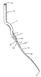
Fig. 1 is a perspective view of the stoma creator device in accordance with
the present invention.
Fig. 2 is a vertical cross-sectional view on a greatly enlarged scale taken
along line 2-2 of Fig. 1.
Figs. 3-12 are schematic views showing the method of utilizing the stoma
creator of the invention.
Having a reference to the drawings, attention is directed first to Fig. i
which illustrates a stoma creator embodying this invention designated
generally
_ 2o~os~o
7
by the numeral 10. The basic components of this device 10 are a flexible tube
11, a barbed union 12 and a tapered dilator 13. The stoma creator 10 has a
first
opening 15 at its proximal end and a second opening 18 at its distal end with
the
diameter of the first opening 15 being greater than the diameter of the second
opening 18. As can be seen, the first opening is at the one end of the
flexible
tube while the second opening 18 is at the tip of the tapered dilator 13. At
its
tip, the tapered dilator is approximately 5 French and gently tapers to the
proximal end of the dilator 20 to approximately 14 French.
Preferably the flexible tube is fabricated from a silicone material with
an interior diameter sufficiently large to accommodate a 22 French gastrostomy
tube. The barbed union 12 preferably is fabricated from nylon. The tapered
dilator which is semi-rigid, yet flexible, preferably is fabricated from
polyethylene.
As can be seen in Fig. 2, tube side wall 25 of the distal tube end 27 is
in frictional contact with union side wall 28. That portion of barbed union 12
features a first securing means 29 in the form of a gently undulating portion
of
the barbed union. The first securing means 29 is associated with the union
first
end portion 31, with union first end portion 31 having a first union aperture
32.
The distal tube end 27 of flexible tube 11 is shown as being secured to the
union
first end portion 31 by an adhesive bond 34. That particular juncture can be
subjected to corona discharge or plasma treated to enhance bond strength.
The barbed union 12 also has a tapered section 36 intermediate to the union
first end portion 31 and a second end portion 37. Second end portion 37 has a
second union aperture 38 in addition to barb 41. The second end portion 37 of
tapered dilator of barbed union 12 is preferably subjected to an adhesive bond
34 between the dilator side wall 48 and the barb 41.
20~08~.~
8
Figs. 3-12 disclose the method of utilizing the stoma creator of this
invention to place a feeding tube utilizing a PEG procedure. As can be seen in
Fig. 3, the body 50 is rolled into the supine position and an endoscope 52 is
passed into mouth 55 and down esophagus 57 into the stomach 60 which
previously
has been insufflated with air. At this time the room lights should be
relatively
dimmed and the endoscope 52 should be deflected to the interior surface of the
stomach 60. An insertion site for a slotted needle should be chosen that is
free
of major vessels, viscera and scar tissue. This site is usually one third the
distance from the left costal margin at the midclavicular line to the
umbilicus.
The intended insertion site should be depressed with a finger 65. The
endoscopist should clearly see the depression as the finger presses on
abdominal
wall 68.
As can be seen in Fig. 4, the insertion site having been prepared with a
local anesthetic has hand 69 insert slotted needle 70, having at is tip a slot
71 into which fits a T-fastener 72 with a string 73 depending from T-fastener
72
upwardly along slotted needle 70, through the insertion site in the abdominal
wall 68. A grommet 74 is located atop a T-fastener stylet 75 which depends
downwardly through the interior of slotted needle 70. As can be seen in Fig.
4,
the grommet is disposed a short distance above the slotted needle such that
the
bottom portion of the T-fastener stylet is positioned just above slot 71.
As can be seen in Fig. 5, finger 65 depresses grommet 74 such that T-
fastener stylet 75 passes downwardly through the slotted needle 70 so as to
dislodge T-fastener 72 from slot 71. At the opposite end of the string 73 from
the T-fastener 72 is a cotton pledget 76 atop which fits a nylon washer 77 and
above which is positioned an aluminum crimp 78 which when crimped restricts
the
upward movement of both the nylon washer and cotton 77 pledget 76.
9
Fig. 6 shows the body 50 with endoscope 52 inside the stomach 60 following
the placement of a plurality of T-fasteners preferably four in number. When
the
T-fastener is in its operative position, the T-fastener 72 is pulled upwardly
towards abdominal wall 68 until the T-fastener is in contact with the interior
of stomach wall 80. The stomach wall is 'then pulled slightly toward the
abdominal wall 68 until the distance between the two is relatively minimal. At
that point, the cotton piedget 75 is securely positioned above the opening
since
the slotted needle has now been withdrawn. The nylon washer 77 is placed on
top
of the pledget and the aluminum crimp 78 is secured in place. Once the T-
fasteners are in place against the inner wall 82 of stomach wall 80, a stoma
84
is formed.
Following local anesthesia, an adequate skin incision is made to the
anterior abdominal wall. Aiming slightly cephalad, a non-slotted needle 85
preferably a Seldinger needle, is inserted into the skin incision then through
the abdominal wall into the stomach. When the endoscopist sees the Seldinger
needle in the stomach, the inner stylet of such a needle is removed leaving
the
outer cannula or needle hub 87 in place. To assist in the procedure the
polypectomy snare, which should have been previously passed through the
endoscope's accessory channel, is loosely looped over the outer canula.
As can be seen in Fig. 7, a guidewire 90 is inserted through needle hub 87
of the non-slotted needle 85. When the guidewire is visualized in the stomach,
the polypectomy snare is moved down the outer cannula so as to snare the
guidewire. The endoscope and the polypectomy snare are then withdrawn from the
stomach 60 as the guidewire 90 is freely fed into the cannula 87. The
guidewire
is then pulled through the esophagus and out of the mouth.
As can be seen in Fig, 8, the stoma creator 10 of this invention is
~05(~Q~~
threaded over guidewire 90, and in the preferred method is then passed into
the
oropharynx through the esophagus and into the stomach. Once in the stomach,
the
1 eadi ng end of the tapered di 1 ator 13 wi 11 meet the outer cannul a 87 of
the non
slotted needle 85 and will follow the tract of the cannula as it pushes the
5 cannula back through the interior abdominal wall.
The leading end of the stoma creator 10 emerges from the abdominal skin,
the outer cannula 87 of the Seldinger needle 85 may be grasped, removed from
the
guidewire, and discarded. The tapered dilator 13 of the stoma creator, which
is
approximately 27" in length should then be grasped to assist in pulling the
10 gently tapered portion of the dilator through the abdominal skin. After the
tapered dilator as well as barbed union 12, is completely through the skin
approximately another 2-3" of the preferably white silicone tube should be
pulled
through the stoma.
With the guidewire still in place, the tapered tube 11 should then be cut
so as to sever the tapered dilator 13 and barbed union 12 from the rest of the
stoma creator 10. In addition to having a portion of the flexible tube project
through abdominal wall 68, the portion of the tube with first opening 15 also
projects from mouth 55. Once the tube has been cut thereby forming a second
tubular opening 95, the tapered dilator may then be removed from the guidewire
and discarded.
Figs. 9 and 10 also disclose a gastrostomy tube 100, preferably one which
is relatively flexible and having a gastrostomy tube stylet 103 inserted
therethrough passed over guidewi re 90 and moved toward the second tubul ar
opens ng
95. The skin disk 105 associated with this flexible gastrostomy tube. The
tapered distal tip of the gastrostomy tube stylet 103 should protrude from the
lower most portion of the gastrostomy tube 100.
CA 02050816 2002-05-27
11
A water soluble lubricant is preferably used to lubricate the outside surface
of
gastrostomy tube 100. The gastrostomy tube stylet 103 features a stylet hub
106 at its
upper-most portion which should be moved adjacent feeding lumen 107 of the
gastrostomy
tube 100. Simultaneously then the gastrostomy tube and stylet should be pushed
and the
flexible tube 11 pulled so that the junction of the two tubes passes through
the abdominal
wall 68 and enters the stomach 60. When the junction is in the stomach, the
balloon portion
100 of gastrostomy tube 100 is filled with approximately 20cc of sterile
saline or water.
Once the balloon is filled to prevent the unintentional removal of the
gastrostomy tube 100,
the portion of the flexible tube 11 is pulled away from the tip 112 of the
gastrostomy tube
100. The flexible tube portion 11 of stoma creator 10 may then be pulled out
of the stomach
and removed from the guidewire. It can be seen in Fig. 9 that the balloon
portion 100 of the
gastrostomy tube is filled through the use of a syringe 115, with the liquid
being injected
through a valve component 116 of the gastrostomy tube 100.
As can be seen in Fig. 11 the endoscope is passed into the stomach and under
endoscopic guidance the proper positioning of the balloon 110 in relationship
to the gastric
mucosa is assured. The gastrostomy tube is secured in position by positioning
of the skin
disk 105 against the abdominal wall 68. The guidewire 90 may then be removed
from the
abdominal site.
Fig. 12 discloses the feeding tube in position once the endoscope has been
removed. Once scar tissue forms through the stoma, and a mature stoma tract is
formed,
the T-fasteners are no longer required and may be passed through the system by
cutting
suture 73.
CA 02050816 2002-05-27
12
The enteral feeding industry has sought ways to minimize the trauma and cost
associated with the placement and removal of gastrostomy devices. This
invention solves
this long-felt need. A less traumatic placement and easier replacement of
these devices is
provided.
While the form of apparatus and method herein described constitute a preferred
embodiment of this invention, it is to be understood that the invention is not
limited to
precise form of apparatus or method and that changes may be made therein
without
departing from the scope of the invention which is defined in the appended
claims.