Note: Descriptions are shown in the official language in which they were submitted.
WG 90/12534 ~ 0 ~ ! ~ ~ ~ PCT/GB90/00648
-1-
Device for monitoring body functions
TECHNICAL FIELD
This invention relates to a device for use in real-time monitoring of human
or animal bodily functions in vivo and more particularly to a device for
monitoring these functions via the eye.
It is necessary to monitor the condition of a human or animal body in many
different situations. One particularly important time is during anaesthesia
when surgery or other treatment is being carried out, The status and
condition of the body is assessed by monitoring the level or variation of a
number of parameters such as pulse rate, blood pressure, temperature etc.
A range of instruments and devices are available to assist a doctor in
monitoring the condition of a patient in these situations and some of those
available with reference to anaesthesia will be described below.
BACKGROUND ART
The most important function to monitor is oxyg=n supply to the brain, eg
for example during anaesthesia. The measurement of this is known es pulse
oximetry. The brain can only survive on oxygen and glucose, le by aerobic
metabolism, whereas most other tissues can survive for a period without
oxygen, ie by anaerobic metabolism. The oxygen supply !.s a function of
delivery, ie cardiac output which governs all blood flow, and the amount of
oxygen available in the blood, which is governed by the 0= haemoglobin
dissociation curve.
The blood flow is assessed by monitoring blood pressure and by assuming
the resistance is constant, so the pressure is directly related to flow
(Ohm's law> end by pulse rate which is generally measured with an
electro-cardiogram (ECG) and/or a pulse oximeter. The amount of oxygen
available is measured with a pulse oximeter which calculates the percentage
saturation of the haemoglobin by using one, two or three mono-chromatic .
light sources. The amount of light passing through tissue of the body is
recorded to determine the quantity of light absorbed by the haemoglobin.
These devices usually measure the saturation in the periphery, eg in the
toe, finger or ear, and this is assumed to be related to the oxygen
saturation of blood delivered centrally to the brain.
WO 90/12534 Z Q ~ ~ 4 ~ ~ PCT/GB90/00648
_2_
The mayor limitation of pulse oximetry is that the measures are of
peripheral and not central blood flow. Other limitations of pulse oximetry
are;
<i) The calculations are based on the Beer Lambert -law and it is known
that due to the scattering of light by skin tissue and other structures the
application of this law is not valid under these conditions.
<ii> The wavelengths of light used are usually determined by the available
light emitting diodes and these may not be ideal. Existing machines use
only one, twa or three wavelengths which further limits the accuracy of the
oxygen saturation calculation.
<iii> Ambient light may interfere with the signal detection and hence reduce
its sensitivity.
<iv> The spectral absorbance of the tissue through which the light is
transmitted affects the results, eg with non-Caucasians.
<v) Other chemicals normally found in the blood, eg tiilirubin, may also
interfere with the transmission of the light and have en unlasown influence
on the results.
The electro-retinogram IERG> is also used to assess the function of the
retina. This device uses a contact lens provided with a circular electrode
for detecting electrical activity of the retina. Stimulation is provided by
en external light source directed into the eye.
Contact lenses provided with mirrors ar lenses have also been used for
observing the angle of the anterior chamber and the peripheral fundus of
the eye. A high-negative-powered contact lens has also been used to assist
observation of the retinal fundus during ophthalmoscopy and n telescopic
device has been used on a contact lens for the study of stabilised retinal
images.
,,-.._., ~ ;, _:: y.
~.~;>:.~;~,_ .. .~
., .:.,., :;-~ay~ .,=-. ,~;i~. ~ i.s :, a
CA 02051419 1999-03-04
- 3 -
Visual evoked potentials (VEP), have also been used
to monitor the condition of a patient during anaesthesia and
at other times. The potentials are measured by recording
changes in the electrical activity of the brain following a
measured visual stimulus or series of measured visual stimuli.
An electro-cardiogram (ECG) is used to measure the electrical
activity of the heart and as such provides a measure of heart-
rate, heart rhythm, muscular contraction and, indirectly,
oxygen supply to the muscle.
One of the aims of the present invention is to
provide a device for non-invasive, real-time monitoring of
cardiovascular, respiratory, neuro-muscular and other bodily
functions whilst avoiding soma of the disadvantages of the
prior art.
DISCLOSURE OF INVENTION
According to a first aspect of the invention there
is provided a device a device for use in real-time monitoring
of human or animal bodily functions in vivo comprisings a
scleral contact lens for locating and supporting the device on
an eye including a pupil having a size determined by dilation
of the pupil, a retina and an iris, of a human or animals and
a first optical system supported by the contact lens having at
least one discrete light input means and at least one discrete
light receiving means, wherein the light input means is
arranged to direct light through the contact lens and the
pupil of the eye so as to illuminate at least a portion of the
retina of the eye substantially independently of the pupil
size and the light receiving means is positioned so as to
27954-1
CA 02051419 1999-03-04
- 4 -
receive light returning through the contact lens directly from
a portion of the retina which is illuminated directly by the
light input means.
A preferred form of the device avoids the
shortcomings described above of conventional pulse oximetry,
as well as providing means for monitoring other bodily
functions, by using a first optical system for visible, ultra-
violet, and infra-red spectrophotometric or fluo-
spectrophotometric analysis of retinal blood flow.
Another preferred form of the device enables the
size of the pupil to be monitored, even when the eyelids are
substantially closed, by using a second optical system for
infra-red pupillometry, eg during anaesthesia.
According to a second aspect of the invention there
is provided a disposable component of a device for real-time
monitoring of human or animal bodily functions in vivo, the
component comprising a scleral contact lens for locating and
supporting the device on an eye including a pupil having a
size determined by dilation of the pupil, a retina and an
iris, of a human or animal and mounting means on the contact
lens configured for detachably mounting and supporting an
optical system on the lens in such a manner that, When mounted
thereon, the optical system is positioned to direct light
through the contact lens and the pupil of the aye so as to
illuminate the retina of the eye substantially independently
of the pupil size and to receive light returning through the
contact lens from that portion of the retina illuminated by
the optical system.
27954-1
CA 02051419 1999-03-04
- 4a -
According to another aspect of the invention there
is provided a device for use in real-time monitoring of human
or animal bodily functions in vivo comprisingi a scleral
contact lens for locating and supporting the device on an eye
including an iris, of a human or animal, the eye having a
pupil defining an optical axis, and an optical system
supported by the contact lens arranged to direct light towards
the aye and to receive light reflected from the iris of the
eye, characterized in that the optical system comprises an
array of discrete light input means for providing light and
for directing that light towards the eye at a series of
different distances from the optical axis and an array of
discrete light receiving means for receiving light reflected
from the iris of the aye at a series of different distances
from the optical axis.
According to a further aspect of the invention there
is provided a method of real-time monitoring human or animal
bodily functions in vivo using a device as described above.
Other preferred features of the invention will be
apparent from the following description and the subsidiary
claims of the specification.
BRIEF DESCRIPTION OF THE DRAWINGS
The invention will now be further described, merely
by way of example, With reference to the accompanying
drawings, in which:
Figure lA is a cross-sectional view of a special
adaptation of a scleral contact lens used in an
embodiment of the invention and Figure 1B is a
27954-1
CA 02051419 1999-03-04
- 4b -
perspective view thereof
Figure 2A is a cross-sectional view showing components of
a first optical system used with the contact lens shown
in Figure 1 for spectrophotometry and Figure 2B is a
perspective view thereof
Figures 3 and 4 are perspective views of further parts of
the first optical system shown in Figure 2i
27954-1
WO 90/12534 PC?/GB90/00648
2~~1419
-5-
Figure 5 is a cross-sectional view corresponding to Figure 2 showing an
embodiment of a second optical system of the invention used with the a
scleral contact lens for infra-red pupillometry;
Figure 6 is an end view of part of the second optical system shown in
Figure 5;
Figures 7A, B and C are further views of the contact lens showing
electrodes used therewith; and
Figures 8, 9 and 10 are cross-sectional views showing the use of a
scleral contact lens such as that shown in Figure 1 on the eye and the
optical paths of incident light in three different embodiments of the
first optical system and Figure 11 is a corresponding view showing the
path of light rays showing the limits of the field of view of the
retina as seen by the light receiving menus.
BEST MODE OF CARRYING OUT THE INVENTION
The device to be described uses a scleral contact lens as a vehicle for an '
optical system to monitor human or animal bodily functions via the eye and
is based on the realisation that since the pre-retinal structures of the
eye are designed to transmit light, it is well suited to optical methods of
examination. Furthermore, the eye provides a more direct method of
assessing the condition of the brain then some of the prior art methods
described. This is because the ma,~or blood supply to the eye via the
ophthalmic artery is a branch of the internal carotid artery. The eye,
therefore, is sometimes aptly referred to as the 'window' of the braid.
Preferred forms of the device enable several different functions or
parameters to be monitored, both in con,~unction and independently of the
optical system of the device, using the same equipment.
As mentioned above, the device offers, a non-invasive method of reel-time
monitoring of the cardiovascular, respiratory and neuro-muscular functions
in man. In addition, it offers a method of in vivo measurement of the
various cellular end chemical constituents found normally in blood. It is
also possible to measure foreign chemicals and their metabolites, e.g, drugs,
once their individual absorbence/refleetanee spectrum has been identified.
y,, .~
WO 90/12534 2 ~ ~ ~ ~ ~ ~ _ PCT/GB90/00648 _
-s-
Before the device is described in detail, some of the different functions it
can be used to perform will be briefly described:-
1. Measurement of the amount and change of any or all cells and chemicnle
found in the blood using light, in the visible, infra-red and ultra-violet '
range to detect the unique absorbnncelreflectance spectrum of each call or
chemical by spectrophotometry. This applies to naturally occurring
substances as well as foreign substances.
2. Measurement of changes in the oxygen saturation of haemoglobin by
spectrophotometry to assess oxygen delivery to the retina and so provide a
measure of oxygen delivery to the brain. Similarly, changes from one
haemoglobin to another, eg o~yhaemoglobin to reducsd. haemoglobin, can be
measured es well as percentage changes in abnormal haemoglobin such as Hbs,
the cause of sickle cell disease.
3. Measurement of arterial carbon dioxide tension using the reflectance of
infra-red light from blood vessels in the retina.
4. Measurement of retinal blood flow (which is related to cardiac output
and to cerebral blood flow) using spectrophotomatry to measure the
transport of specific substances in the blood.
5. Measurement of the pulse rats by detecting blood volume changes
consequent to changes in the width of blood vessels on the retina or the
cycles of oxygen saturation of haemoglobin during diastole and systole by
spectrvphotomstry.
6. Measurement of the depth of anaesthesia and/or level of consciousness
by infra-red pupillometry.
7. Messurement of pressure changes in the eye to measure or monitor
arterial blood preaeure and/or pulse rate. ~.
8. Measurement of the change in pH of artificial tears used in this
pressers measurement es a measure of the pH changes in the blood.
~: ,
i:.:
:.:
WO 90/12534 '~ O 3 ~ 4 ~ ~ PGT/GB90/00648
_'_
9. Measurement of the increase in volume of these artificial tents as n
measure of the depth of anaesthesia.
10. Measurement of integrative retinal activity to measure the electrical
activity of the retina to assess the depth of anaesthesia and the level of
consciousness by using the device as part of an electroretinogram.
11. Measurement of changes in visual evoked potentials to assess the depth
of anaesthesia and the level of consciousness.
12. Measurement of electrical changes recorded by the electrodes used for
measurement of the integrative retinal activity sad visual evoked potentials
._ .
to monitor the electrical activity of the heart and to record nn ECG.
,
13. Measurement of neuromuscular blockade during anaesthesia by use of ,
electromyograms tEMGs> of all four recd eye muscles.
14. Measurement of neuro-museuler blockade during anaesthesia by electrical
stimulation of the lateral rectus muscle, with train of four stimulation,
and using infra-red pupillometry to record the movement or lack of
movement of the eye.
15. Measurement' of body temperature using either a thermistor
(thermocouple> or infra-red sensor provided on the device.
16. Measurement of biochemical reactions and cellular respiration in the
retina.
The expression 'human or animal bodily functions' used in this epeeification
is Intended to include all the different functions mentioned above and the
monitoring of any substances and changes in the blood of the retina end any
biochemical (organic or inorganic) changes in the cello of the retina.
It should also be noted that it is possible with the device to monitor in
real time any one or all of these functions separately or together, either
intermittently or continuously.
WO 90/12534 '~ O 5 i 41 ~ PCT/GB90/00648 ..
_g_
The term 'light' used in this specification is intended to include visible
wavelengths and other, non-visible wavelengths such ee infra-red and
ultra-violet light.
The device will now be described in more detail with reference to the
accompanying drawings. The scleral contact lens will first be described end
then the components relating to the different functions mentioned above
which can be grouped under the headings spectrophotometry, pupillometry,
fluid measurements, electrophysiology and temperature measurements.
Finally, the analysis and display equipment used with the device will be
briefly described. ' ,
'P1~ CONTACT LENS
A 6cleral contact lens 1 formed of polymethylmethncrylate (PMMA> or other
suitable eterilisable material is preferaply used. The ecleral contact lens .
1 is designed to fit onto the sclera and bulbar conjunctiva of the eye so
if the eye moves the contact lens 1 souse with it. The ecleral contact
lens was first used in 1888 ns a protective device for the cornea end there
have been various clinlenl and therapeutic applications since then, such as
those mentioned above.
To protect the cornea, sclera and bulbar conJunctive, a hard haptic ecleral
contact lens nay be ensheathed in a low or high water content, hydrophilic
material such as that used in the manufacture of soft contact lenses. Such
a sheathing also allows tho contact lens to be made of other materials as
only the sheathing material is in contact with'the eye.
The ecleral contact lane shown in Figure 1 has a central optical portion 2
provided with a converging lens for focussing light through and
substantially in the plane of the pupil so as to illuminate the retina of
the eye. Figure 1 shown the lens 1 and the eantral optical portion 2 <alsa
Imown as the corneal portion). The optical portion 2 hoe a smaller radius
of eurwature than the reaainder~ of the lens <the haptic portion>. The
radius g of the optical portion 2 is made shorter than the 98th percentile
WO 90/I2534 '~ ~ ~ I ~ ~ g . PCT/GB90/00648
_g_
of human cornea and is therefore typically around 6.5ma. When the lens i
is fitted to the eye, the optical portion 2 ie thus raised from the cornea.
The diameter ~ of the optical portion 2 is typically l3nm or more.
The back haptic radius g of the ecleral contact lens 1 is typically between
12 and l5mm and the back haptic size ~ between 20 and 25mm (vertically)
with the horizontal size usually being 1 to 2 mm larger. A selection of
three different size lenses 1 1s usually provided to fit cost human eyes.
At the centre of the optical portion 2 le a converging lane 3 in the form
of a high powered positive lenticulsr with a diameter ~ of about 3mm
provided on a plano carrier. The optical axis of the lens 3 is
substantially coincident With the optical axis~of the eye.
The lenticular 3 is arranged to focus colliaated light in the plane of the
pupil 30 for an average anterior chaober depth of 4am (see Fig 2A> to
produce an image of the illumination source (see below) substantially in
the plane of the pupil. This is laiown ae Mexwellisn view. Illuninntion is
thus provided along the optic axis of the eye and the lentieular 3 focusses
the light substantially in the plane of the pupil 30. In this say, the
retina is illuminated substantially independently of pupil size so the need
to use a mydriatic to dilate the pupil is miniaised.
As problems can be encountered with light reflected from the cornea if the
illumination is directly along the optic axis, it nay be preferable to
.:
~.,....
provide the illumination slightly off axis or nt a small angle thereto but,
nevertheless, substantially along the optic axis. Alternatively, nn
anti-reflective coating say be applied to the surfaces of the optical
components.
In the arrangement shown, the focal length of the lenticular 3 is around 5
mm, ie it hna a strength of about 200 dioptres. As the optical portion 2
of the sderal contact lens 1 is raised from the cornea, it increases the
distance between the lenticular 3 and the eye so avoiding the need for the
lenticulnr to be of even higher power in order to focus light substantially
in the plane of the pupil 30.
WO 90/12534 ~ U '~ ~' ~ ! ~ PCT/GB90/00648
-10-
Between the optic and haptic portions of the contact lens 1 there is a
transition curve where the two portions blend together. At this point n
small <eg about lmm in diameter) fenestration or aperture 4 is provided
(both medially and laterally) so that a fluid such es artificinl tears may
be passed through the lens 1 to the eye to prevent the cornea becoming dry
beneath the lens. Artificial tears are provided along a tube or cannula 5A
connected to one of the apertures 4 and a tube or cannula 5B connected to
the other aperture 4 receives fluid returning from the eye. ,
In an alternative arrangement (not shown>, the tubes 5A and 5B may be
included within the wail thickness of a carrier 6 (see below) mounted on
the contact lens 1 with countersunk apertures on the corneal side of the
lens 1.
In either arrangement, the tubes 5A and 5B are preferably connected in a
closed system tnot shown>, e.g, by flexible tubing leading to other devices
(see below>.
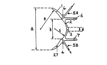
Extending from the scleral contact lens 1 is a carrier 6 for receiving
components of one or more optical systems such as those described below. A
rubber O-ring locking device 7 or other attachment means ie provided for
detachably securing these within the carrier 6. These further components '"
will be descnihed below in relation to the different functions of the
device.
Both the carrier 6 end the haptic portion of the contact lens 1 are
preferably provided with en opaque lamination, or manufactured from opnque
material such ns black perspex, to prevent light from outside the device
interfering with its function.
The scleral contact lens 1 and carrier 6 may be formed of relatively
inexpensive material so they can be disposed of after use. Components of
the optical system can thus be detached from the carrier 6 ae described
above and then installed in the carrier 6 of a new contact lens unit to be
used with another patient.
K;~i ~ ~ , .
wo 90~ 1 Zs34 ~ p ~ ~ 4 ~ g PCI'/GB90/00648
-ii-
SPECTROPHOTOMETRY AND FLUO-SPECTROPHQTOMETRY . ..
For measurements involving illumination of the retina and the detection of
light returning therefrom to determine the absorbancelreflectance spectrum
of the retinal blood supply, a first optical system comprising optical
components such as those shown in Figure 2 (end Figures 8 to 11 ) is used
within the carrier 6. The arrangement shown in Figure 2 comprises n
support 17 which fits within the carrier 6 and which houses a coherent or
semi-coherent fibre optic bundle 8 positioned in front of the lenticular 3
and directed towards the pupil of the eye along the optic axis thereof.
Figures 3 and 4 show the parts of this optical system in more detail. The
fibre optic bundle 8 comprises a central illumination fibre or fibres 9 for
directing light through the lenticular 3 and one or more paraxial receiving '
fibres 10 grouped around the central fibre 9 for receiving light returning
from the retina. A light absorbent outer sheathing 11 is provided around
the fibres to prevent interference from ambient stray light and to prevent
light loss from the fibres. The illumination fibres 9 are also masked from
the receiving fibres 10. The central fibre 9 thus provides a discrete light ,
":"
input means and the fibres 10 a plurality of discrete light receiving means.
A light source is provided remote from the device at the other end of the
illumination fibre 9 and preferably comprises a halogen bulb 13 positioned
behind a rotatable wheel 14 provided with a range of different,
monochromatic interference filters 15 with a condenser lens 16 between the
bulb 13 and the wheel 14. Thus, by positioning the appropriate filter 15 in
front of the beam of light from the bulb 13, light of a selected wavelength
is transmitted through the illumination fibre 9 to .the lenticular 3 which
focusses the light through the pupil of the eye to illuminate the retinal
fundus. Different wavelengths can be used in succession by rotation of the
filter wheel 14.
Alternatively, the light source may be provided either by reflected,
coherent monochromatic light <eg from a laser) or selected light emitting
diodes.
WO 90/12534 a PCT/GB90/00648
~fl5 X419
-12-
Light returning from the retina passes out of the eye through the pupil,
through the optical portion 2 of the contact lens 1 towards the ends of the
receiving fibres 10 which transmit the returning light to a remote
light-sensing means, eg a photo-multiplier (not shown), provided at the
other ends of the receiving fibres 10, to determine the intensity of the
returning light. Thus, by using a series of different wavelength selective
filters 15, the ahsorbance/reflectance characteristic of the retina and its
blood supply can be determined.
The first optical system may be arranged in a variety of ways to provide
spectrophotometry of the retina. When optical fibre are used, as described
above, the illumination may be provided by means of spectral light emitting
diodes (not shown>, eg emitting red, green, yellow and blue light.
Alternatively, white light may be used to illuminate the retina and the
returning light passed,through monochromatic filters before being passed to
the light detector. The fibres themselves could also act as filters if they ,y
are formed of selectively, spectrally absorbing material. It would also be
possible to monitor simultaneously the different wavelengths in the
returning light.
In a further alternative, the wheel 14 may be replaced by a coloured liquid
crystal charged couple device <CCD> for direct colorimetry. With white light
illumination vin the fibres 9, light returning from the eye is directed by
the receiving fibres 10 onto the charged couple device. Ultra-violet and
infra-red light sources may also be used.
In some cases, it mny also be convenient for the same optical fibre or
fibres to act ns both light input means and light receiving means.
In a further alternative arrangement (not shown>, a number of photodiodes
(spectral light emitting diodes) or other discrete light emitting means of
specified wavelengths may be carried by the device to provide a light
source on the device end so avoid the need to use optical fibres. Such
diodes may be mounted on a separate unit for insertion into the carrier 6
or mounted directly on a support attached to the scleral contact lens 1.
WO 90/12534 2 ~ J ~ ~ ~ 9 . PCTlGB90/00648
-13-
It is also possible to provide a photodetector <not shown) or other discrete
light sensing means, carried by the device to provide a light receiver
without the need for optical fibres. This may, for instance, be annular in
shape and positioned on the optical portion 2 of the contact lens to
receive light returning from the fundus of the eye.
The light source and light sensing means mounted in the device would be
provided with electrical connections to enable them to be connected to a
suitable power source and other electrical equipment. A wide variety of
other optical systems using one or more discrete light input means and
discrete light receiving means arranged to determine the
absorbance/reflectance characteristic of light returned by the retina can be
used and will be apparent to~those skilled in the art.
The light input means and light receiving means, whether in the form of
optical fibres or discrete devices mounted on the device, are sufficiently
small and lightweight to allow the device with its optical system to be
supported on a patient's eye so avoiding the need to position and support w
heavy, bulky equipment in front of the eye. The device is therefore easy
and convenient to use, particularly for continuous monitoring of a patient's
condition, eg when anaesthetised.
Figures 8, 9 ana 10 show the use of the device described above in relation
to the eye and illustrate different arrangements of the first optical
system.
The device shown in Figure 8 corresponds to that shown in Figures 1 end 2,
but includes an additional converging lens 28 on the end of the central
illumination optical fibre 9 as this helps provide an approximately
collimated beam of light from the fibre 9 towards the lenticular 3. It also
helps minimise the effect of unwanted reflections from stray light reaching
the iris 29 through the optical portion 2 of the lens around the
lenticular 3.
As shown in Figure 8, the lenticular 3 focusses the light in the region of
the eye's papillary plane 30 <shown by dotted lines) so as to minimise the
WO 90/12534 ~ ~ ~ ~ ~ ~ ~ PCT/GB90/00648
-14-
effect of pupil size on the level of illumination of the retinal area 31.
The optical portion 2 of the scleral contact lens asy have a smaller radius
of curvature than the cornea <as shown in Figure 1> or may have a similar
radius of curvature to the cornea but not in contact therewith as shown in
Figures 8 to 11. In either case, the optical effect of tears between the
lens and the cornea will have little effect on the focussing of light in the
plane of the pupil due to the depth of field of the lenticular 3 <or the
other lenses described below>.
In a further arrangement (not shown>, the contact lens may be shaped so
that tear fluid between the lens and the cornea forms part of the optical
system for focussing the light in the plane of the pupil or acts as the
sole converging lens in the optical system.
In the arrangement shown in Figure 9, a high powered converging lens 32 is
provided on the end of the central illumination fibre 9 and replaces both
the lenticular 3 and the converging lens 28 of the arrangement described
above. The optical portion 2 of the contact lens may thus be of zero
strength. The lens 32 produces a convergent beam of light which passes
through the optical portion 2 of the contact lens 1 and is brought to a
focus in the region of the eye's papillary please 30. ~ The distance between
the lens 32 end the contact lens 1 can be nd~usted by sliding the bundle of .
optical fibres .8 in and out of the carrier 6 to provide optimal focussing
of the incident light beam in the eye's papillary plane 30.
The arrangement shown in Figure LO is similar to that of Figure 9 but, in
this case, the convergent lens 33 is provided in the carrier 6 between the
illumination optical fibre 9 and the contact leas 1 rather than on the end
of the fibrs 9. As indicated diagramatically in Figure 2A, a converging
lens may, in fact, be provided at any position between the light source and
the contact lens so long as it is arranged to focus light substantially in
the papillary plane of the eye. If desired, the optical system may comprise
a plurality of lenses mounted on the light source, within the carrier 6 or
on the contact lens 1 or any combination thereof to focus light in the
PuP~r'Y Pie.
WO 90/12534 2 ~ C~ ~ ~ ~ 9 PCT/GB90/00648
-15-
Figure I1 illustrates the limiting light rays of diffusely reflected light
from the retinal area received by the paraxial optical fibres 10 which then
transmit the light to photodiodes or other light sensing means (not shown>.
As described above, in any of these arrangements, the illuminating optical
fibre may be replaced by a light emitting diodes or other light emitting
means end the receiving optical fibres 10 may be replaced by miniature
photodiodes, charged couple devices or other light sensing means. In each
case, the light emitting 'end light receiving means would be provided with
electrical connections to enable them to be connected to a power source end
other electrical equipment.
The first optical system may thus be used to measure changes in the
reflectance/absorbance of the blood vessels of the retina. Any combination
of mono-chromatic lights or white light, as well as wave-lengths in the
infra-red and ultra-violet spectra can be used. Specific, selected
wavelengths permit optimal discrimination of the various blood components.
In this way, it is, for instance, possible to provide an accurate
measurement of the oxygen saturation of the retinal blood flow and, as this
is closer to cerebral blood flow than the toe, finger or ear blood flow
previously measured, it provides a more accurate assessment of blood
delivered to the brain.
The spectrophotometric system described above may also be used for
fluo-spectrophotometric analysis in which the retina is irradiated with one
wavelength and it emits light of a different wavelength which is detected
by the optical system. Most natural substances haNS auto-fluorescent
properties. Fluorescent dies can also be used as appropriate.
As indicated above, this system also enables the device to monitor pulse
rate by measuring changes in light nbsorbance/reflectance of blood vessels
on the retina between diastole end systole.
The measurement of the retinal blood flow also gives an indication of
cerebral blood flow as the retina and its blood supply are part of the
W0 90/12534
PCT/GB90/00648
-16-
brain and changes in retinal blood flow give en indication of changes in
cerebral blood flow ea well as changes in the cardiac output.
It is also possible to calculate changes in the resistance to blood flow
using Ohm's Law and so provide another measure of the depth of anaesthesia.
Such measurements of cardiovascular and respiratory functions ere always
monitored in intensive and coronary care units es well es in any
unconscious patient.
Another of the functions which can be monitored using the first optical
system described above is the change in the saturation of haemoglobin, both
adult and foetal, with various chemicals over time; for instance of oxygen
(oxyhaemoglobin>, carbon dioxide <cnrboxyhaemoglobin>, haemoglobin without
any gases (reduced haemoglobin) and sulphur <methaemoglobin> or any of the
other hsemoglobins.
A measurement of the percentage saturation of haemoglobin enables the
haemoglobin dissociation curve to be calculated and so provide real time
monitoring of this curve ns well as changes in the curve with time.
It is also possible to measure in vivo abnormal haemoglobins such as HbS
which causes sickle cell disease. If characteristic ebsorbance/reflectance .
spectra for each of the abnormal haemoglobins are defined, these can be
held in a computer memory and then compared with in vivo measurements and
their changes with time can be monitored.
It is also possible to use the device for real-time monitoring of all the
many and varied biochemical substances found in the blood. For example,
bilirubin has a specific light absorbencelreflectance spectrum es do other
chemicals found in the blood, including the many end varied amino-acids end
proteins. The limiting factor is, of course, the degree of difference
between the absorbence/reflectance spectra for each of these chemicals.
If the absorbance/refleetance spectra of drugs, chemicals and their
metabolites are known, it will also be possible to monitor real time changes
WO 90/12534 2 0 51419 ' p~/GB90/00648
-17-
in the bloodlplasma concentration of these as they are edminietared to a
patient. The characteristic curves of spectral abaorbanee/reflectance can
be held in a computer memory for automatic comparison with the test data.
Selected wavelengths in the infra-red may also be used to illuminate the
retina and the amount they are absorbed/reflected used to provide a measure
of the carbon dioxide pressure in the blood.
It is also possible to use the ebsorbance/reflectence of light from the
retina to monitor in real time all biochemical changes pccurring in the
cells of the retina, for example, cellular respiration of the retina. It is
also possible to combine the measurement of fluorescence with that of
absorbance/reflectance tQ improve further the sensitivity of the system.
Measurement of the biochemical activity of retinal cells provides an
indirect measurement of the biochemical activity of the brain and so
provides a measure of the oxygen demand and utilisation thereof.
Figure 5 shows components of a second optical system that may be attached
to the carrier 6. The second optical system is arranged to direct light
towards the pupil and iris of the aye and to receive light reflected
therefrom. In., the illustrated arrangement, the second optical syste,
comprises a further coherent fibre optic bundle 18 the ends of which are
arranged in a cruciform array as shown in Figure 6. M outer protective
casing 19 of PMMA or other similar material secures the bundle 18 onto nn
optical mount on the side of the carrier 6. A mirror 20 is provided in the
carrier 6 inclined at an eagle of 45 degrees so that light from the bundle
18 is reflected towards the eye through the optical portion 2 of the
aclernl contact lens 1 and so that light returning from the eye ie '
reflected back to the ends of the fibres arranged in the cruciform array.
The array of fibres is, in fact, made up of two interleeed coherent arrays,
a first array 21 (shown shaded) for illumination and n second array 22
(shown unshaded> attached to a charged couple device tCCD) detector (not
WO 90/12534 r PG?/GB90/00648
~0~1419
-18-
shown) for receiving light reflected from the eye. As shown in Figure 6, no
fibre is necessary in the centre of the array.
In use, infra-red light is passed to the first array of fibres 21 and is
directed at the iris of the eye by the mirror 20 and the optical portion 2 ' '
of the contact lens 1. Depending on the size of the pupil, the light from a
particular fibre either posses through the pupil into the eye where it is
absorbed or is reflected by the iris and transmitted back to<the second
array of fibres 22 by the optical portion 2 of the lens 1 and the mirror
20. The CCD detector measures the intensity of any light received by the
fibres in the second array 22. The diameter of the pupil can thus be
measured by determining which of the second array of fibres 22 receives
light reflected from the eye. If the pupil is large, only the outermost
fibres will receive light reflected from the iris whereas if the pupil is
small all but the innermost fibres will receive reflected light.
The illustrated array of fibres senses the size of the pupil across two
diameters and the array preferably includes further fibres arranged across '
other diameters of the eye, eg along the dotted lines 23 shown in Figure 6.
Other forms of array could also be used, for instance a series of
concentric rings with alternate fibres in each ring directing light towards
the eye and detecting light reflected therefrom. The resolution of the
pupil diameter ~is a function of the number of fibres across the array. It
would also be possible to use the same fibres as both illuminating means
for directing light towards the eye and receivers for receiving light
reflected therefrom.
As for the spectrophotometric optical system described above, the light
input optical fibres may be replaced by an array of discrete light emitting
means, such es light emitting diodes, and the light receiving fibres may be
replaced by an array of discrete light sensing means, such as photodiodes.
In an alternative arrangement (not shown>, the second optical system may be
provided without the first optical system. In this case, the lenticuler 3
or other lens system may be omitted and the first and second arrays 21 and
.::;
::;
r?:r
WO 90/12534 ~ O ~ ~ 4 ~ ,~ PC1'/GB90/00648
-19-
22 of fibres arranged in the support 17 in place of the optical fibre
bundle 8 so as to direct infra-red light towards the eye without the need
for the mirror 20.
It is possible to monitor continuously the depth of anaesthesia by
measuring the dilation of the pupil using the second optical system
described above, the reflected infra-red light being used to measure the
area of the pupil. Observation of changes in pupil size is the original way
of clinically assessing the depth of anaesthesia and is the bench-murk
against which all other techniques are measured. It is also possible to
measure the papillary light response as an assessment of the depth of
anaesthesia.
FLUID MEASUREMENTS
As mentioned above, the haptic scleral contact lens 1 is provided with
means for supplying artificial tears to the eye to prevent the cornea from
becoming dry. This provides another way of measuring blood pressure and
pulse refs since the pressure within the eye unties ae the blood vessels
therein expend end contract with each pulse. The liquid interface between
the haptic shell 1 and the cornea of the eye acts ns a pressuro transducer
for sensing 'these pressure changes. The pressure changes in this interface
are transmitted to the liquid within the tubes 5 connected to the apertures
4. Thus, by using a closed system connected to the tubes 5A and 5B with , .
monitoring apparatus (not shown) such as a pressure sensor, it is possible
to measure pressure changes or movement of the liquid within the tubes 5
to provide a measurement of arterial blood pressure and pulse rate.
Changes in the hydrogen ion concentration <pH> of artificial tears supplied'
to the eye through the tubes 5A and 58 in a closed system een also be
measured. The pH of natural tears produced by the eye is related to pH
changes in the blood end es these mix with the artificial tents supplied
through the tubes 5, the pH of the fluid withdrawn from the tubes 5 can be
measured by colorimetry or a pH electrode to monitor these changes.
WO 90/12534 ~ .~ PCT/GB90/00648
~0~~41~ _
-20-
The volume of natural tears is also related to the depth of anaesthesia and
this can also be monitored by measuring the changes in the volume of fluid
within the tubes with a volumetric measure.
ELECTROPHYSIOLOGY
Two sets of electrodes are provided on the scleral contact lens 1, one to
apply electrical stimulation to the extra-ocular muscles or to reeord the
electrical activity therefrom, the other to detect electrical changes in the
retina of the eye. These electrodes are shown in Figure 7.
The first set comprises electrodes 24 of gold or other suitable conducting
material embedded in the haptic portion of the lens 1 at points around its
periphery. The electrodes 24 are embedded in the haptic portion between
6mm and l0mm from the centre of the optical portion 3 so as to lie over
the recd muscles insertions when the lens 1 is placed on the eye. The
second set comprises a ring electrode 25, again preferably of gold,
extending around the optical portion 2 of the lens 1. Connections to the
two sets of electrodes 24 end 25 are provided by electrode pin contacts
(not shown>. Screened connections from the electrodes 24 and 25 extend
towards sockets 26 in the haptic lens 1 as shown in Figure 7B where they
connect with' the contacts provided on the pin contacts inserted therein.
The enlarged view in Figure 7C shows the separate contacts within each
socket 26.
The first set of electrodes 24 are used to record the activity of the eye
muscles to provide an electromyogram (EMG>. The second set of electrodes
25 can be used in connection with a conventional electroretinograph <ERG>.
The lateral rectos muscle may be stimulated percutaneously end the
resultant EMG recorded via the electades 24. Any significant contraction of
the lateral rectos muscle may be meas.u-ed by changes in electrical
potentials using a further electrode <not shown) for instance attached to
the face above the eye. It is thus possible to use the standard train of
WO 90/12534 ~ ~ ~ ~ ~ ~ 9 PGT/GB90/00648
-21-
four stimulation to measure the degree of muscle relaxation with this
system and so monitor the degree of muscle paralysis.
As mentioned above, the device shown in Figure 7 may also be used to
measure potential changes in the retina in the same manner as a
conventional electroretinogram. However, the light used to stimulate the
retina may be provided by the first optical system shown in Figures 2-4 or
Figures 8-11 rather than a separate, external light source.
Electrodes placed elsewhere on the heed may also be used to record visual
evoked responses in a conventlonal manner but using the first optical.
system described above to provide direct stimulation of the retina so
allowing peroperative vl5ual evoked potentials to be recorded because the
amount of light reaching the retina is independent of pupil size. .
The electrical activity of the heart can also be recorded by suitable
electrodes placed anywhere on the body so either of the electrodes 24 or 25
provided on the haptic porton of the lens 1 may also be used in con3unction
with other electrodes to record an ECG.
TEMPERATURE SENSING
The lens 1 may also be provided with a thermistor tthermo-couple) or
infra-red sensor 27 <see Figs lA and 7B> to measure the temperature of the
eye.
Temperature measurements are necessary during anaesthesia to diagnose
- malignant hyperexia which is an unusual but potentially fatal condition.
The temperature is also important in calculating the amount of oxygen
eveilnble to the tissues tag the brain) as it effects the haemoglobin
dissociation curve.
It will be appreciated that the contact lens 1 locates and supports the ,
. device on the eye and, when using discrete light emitting and discrete light
'
receiving menus as described above, the contact lens is able to support the
opticnl system end other sensors attached thereto. There is, therefore,
..: , . - ., : -
r::. ; . ~ ,. ,
WO 90/12534 PCT/GB90/00648
-22-
no need to hold or support larger or heavier equipment in front of the eye.
Instead, optical fibres or electrical wires transmit light and/or information
between the device and a remote light source and remote light sensing
means which may be mounted alongside or form part of analysis and display
equipment located at the patient's bedside or beside the operating table.
The analysis and display equipment connected to the various sensors of the
device is arranged to provide a variety of different displays and outputs.
The importance of real-time monitoring during anaesthesia and in medicine.
generally is to record changes in a patient's physiological state with time.
The device will enable the doctor to monitor heart rate, blood pressure
(both of which are important in cardiac output>, and the amount of oxygen
being carried by the blood with the saturation percentage. Thus, the mayor
components of oxygen delivery, transport and content can be monitored. The
Hb dissociation curve can be calculated from the data obtained end any
change noted in addition to changes consequent to any clinical intervention,
the time of which may nleo be recorded. A statlnticnl analysis can be
automatically carried out to calculate the mean end the standard deviation
of each fenttwe meneured as well as changes in measurements over time to
provide an alert or signal an alarm system if significant changes ere
recorded.
The pupil diameter and the electroretinogram are both measures of the depth
of eneasthesie end reel-time monitoring enables en indication to be given
if this is becoming too deep or too light.
The EMG provides reel time monitoring of the degree of muscular paralysis
which is particularly important during microscopic surgery.
filsetroaic control aeons are provided to monitor, display and record
signals provided by each of the different monitoring systems described
above. The electronic equipment may be arranged to provide a wide variety
WO 90/12534 ~ ~ j j ~ ~ ~ PC1'/GR90/00648
-23-
of information depending on the intended use end the requirements of the
doctor. Further details of the construction and operation of the electronic
control means is not provided since a wide variety of systems will be
apparent to those skilled in the art or are already available.
Features which may be displayed on the instrument include: the saturation
percentage of oxygen, the haemoglogin dissociation curve, pupil diameter,
pulse rate, blood pressure, temperature and pH as well as the displays
conventionally provided by an electrocardiograph (ECG>, electroretinogrnph
<ERG>, and electromyograph (EMG>. The displays may be presented in graphic
or numerical format <or both).
All this information is stored and a hard copy may be produced at the end ' ,
of the procedure for placing in the patient's records.
It will be appreciated that the device described above may be used in a
wide variety of diagnostic, monitoring end examination techniques which may
be carried out in vivo, non-invasively end in real time. In view of the
small size and lightness of the optical system carried by the scleral
contact lens, the device can be conveniently located and supported on the
eye of the patient and simply connected to other monitoring equipment by
the appropriate optical and/or electrical connections. No heavy or bulky
equipment needy to be positioned or supported in front of the eye so the
device can be used to monitor a patient's condition, eg on an operating
table, without obstructing access to the patient.
INDOSTRIAL APPLICABILTTY
The device described above can be manufactured for use in hospitals and w
surgeries. The disposable component described may be manufactured and
supplied as a spare port for use with such devices. The methods described :.
mny be used by the medical profession to monitor human or animal bodily
functions.
x
Y.
i