Note: Descriptions are shown in the official language in which they were submitted.
PATENT
DOCKET 991
CASE 162
TEST SYSTEM VIEWER FOR FLUORES OE NCE EVALUATION
BACKGROUND OF THE INVENTION
1. Field of the Invention
This invention relates to a test system viewer for
fluorescence evaluation designed as an adjunct to single
or multiple test sampling.
2. Description of the Prior Art
Evaluating a test sample for ultraviolet fluorescence
simply requires exposing the test sample to a source of
ultraviolet light and observing the test sample for
fluorescence. An accurate quantitative determination of
the sample fluorescence can be made with the use of a
spectrophotometer. However, the cost of the spectro-
photometer can be prohibitive and the time for individual
sample preparation can be lengthy. At the other end of
the scale, one can observe the presence or absence of
sample fluorescence by exposing the test sample to an
ultraviolet light source. This relatively simple proced-
ure is inexpensive and rapid. However, the procedure is
not quantitative because of the lack of consistency in the
evaluation, i.e., variable distance of light source to
sample, vari.able viewing angle of observer to sample,
variable distance of observer to sample, variable distance
between sample and standard, etc. In addition, the pro-
2 ~ 3 ~
cedure provides little protection for the eyes of theobserver from the potentially harmful effects of the
ultraviolet radiation.
The above limitations have been overcome by the
devPlopment of a relatively inexpensive test system viewer
for the semi-quaniitative determination of sample
fluorescence while still protecting the human eye from
ultraviolet radiation.
SUMMARY OF THE INVENTION
A test system viewer for fluorescence evaluation has
been designed as an adjunct to single or multiple test
sampling. Use of the viewer facilitates the simultaneous
evaluation of multiple test samples, provides consistency
in the evaluation of the fluorescence and protects the
operator from the harmful effects of ultraviolet radi
ation.
The viewer is comprised of a base and a viewing
shield. The base contains a ~luorescent light source and
an internal fluorescent standard. The viewing shield has
an internal ledge and a viewing area with a fixed viewing
angle to the internal fluorescent standard. The internal
ledge and the viewing angle cooperate to b]ock ultraviolet
rays from the fluorescent light source and to prevent the
operator from directly viewing the light source during
operation of the viewer.
In a preferred embodiment, the base and the vlewing
2 ~ 3 ~
shield are connected by a hinge and the viewer has a
safety switch to cut power to the fluorescent light source
when the viewing shield is opened during operation. In
addition, the internal fluorescent standard and the test
samples being evaluated for fluorescence can be positloned
so that each test sample is located directly adjacent the
internal standard for the evaluation.
BRIEF DESCRIPTION OF THE DRAWINGS
Further details are explained below with the help of
the examples illustrated in the attached drawings in
which:
Fig. 1 is a side elevation section of the viewer with
the facing wall cut away.
Fig. 2 is a side elevation section of the viewer with
section being just short of the mid line through the
viewer.
Fig. 3 is a top plan view of the viewer.
DESCRIPTION OF THE PREFERRED EMBODIMENT
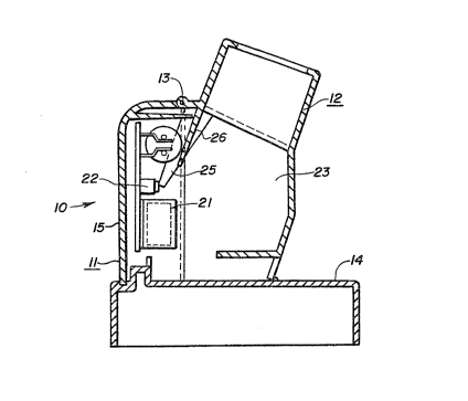
Fig. 1 shows that in the preferred embodiment the
viewer 10 has a base 11 and a viewing shield 12 attached
to the base by a hinge 13. The base has a platform 14
upon which the sample to be analyzed rests and an upright
back 15 which forms a right angle with the rear of the
platform.
Referring to Fig. 2 it will be seen that the back 15
contains a shelf 16 that has an internal positive standard
$
17 attached thereto, a longwave ultraviolet lamp 20, a
power switch 21 (Fig. 1), a safety switch 22, and a view-
ing area 23. The shelf 16 is designed so that an internal
standard 17 may be attached to the shelf and compared to
the test sample 24. In addition, the shelf 16 is above
and over the platform 14 enabling a test sample or samples
24C and 24D to be positioned directly adjacent the
internal standard during the analysis, see Fig. 3.
The longwave ultraviolet lamp 20 is controlled by the
power switch 21 located on the side of the back 15. The
power connection is shown at 19 in Fig. 3O Directly above
the power switch 21 and interior thereto is a safety
switch 22 design~d to cut power to the lamp when the
viewing shield 12 is opened during operation. The safety
switch 22 is released to shut off the power when the
finger extension 25 which is an integral part of the
shield 12 is moved from the safety switch 22 as the shield
is opened. In this way, the operator will not be directly
exposed to the ultraviolet rays during operation of the
viewer. The viewing shield has also been designed to
contain an internal ledge 26 that blocks the ultraviolet
rays and prevents the operator ~rom directly viewing the
ultraviolet lamp during analysis. In addition, the shield
has a viewing area 23 which has been designed at an angle
in cooperation with the viewing shield ledge to prevent
the operator Prom directly viewing the ultravio:Let lamp.
~ 3
In addition to the safety features incorporated with
regard to the ultraviolet rays, the fixed angle of the
viewing area and the permanent location of the internal
standard result in a consistent measuring system whereby
the operator's eyes are always located at this same angle
and approximate distance from the test sample during the
analysis. As mentioned above, the base and viewing shield
are attached by a hinge at the top of the base and viewing
shield. It is further note worthy that lenses are not
required or shown in the preferred embodiment although
they could be incorporated.
EXAMPLE
This example describes how the view~r is used in
conjunction with a test for the presence of the enzyme
elastase in a given solution. The test employs enzyme
test strips, a test strip carrier or incubator box and a
timer. The test strips and carrier are the subject of a
separate patent application, attorney docket No. 157g-
2CIP, the contents of which are hereby incorporated by
reEerence. The timer is the subject of patent
application, attorney docket No. 990, the contents of
which are also hereby incorporat~d by reference.
Test strips were prepared from filter paper. Whatman
541 filter paper (0.16 mm in thickness) was sandwiched
between two plastic portions so that more than one mm of
the Pilter paper was exposed and one mm oP the filter
2 ~ 3 ~
paper was between the two plastic portions. The filter
paper was then impregnated with an elastase enzyme sub-
strate and the test strips cut to size.
The filter paper was impregnated with substrate by
wetting the filter paper exposed tips of the test strips
in 0.85 millimolar methoxysuccinyl alanine alanine-pro-
line-valine-7-amino-4-trifluoromethyl coumarin (Lot # AP65
from Enzyme Systems Products, Livermore, California) in
elastase substrate buffer (0.5 M NaCl, 0.1 M HEPES(N-2-
hydroxyethyl-piperazine N-2-ethanesulfonic acid), pH
8.14). The impregnated filter paper was allowed to dry
overnight. Following drying, test strips of eight mm in
length and two mm in width were cut so that one mm of
impregnated filter paper was exposed and available for
absorption of biological fluid.
The impregnated test strips were used to test for
elastase as follows. An elastase test solution was
prepared by dissolving 1.2 mg of elastase (Biozyme, San
Diego, CA) in elastase substrate buffer described above so
that a final concentration of 2 mg/ml elastase enzyme was
obtained. The stock solution and serial dilutions (50 to
5 ug/ml elastase) were tested.
The test strips were tested in an assay in such a way
that final evaluations of a maximum of six test strips
were made no less than four and no more than eight minutes
after exposure to the test enzyme. Thus, a first test
~ ?
strip, impregnated with methoxysuccinyl-alanine-alanine-
proline-valine-7-amino-4-trifluoromethyl coumarin was
inserted into an elastase test solution for 15 seconds.
When the first test strip was inserted into the solution,
the start button of the timer was depressed, a short beep
was sounded, the strip indicator flashed the character #
and the number 1 was displayed on the display panel.
Simultaneously with the depressing of the start buttQn,
the running clock displayed 3 minutes and 45 seconds, the
time remaining in the test mode for the collection of test
samples. When the running clock reached 3:30, a single
beep sounded, the character # disappeared from view and
the number 1 ceased to flash but was continuously
displayed. At this time, the first test strip, containing
approximately 0.9 ul of elastase test solution, was
removed from the test solution, attached to the adhesive
layer in an incubator box and allowed to incubate at room
temperature. The incubator box had been previously
attached to the timer via the timer's carrier attachment
plate side slots and protuberances.
A second test strip was inserted into the elastase
test solution for 15 seconds. When the second test strip
was inserted into the solution, the start button was
depressed, a short beep was sounded, the strip indicator
~lashed the character ~ and the number 2 was displayed on
the display panel. When the second test strip was in-
,
serted into the solution and the start button depressed,the running clock displayed 3 minutes and 15 seconds, khe
time remaining in the test mode for the collec~ion of test
samples. When the running clock reached 3:00, a single
beep sounded, the character # disappeared from view and
the number 2 ceased to flash but was continuously
displayed. At this time, the second test strip was
removed from the test solution, attached to the adhesive
layer in an incubator box and allowed to incubate at room
temperature.
The collection and timing procedure was repeated for
test strips 3, 4, 5 and 6. When the sixth test strip was
inserted into the solution and the start button depressed,
the running clock displayed 1 minute and 15 seconds. When
the clock reached 1:00, a double beep sounded, the
character # disappeared from view and the number 6 ceased
to flash but was continuously displayed. The maximum num-
ber of samples had been collected with 1 minute remain-ing
in the test or collection phase and the timer automat-
ically switched to the view phase. The running clock dis-
played 3:45 which was the time remaining for the incuba-
tion of the sixth test strip so that all six strips would
have incubated for at least four minutes and be evaluated
in less than eight minutes. At the end of the 3 minute
and 45 second view phase, a beep and chime alarm sounde.d
and the runnlng clock displayed 1:30, the time remaining
in the evaluation phase so that all six strips would be
evaluated between six and one-half and eight minutes,
i.e., within the predetermined four to eight minute
window.
The six test strips attached to the adhesive strip in
the incubator box were evaluated for fluorescence in a
viewer or viewing chamber equipped with a longwave
ultraviolet lamp ~General Electric F4T5/BLB). The lamp
was mounted so that it illuminated equally the test strips
and a positive internal standard.
Reactive elastase was determined by measuring a
fluorescing leaving group which was released by th~ hydro-
lytic action of elastase upon the substrate and visually
assayed in the viewing chamber. After the incubation
period each test strip was evaluated for fluorescent
intensity. A bright green fluorescence, indicative of
substrate cleavage with release of 7-amino-4-trifluoro-
methyl coumarin, indicated the presence of the elastase
enzyme. Thus, a test strip which fluoresced with the
brightness and intensity equal to or greater than the in-
ternal standard was recorded as a positive response, indi-
cative of the elastase enzyme. In the absence of elas-
tase, the test strip fluoresced a dull blue-purple, indi-
cative of întact substrate. After one minute and 30 sec-
onds, the alarm of the timer sounded a continuous alarm
indicating the end of the evaluation phase and shut itself
off.
It should be noted that the semi-quantitation of test
strip fluorescence was possible according to the following
scale. A zero concentration of elastase was indicated by
a dull blue-purple color. Low concentrations of elastase
caused the paper strip to show light blue under the ultra-
violet light, moderate concentrations of elastase caused
a green-blue color to develop, higher concentrations
caused a green color, and even higher concentrations
caused a bright green color.
While the present embodiment of the invention and
method of practicing the same have been illustrated ancl
described, it will be recognized by those skilled in the
art that this invention may be otherwise variously
embodied and practiced within the scope of the following
claims.