Note: Descriptions are shown in the official language in which they were submitted.
CA 02051683 1999-04-27
-1-
CEREBRAL BIOPOTENTTAL ANALYSTS SYSTEM AND METHOD
CROSS REFERENCE TO RELATED PATENT
The subject matter of this application is related to
that of United States Patent 4,907,597, issued March 13, 1990,
which is also assigned to the assignee of the present applica-
tion.
BACKGROUND OF THE INVENTION
The present invention relates to a real-time, high-
frequency, high-resolution cerebral biopotential analysis
system and method, and more particularly to a computer-based
biopotential diagnostic system and method for quantitatively
determining, in a noninvasive manner, cerebral phenomena that
can be ascertained by analyzing cerebral electrical activity.
Despite a considerable expenditure of time and effort,
current approaches to the quantitative, noninvasive assessment
of cerebral electrical activity, as displayed in an "EEG"
waveform, have not been successful in fully extracting all of
the information which is present in this complex waveform. A
great need remains for an accurate, sensitive, reliable, and
practical neurologic profiling technology. In particular,
contemporary intra-operative EEG monitoring techniques have not
been widely adopted due to their inherent limitations. Indeed
eighty percent (80$) of all medical malpractice suits are
believed to be related to post-anesthesia morbidity and
mortality, and if such EEG monitoring techniques were reliable,
they certainly would have been adopted.
69675-99
WO 90/i 1718 PCT/US90/01378
2~J~.6~s
A number of devices known in the prior art ars capable
of tracking cerebral activity qual:.~atively. Techniques in-
volving the use of the "classical", conventional analog EEG
are restricted to analyses in the time domain, and require
considerable training for adequate interpretation.
Moreover, since the frequency resolution of the human eye
at standard speeds and gain is 30 - 60 Hz, much high fre-
quency content is invisible. Thus visual EEG assessment is
better characterized as being an art rather than a science.
Zn fact, it has been shown that the average correlation be- '
tween seven experienced readers did not exceed 56 per cent.
The use of frequency (power spectrum) analysis of the
EEG in the 1960's introduced the notion of some basic
processing of the signal prior to visual inspection and led
to the application of frequency aaalysis of the EEG to
various cerebral monitoring problems. In the past 25 years
at least 100 papers have been published in the medical
literature describing applications of power spectral '
analysis for purposes such as assessing the depth of anes-
thesia and cerebral ischemia under various intraoperative
conditions. United States Patent No. 4,557,270 issued to
John also describes the use of power spectral analysis to
evaluate cerebral perfusion during open heart surgery.
Several recent studios, however, have shown many deficien-
cies in the use of power spectral analysis to monitor
cerebral perfusion and to determine post operative
neurologic outcome. In addition, neither power spectral
analysis nor any other monitoring technique has been shown
to be reliable, and this is demonstrated by the fact that
the well-accepted Hasvard Medical School Anesthesia Monitor-
ing Standard does not include any type of intraoperative
neurologic monitoring due, in all likelihood, to the com-
plexity of interpreting raw EEG data and the unreliability
WO 90/11718
PCT/U590/01378
-3- ..
of existing automated systems utilizing power spectral or
time-domain analytic techniques.
The discharge of thousands of bio-electrically active
cells in the brain, organized in larger, interacting neural
centers contributes to the formation of an electrical sig-
nal with a wide frequency spectrum and extremely complex
dynamics. Embedded in that signal is information regarding
frequency content, non-linearities, and phase relationships
arising from the complex neuronal firing patterns that take
place. Because of the complexity of the EEG signal, conven-
tional time and frequency modes of analysis have not been
adequate to fully profile its behavior. In the Fourier
transforsa of the second order autocorrelation function (the
power spectrum) processes are represented as a linear summa-
tion of statistically uncorrelated sine-shaped wave com-
ponents. Contemporary approaches to monitoring the EEG by
means of the power spectrum have thus suppressed informa-
tion regarding non-linearities and inter-frequency phase
relationships and are of limited utility in representing
the EEG's dynamic structure. Furthermore the high frequency
low amplitude elements of the EEG have bees discarded to
data by the filtering and sampling characteristics of known
analysis techniques.
Because the EEG has a wide spectrum and is highly
dynamic and non-linear, the phase relationships within the
EEG, especially is the higher frequencies, must carry a
grant deal of diagnostic information regarding cerebral
!unction. The Fouriar transform of the third order autocor-
relation function, or autobispectrum, is an analytic
process that quantifies deviation from normality, quadratic
eon-linaaritiss and inter-frequency phase relationships
within a signs!. Ths Fourier transform of the third order
crosscorrelation function, or crossbispectrum, is an
WO 90/11718 PCT/US90/01378
~~J~~~a.~
analytic process that provides similar infosznation between
two signals.
Autobispectral analytic techniques have been applied
to the EEG signal and the basic bispectral properties of
the conventional EEG focusing on frequencies below 32 Hz
have bees investigated. Such studies have also bean con-
ducted to search for changes between waking and sleeping by
means of autobispectral analysis. Autobispectral analysis
and power spectral analysis have also been used in an at-
tempt to show that the EEGs of monozygotic twins are
similar in structure.
To date, no previous study has examined the high fre-
quency (greater than 32 Hz) content of the EEG and found in-
formation of diagnostic value. It also does not appear that
any study has shown autobispectral or crossbispectral
analysis to be of any value for any diagnostic purpose and
certainly neither of these anal~rtic techniques have been
shown to have any value in quantifying depth and adequacy
of anesthesia, pain responses induced by surgical stress,
cerebral ischamia, consciousaess, degrees of intoxication,
ongoing cognitive processes or interhemispheric dynamic
phase relations.
It is therefore a principal object of the present in-
vention to provide a noninvasive high resolution high fre-
quency electro-encephalographic system and method capable
of recognizing and monitoring physical phenomena that are
reflected in cerebral electrical activity.
Another object of the present inveatioa is to provide
a aoaiavasive electroeacephalographic system and method
capable of datermiaiag sad monitoring depth sad adequacy of
anesthesia, pain responses during surgical stress, acute
W090/11718 Z~ ~~-~''~~' PCT/US90101378
-5-
cerebral ischemia, lev~1 of consciousness, degrees of in-
toxication and normal or abnormal cognitive processes.
SUMMARY OF THE INVENTION
Accordingly, the system and method of the present in-
vention utilizes a suitable electrode and amplifier system
to obtain 19 unipolar EEG signals from regions of interest
on both left and right hemispher~s of a subject's brain.
Band-pass filtering of 2 - 500 Hz is ust~d to obtain signals
with a high frequency content. High gain amplifiers maxi-
mize the dynamic range for the high frequency, lorr energy
gave components of the signals. The system applies digital
sampling techniques to the signals and transmits digitized
data over a high speed serial limo to a host computer. The
system divides a 32 second long data aegmant from each lead
into 128 consecutive 0.25 second intervals. The system nor-
malizes all 19 unipolar leads by the standard deviation,
and then characterizes tho dynamic phase relations within
the signal by processing for autobispectral variables using
either a Fast Fourier Transform (FFT) based approach, or a
parametric cubic fitting approach. Similarly three cor-
responding left and right hemisphere data pairs are normal-
ized in the same manner and dynamic phase relations between
two hemispheres are then characterized by processing for
crossbispectral estimates utilising either the FFT or
parametric based techniques. The outcome is a set of two
dimensional arrays representing the dynamic interactions be-
tween all the possible combinations of frequencies (frequen-
cy pairs) in the spectrum of interest. For each unipolar
lead, three arrays are produced: autobicoharence,
autobispectral density and autobiphase. Three arrays are
also generated for each bipolar data set: crossbicoherence,
crossbispectral density and crossbiphase.
CA 02051683 1999-04-27
-6 -
Each of the autobispectral and crossbispectral arrays
contains 16,512 data points. Although all, or nearly all, of
these values can be expected to change from normal during
different interventions or due to differing disease states, in
the preferred embodiment only those points which show the great-
est fidelity in tracking the particular diagnostic determination
in question are utilized to create a diagnostic criterion. The
ensemble of points most sensitive to a particular intervention
or ongoing physiologic process can be used to create a
clinically useful single-number index from the computed
bispectral arrays. The system uses these indices as a
diagnostic figure of merit for the assessment of depth and
adequacy of anesthesia, pain responses during surgical stress,
acute cerebral ischemia, level of consciousness, degree of
intoxication and normal or abnormal cognitive processes. This
approach makes it possible for any, even unskilled, operator to
meaningfully interpret the output of the diagnostic device.
In situations where continuous monitoring is required,
indices can be continuously displayed on a video terminal
thereby enabling the operator to interactively evaluate regions
of interest. For record keeping purposes, index values and
other pertinent variables can be sent to a hard copy output
device or stored on a disk.
In accordance with the present invention, there is
provided a method of noninvasively detecting cerebral phenomena
comprising the steps of: acquiring electroencephalographic
signals through at least one electrode from a body surface of
a subject being analyzed; filtering said electroencephalo-
graphic signals to obtain filtered signals having frequencies
69675-99
CA 02051683 1999-04-27
-6a-
between 2 and 500 hertz; dividing said filtered signals into a
plurality of equally sized data records; characterizing dynamic
phase relations within said filtered signals by processing said
filtered signals to generate bispectral values; comparing said
generated bispectral values to reference values to derive a
diagnostic index that quantifies the detected cerebral
phenomena.
In accordance with another aspect of the invention,
there is provided the method of noninvasively detecting cerebral
phenomena as defined above where said step of generating said
crossbispectral density values comprises the steps of: comput-
ing fast Fourier transforms Xi(f) and Yi(f) of said data
records i; computing power spectra PXi(f) and PYi(f) of said
data records by squaring the magnitude of elements of said fast
Fourier transforms Xi(f) and Yi(f) respectively; computing for
at least one electrode pair an average complex triple product
of all data records acquired by said at least one electrode
pair; computing for said at least one electrode pair an average
real triple product of all data records acquired by each of
said at least one electrode pair; computing for said at least
one electrode pair a crossbispectral density value as the
absolute value of the average complex triple product for said
electrode pair.
In accordance with a further aspect of the invention,
there is provided a system for noninvasively detecting cerebral
phenomena comprising: means for acquiring electroencephalo-
graphic signals through at least one electrode from a body
surface of a subject being analyzed; means for filtering said
69675-99
CA 02051683 1999-04-27
-6b-
electroencephalographic signals to eliminate those signals
having frequencies less than 2 hertz or frequencies greater
than 500 hertz; means for dividing said filtered signals into
a plurality of equally sized data records; means for generating
bispectral values capable of characterizing dynamic phase
relations within said filtered electroencephalographic signals;
means for comparing said generated bispectral values to refer-
ence values in order to derive a diagnostic index that
quantifies the detected cerebral phenomena.
These and other objects and features of the present
invention will be more fully understood from the following
detailed description which should be read in light of the
accompanying drawings in which corresponding reference numerals
refer to corresponding parts throughout the several views.
69675-99
wo 9oimns 2~ ~~~~~ ~ PCT/US90/01378
BRIEF DESCRIPTION OF THE DRAWING,
Fig. 1 is a schematic vier of the system of the
present invention for detecting cerebral phenomena in a non-
invasive manner;
Fig. 2 is a schematic view of a 19 channel EEG data ac-
quisition system including a serial interface utilized in
the system of Fig. 1;
Fig. 3 is a schematic viarr of the microcomputer used
to calculate and display the EEG bispectrum in the system
of Fig. 1;
Fig. 4 is a schematic view of the processing opera-
tions pesformed by the system of Fig. l:
Fig. 5 is a flow chart of the operations of the
monitor module shops in Fig. 4;
Fig. 6 is a view of a sample display represaatation of
bispectral values generated by the system of Fig. 1;
Fig. 7 is a flog chart of the operations of the ac-
quisition and EEG rax data management module of the system
shoxa in Fig. 4;
Fig. 8 is a floe chart of frequency domain based
method for producing autobispectrum or crossbispectrum used
by the system of Fig. 1;
Fig. 9 is a flow chart of a parametric based method
for producing autobispectrum or crossbispectrum in the sys-
tem of Fig. 1;
WO 90/11718 , ' PCT/US90/01378
~~J~ ~~~ ~ -8-
Fig. 10(a) is an illustration of s graph showiag a
bispectral density array generated by the system of Fig. 1;
Fig. 10(b) is an illustration of a graph showing a
biphase array generated by the system of Fig. 1;
Fig. 10(c) is an illustration of a graph showing a
bicoherence array generated by the system of Fig. 1;
Fig. 11 is a flow chart of the diagnostic index genera-
tion module shown in Fig. d;
Figs. 12(a) - 12(c) era illustrations of arrays of
bispectral density values for three different states of one
patient;
Figs. 13(a) - 13(b) are graphs of statistical arrays
generated by the system and method of the present invention;
Fig. 14 is an annotated continuous autobispectral den-
sity diagnostic index graph for ono load generated by the
system of Fig. 1.
W090/11718 ~~ ~~ f~~~ PCT/US90/01378
-9- ,
. I 1. ,.
DETAILED DESCRIPTION OF THE PREFERRED EM80DIM
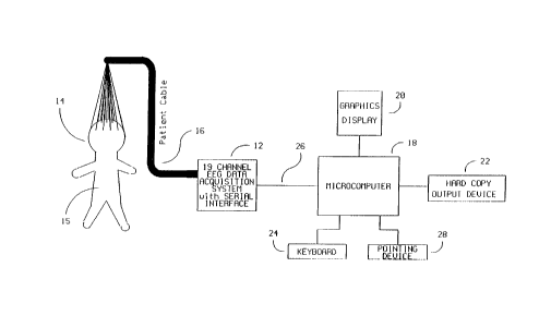
Referring to Fig. 1 the apparatus of the present inven-
tion includes a 19 channel EEG data acquisition system 12
connected to a microcomputer 18 through a high speed serial
interface 26.
The EEG leads are connected to a patient's head 14 by
a set of surface electrodes. The International 10/20
electrode system and nomenclature is preferred. The EEG sig-
nals are picked up by the electrodes and transmitted over a
patient cable 16 to the EEG data acquisition system 12.
The data acquisition system 12 filters, amplifies and
digitizes the EEG waveforms and sends the digitized data to
the microcomputer 18 via a high speed synchronous serial
line 26. In addition, the serial lima 26 can be used to
download filtering, gain and sampling rata instructions to
the data acquisition unit 12.
The microcomputer 18 processes the serial data stream
in order to generate all computed data arrays. Those arrays
are then used in conjunction with predetermined reference w
arrays derived from clinical studies to produce diagnostic
indices which indicate tho status of tho patient. These in-
dices are displayed oa the graphics display 20. Printed out-
put of the diagnostic index is also available on the hard
copy output device 22 which is connected to the microcom-
puter 18. Interaction between the operator and the acquisi- '
tion arsd analysis components of the system is provided by
mesas of a keyboard 24 and pointing device 28 with feedback
on the graphics display 20.
The 19 channel data acquisition system 12 is shown is
greater detail in fig. 2. The EEG surface potential,
WO 90/11718 PCT/US90/01378
'~~rJ~.E~~
-10-
detected by a surface eleetrode mounted on the patient head
14, passes through an electrosurgery protection circuit 30,
a defibrillator protection circuit 32, and an amplifier/
filter circuit 36 before being passed on to the multi-chan-
nel analog to digital convertor 38.
The electrosurgery protection circuit 30 includes a
radio frequency (rf) filter, which limits the rf current
through the patient leads 16 to less than 100 microamperes
and thus protects the patient 15 from rf burns and proteets
the amplifiers 36 from damage resulting from exceeding the
absolute maximum input voltage specified by the manufac-
turer. This circuit can be an LC section circuit consist-
ing of a generic inductor connected in series to a generic
capacitor which is then connected to ground.
The defibrillator protection circuit 32 limits the
voltage to the amplifiers 36 to a safe level when a
defibrillator is applied to the patient 15 and discharged.
This circuit can consist of a neon light bulb and or a '
parallel variable resistor connected in series to a
grounded resistor. ..
The amplifier/ filter circuitry 36 is controlled by
the microprocessor 34 for default gain and filtering levels
or alternate gain and filtering levels as requested by the
operator. Preferred gain and filtering settings are dis-
cussed later. This circuit section consists of three
stages: the first is a pre-amplifier stage that can be as- , .
sembled using a wide variety of high impedance pre-
amplifiers such as those sold by National Semiconductor,
Sunnyvale G; the second is a programmable filters stage
which can utilize components from Frequency Davicas, Haver-
hill 1~1; the third stage is a programmable amplifiers stage
xhich can be assembled from operational amplifiers used in
wo 9oimn8 2~~~fif~3;~ PCT/US90/0137$
-11-
conjunction with a multiplying digital to analog (D/A) con-
verter both components can be supFlied by National Semicon-
ductor. The multiplying D/A is used to aet the gain to the
appropriate levels requested by the microprocessor 34.
The high impedance pre-amplifier of each channel will
saturate to either the positive or negative supply voltage
if the input of the pre-amplifier is not tezminated. This
will lead to large positive value or a large negative value
at the output of amplifier/ filter section 36. Such value
will be used to identify lead failure.
The output of all 19 channels of the amplifier/ filter
36 is fed to a multi-channel analog to digital converter
(A/D) 38 which is under microprocessor 34 control for sam-
pling rate settings. The analog signals are converted to
digital data format suitable for input to a computer. A/D w
converters sold by Analog Devices, Norwood 1~ can be used
for this purpose.
The multi-channel A/D converter 38 is optically
coupled to data bus 40 by optical isolator 42. All control
lines to the sample and hold circuits, the multiplexer and
the A/D convertor 38 are also optically isolated by optical
isolator 44. Any known optical isolators can be used for
this purpose.
All DC power lines going to the amplifiers 36, sample
and hold circuits, multiplexer and A/D convertor 38 are
also isolated from the AC power line with a DC/DC convertor
46 in order to provide complete patient isolation from
ground. DC/DC converters available from Burr Brown can be
usad.for this purpose.
WO 90/11718 PCT/US90/01378
~~:W.~~;y -12-
The basic instructions for controlling operation o!
the microprocessor 34 are stored in a road only memory
(ROM) 48. The random access memory (RAM) 50 is used as a
buffer memory for data and a portion of the RAM 50 can also
b~ used as program memory when a control program is being
downloaded from the microcomputer 18.
Serial interface 52 operates under the control of the
microprocessor 34. The serial interface 52 is optically
coupled with optical isolators 54 to high speed synchronous
serial drivers 56 to provide a synchronous serial link be-
tureen the 20 channel data acquisition system 12 and any com-
patible high speed synchronous serial interface card on any
computer. The serial lines are isolated by optical
isolators 54 and DC/DC convertor 58 to provide increased
patient safety and to protect the host computer 18 from any
transients.
Tha host or microcomputer 18 of >3'ig. 1 is shown in
greater detail in 13'ig. 3. Tho entire microcomputer system
runs under control of a microprocessor 62 with the program
memory for the microprocessor 62 being stored in ROM 64.
The RAM 66 is used for storage of intermediate data. The ,
mass storage device 84 is used for storing clinical
databases as wall as archiving patient data.
In a preferred embodiment, the microcomputer 18 con-
tains an array processor 68 (such as the Vortez sold by SRY
of Lowall, MA) on which comploz arithmetic calculations can
b~ performed oa entire arrays of data simultaneously. The
protorred embodiment also includes a math coprocessor 70
which is connected directly to microprocessor 62. Tho math
coprocessor 70 is used for scalar and graphic calculations
while the array processor 68 is used to calculate
bispactral and other data vectors.
wo 90~1171s ~~~~~~~ PCT/US90/01378
-13-
,.
A graphics controller 72 operating under program con-
trol of the microprocessor 62 drives a graphics display 20.
A keyboard controller 74 interfaces directly with the
operator's keyboard 24. A serial port 80 interfaces with a
pointing device 82.
Operator control of the entire acquisition, analysis
and display procedure is controlled by the keyboard 24 and
pointing device 82 ~rith feedback on the graphics display
20. One high speed synchronous serial port 76 is provided
to interface with the 20 channel data acquisition system
12. Port 76 can be used to send control data to the system
(e. g., filtering, gain, sampling rate, start/ stop acquisi-
tion, perform self diagnostics) and to receive EEG data
from the system, as well as to download program data to the
system. Another serial or parallol port 78 is provided to
drive a hard copy output device 22 for printing desired
diagnostic indices.
Referring now to Fig. 4, a block diagram of the system
operations and the method of the present invention is
described. As meationed above, the aystam and method of the
present invention computes dynamic phase and density rela-
tions of EEG signals from a preselected number of leads (19
unipolar and 6 bipolar in the described embodiment). Single
number diagnostic indices are then generated from the data
arrays by utilizing predetermined reference arrays. The
results are quantitative iadices useful for analyzing
cerebral electrical activity as it relates to, for example, ;..
the assessment of depth and adequacy of anesthesia, pain
responses during surgical stress, acute and chronic
cerebral ischemia, level of consciousness, degree of '
cerebral intoxication and normal or abnormal cognitive
processes.
~.:. ~ . -z,. : . s -:... ~. _-
.,,~, <~~. . . .
WO 90/11718 PCT/US90/01378
2051~~~y -14-
Tho monitor module 402, handles the overall operations
of the system via integration of data and process informa-
tion from the user interface module 404, acquisition and
rev EEG data management module 406, bispectral processing
module 408 and diagnostic index derivation module 410. A
detailed illustration of module 402 can be found in Fig 5.
The user interface and display management module 404
represents the means through which the operator controls
and interacts with the system during the course of a proce-
dure. This includes, but is not limited to, entry of infer-
mation regarding the patient, typo of diagnostic procedure
being carried out, lead and acquisition settings; con-
tinuous display of acquisition status, lead integrity, and
diagnostic indices corresponding to regions probed by each
electrode; and requests for printing and archiving results
to disk. Module 404 directly interacts with the monitor
module 402. Tho operations handled by module 404 can bo
achieved under ono of many commercially available environ-
ments such as Microsoft's Windows.
The acquisition and raw EEG data management module
406, handles all of the raw EEG data checking and process-
ing prior to bispoctral analysis. This includes, but is not
limited to, continuous acquisition of EEG data and the
verification of the integrity of the data; preparing all
unipolar EEG data for autobispectral processing; preparing _ ..
all bipolar EEG data for crossbispoctral processing. Module
406 directly iatoracta with the monitor module 402. A more
detailed description of module 406 is provided below in con-
nection with fig 9.
The bispoctral processing module 408 controls the
generation of all data arrays measuring dynamic phase and
:~,~ .
W090/1171$ ~~ ~.~,y.E;~si PCT/US90/01378
-15-
density relations within the EEG. This infozmatioa can be
organized in both autobispectral sad crossbispectral arrays
utilizing either an FFT based or parametric based approach.
The tasks performed by thin module include, but are not
limited to: Fourier transformation; and the generation of
power spectrum, autobispectral densiWr, crossbispectral den-
sity, autobicoherence, crossbicoherence, antobiphase, and
crossbiphase. Module 408 directly interacts with the
monitor module 402, and a more detailed description of
module 408 is provided belor in connection with Figs. 8 and
9.
The diagnostic indez derivation module 410 generates
the data values utilized in the diagnostic process. The
task includes, but is not limited to, identifying frequency
pairs of interest through the use of predetermined clinical
reference arrays and creating a diagnostic index from the
values in the bispectral data arrays at the frequency loca-
tions defined by the reference array. Module 410 directly
interacts with the monitor module 402, and a more detailed
description of module 410 is provided below in connection
with Fig. 11.
Referring now to Fig. 5, the operation of the monitor
module 402 will nox be discussed. In step 502, the data ar-
rays used to store the digitized EEG, the 128 0.25 second
EEG data records, and the bispectral data of each lead are
initialized. Tho data files required for storage and files
containing data bases required for the computation of ding-
nostic indices are also opened is the initializing step 502.
Ia step 504 the system requests the information re-
quired to start the acquisition and diagnostic process from
the user via the user interface module 404. This requested
information includes patient descriptive statistics (sax,
;..... ..Z. ,~~.:~. , ,. , ,.Y.., ..... _.:I9':.'~ .. '~A
WO 90/11718 PGT/US90/01378
-16-
2~5~~,~~~
age, clinical symptoms ~tc..), typo of diagnostic procedure
to ba coaductod, and the loads used for autobispectral
analysis and the leads used for crossbispoctral analysis.
The system includes a default mode of operation and in
this default mode the system continuously monitors the
depth and adequacy of anesthesia, and any pain responses
during surgical stress utilizing a default autobispectral
density database. Default band pass filtering is perforsaed
from 2 to 500 Hz; the default sampling rate is set at 2000
Hz; and default gain is automatically adjusted to achieve
maximum dynamic range in each lead. The following discus-
sion of the monitor module 402 will utilize the default set-
tings of the system.
The EEG signals measured by leads Fpi, Fp2, F7, F3,
Fz, F4, F8, T3, C3, Cz, C4, T4, T5, p3, Pz, P4, T6, Ol, and
02 (A1 or A2 for reference) era used for autobispectral
analysis.
The EEG signals measured from the differbnce of leads
F7 and T3 (F7-T3) and the difference of leads F8 and T4 (F8-
T4) originate from the area covered by the frontal left
hemisphere and frontal right hemisphere regions respective-
ly. These signals from F7-T3 and F8-T4 are paired and used
for crossbispectral analysis. In this way, the interhomis-
pheric relationships for the frontal region can be ex-
amined. Similarly, pairing C3-Cz vith C4-Cz and T3-TS with
T4-T6 for crossbispectral analysis purposes allows for the
examination of the interhamispharic relationships of the oc-
cipital and parietal regions respectively.
Zn stop 506, 128 0.25 second buffers of artifact free
raw EEG data are acquired. 7U.1 channels transmitting ar-
y
WO 90/11718 PCT/U590/01378
2~a'~ ~~3
-17-
tifactual data era properly signaled to the operator to cor-
rect the problem.
The system, in step 508, computes autobispectral ar-
rays for leads Fpl, Fp2, F7, F3, Fz, F4, F8, T3, C3, Cz,
C4, T4, T5, P3, Pz, F4, T6, Ol, 02, and crossbispectral ar-
rays for leads F7-T3 paired with F8-T4, T3-T5 paired with
T4-T6, and C3-Gz paired with C4-Cz. Other leads may, of
course, be used in the computation of theca arrays, and two
different approaches for bispectral computation will be dis-
cussed below with reference to Figs. 8 and 9.
In step 510, the single number diagnostic indices from
all generated autobispectral and crossbispectral arrays are
computed. Autobispectral density and crossbispectral den-
sity clinical reference arrays era utilized in these diag-
nostic index computations. The goneration of the reference
arrays is discussed later. The system instantaneously dis-
plays, in step 512, all computed diagnostic indices for all
leads being analyzed. In step 511, the system checks for an
exit request, and if such a request has not been made, the
system, in step 516, acquires a new 0.25 second buffer and
repeats steps 508 through 51'. Ia step 518, requested prin-
touts are produced, results are stored to disk for archival
purposes sad all files era closed. In step 520, the process
is terminated.
A sample display representation generated by the sys-
tem is shows is Fig. 6. Represeatations of the patieat's
head era shows on the graphics display in Fig 6(a) and Fig.
6(b). The first illustration Fig. 6(a) is divided into
nineteen seetions each representing the region probed by an
electrode. The second illustration Fig. 6(b) is divided
into three horizontal sections representing combined left
WO 90/11718 ._ PCT/US90/01378
2~51~;~3;~ -18-
and right hemisphere activity probed by the group of
electrodes in that region.
For head representation Fig. 6(a), each section con-
tains a compressed continuous tracing 602 of the computed
diagnostic index utilizing the unipolar EEG data aequired ' '
from the electrode in that site. For head representation
Fig. 6(b), each section contains a compressed continuous
tracing 604 of the computed crossbispectral diagnostic
index utilizing bipolar EEG data acquired from several
electrodes in that site.
At the request of the operator any site can be dis-
played as an enlarged view 606 for closer examination. The
background of the tracing of each site (such as 602 or 604)
is color coded to reflect the possible values alloyed for
in the range of the selected diagnostic index. The most ' '
currant value of the diagnostic index for that site will
dictate what color is displayed in the background (e.g. Red
= lowest value to Green = highest value). This will '
facilitate the examination of the patient's status at a dis-
tance. Each site will be covered by a large "X" 608 if a
lead fail or an artifact was detected for any of the leads
contributing to the data required to generate the diagnos-
tic index at that site. '
Referring to Fig. 7, the acquisition and raw EEG data
management module 406 will now be described in greater
detail. Ia stop 702, the system checks whether the 0.25
second buffer for which data is to be acquired is the first
buffer being filled for that run, and if it is, the acquisi-
tion system 12 in stop 704 is supplied with requested fil-
tering, gain, sampling rate and load selection information.
The default sottinga are band pass 2 - 500 Hz for filtor-
W090/11718 ~~~~'~ ~~?~~ PCT/US90l01378
-19- . ,..
ing, 50,000 for gain, 2,000 samples/sec for sampling rate
and signals from all 19 leads are acquired.
In step 706, the acquisition system 12 acquires data
for each 0.25 second buffer for all requested leads and
transfers this data to the host computer 18. The system
detects lead fai r during the acquisition cycle in step ?08
by checking for very large positive or negative values.
Also in step 708 a publicly available algorithm is used to
check for artifact in each lead. In step 710, leads
generating failed and artifactual data are marked for the
monitor module 402.
In step 712, the system normalizes the records of data
acquired from all artifact free leads by subtracting the
mean of the samples in each record from each sample in that
record, and then dividing the sample by the standard devia-
tion of the records. This normalization sets the variance
in each record to 1 and has the effect.of weighing each
record equally during bispectral averaging. The process is
therefore lass dependent on the absolute polder spectral den-
sity at any frequency band.
In step 714, each 0.25 second record from each of the
leads Fpl, Fp2, F7, F3, Fz, Fd, F8, T3. C3, Cz, C4, T4, T5,
P3, Pz, P4, T6, Ol, 02 is assigned to an Xi(t), where Xi(t)
are the iadividual time series records provided fos
autobispectral processing. Also in step 714, the froatal
loft hemisphere time series, Xi(t), from F7-T3 and the fron-
tal right hemisphere time series, Yi(t). from FB-T4 era
provided for crosabispectral processing. Similarly, by pair-
ing loads C3-Cz vith C4-Cz and T3-TS with T4-T6, the cross
bispectrum of the left and right occipital and loft and
right parietal regions respectively can be processed. It
should be anted that for autobispectral analysis Yi(t) is
WO 90/11718 PGT/US90/01378
~G~51.fs8
-20-
set to equal X;(t) and in all cases the index i denotes the
record number from 1 to 128.
In step 716, a circular buffer mechanism is used for
storing the appropriate X;(t) and Y;(t) records for each
lead. The buffer is updated by storing the most recently
acquired data record in the location of the least recently
acquired data record. In step 718, the program checks
whether the circular buffer has 128 acquired data records
to start bispectral analysis, and if there are 128 data
records in the buffer, operation of the system returns to
the monitor module 402 in step 720.
Referring now to Fig. 8, the frequency domain based
procedures for producing the autobispectrum or the
crossbispectrum will nov ba discussed. In step 802, the sys-
tem chocks whether the computation to ba performed is an
autobispectral or crossbispectral computation.
Autobispectral analysis is a special case of '
crossbispectral analysis and therefore different rules of
symmetry apply.
In step 804, the system sets the following symmetries
in order to proceed with autobispectral computation:
fl + f2 < N/2
where N ~ 512 (0.25 sacs * 2000 samples in a pieferrad em-
bodimant), and
0 < f2 < fl
gi~t~ a Yi(t) ___~ Xi(f~ = yi(f~ ,
WO 90/11718 2~~~~ ~ y PCT/US90/01378
-21- . ._ ~ .
where f1 and fz (also refasrad to as Fz and Fz or Frequency
1 and Frequency 2) denote the frequency pairs over which
bispectral computation will ba carried out. Xi(t) and Yi(t)
denote the individual time aariaa records used for
bispectral computation. Xi(f) and Yi(f) denote the Fourier
transform of the time series records and i denotes the
record number and in thin embodiment ranges from 1 to 128.
In step 806, the following symmetries are adhered to
for crossbispactral analysis:
fl + f2 < N/2
0 < f1 < N/2
0 < f2 < N/2
-2f2 < f1
Xi(t) * Yi(t) ___~ Xi(f) * Yi(f)
where all variables represent the same values as they do
for autobispactral analysis, except that for
crossbiapectral analysis Xi(t) and Yi(t) represent in-
dividually derived time aeries records from left and right
hemisphere loads respectively.
The fast Fourier transform (FFT) Xi(f) and Yi(f) of
esch record of the 128 selected records for that lead, is
computed using a standard IEEE library routine or any other
publicly available routine in step 808.
WO 90/11718 PCI'/US90/01378
IG~J~.~.2W i
-22-
Zn Step 810, the power spectra Pxi(f) and Byi(f) of
each record of the 128 selected records for that load is
computed by squaring the magnitudes of each element of the
Fourier transform Xi(f) and Yi(f) respectively.
The system computes the average complex triple product
in step 812 by utilizing the following equations where
bci(fl.f2) is an individual complex triple product from one
record in a given lead and BC(fl.f2) is the average complex
triple product for that same lead:
bci (fl. f2) = Xi (fl) * Yi (f2) * Yi (fl+f2) -
where Y~(fl+f2) is the complex conjugate of Yi(fl+f2), and
128
BC (fl, f2) ' 128 ~ bCi (f 1. f2)
i=1
The average real triple product is computed in stop
814 by using the following equations where bri(fl.f2) is an
individual real triple product from ono record in a given
load and 8R(fl.f2) is the average real triple product for
that same load:
bri (fly f2) i pxi (~1) * pyi (f2) * pyi (fl+f2)
128
HR(fl, f2) ' 128 ~ bri (fl. f2)
i = 1
In step 816, the array of auto/crossbispoctral density
values (BD(fl,f2)) is computed using the following equation:
WO 90/11718 ~~ ~~ f,~ ~ y PGT/US90/01378
-23-
BD (t1. f2) = I BC (fl. f2) I
In step 818, the system computes the array of
auto/crossbiphaae values (~(fl,f2)) using the following
equation:
~p(f1. f2) = tan-1 [Im(BC (fl, f2) ) /~ (BC (fi. f2) ) ]
0 < ~ < 2n (radians)
In step 820, the system computes the array of
auto/crossbicoherence values (R(fl.f2)) using the following
equation:
R(f1. f2) = BD (f1. f2) / [BR(f1. f2) ] 1/2
0 < R < 1
In step 822, the system returns the requested
autocross bispectral density, bicoherence, biphase arrays
to the monitor module 402.
Nov turning to Fig. 9, a parametric based method for
producing the autobispectrum and the crossbispectrum will
now be described. In stops 902, 904, and 906 the system
sets the symmetries and time aeries records is the same man-
ner as described above in steps 802, 804, and 806 respec-
tively. The power spectra of Xi(t) and Yi(t) are estimated
is steps 908, 910, and 912. This estimation method includes
txo major stages, the Autoregressive (AR) model order selec-
tion and the power spectrum computation for Xi(t) and
Yi(t). Ia step 908, the system computes two sequences of
autocorrelstions. tR2xim)~ and (R2Y(m)~ using the following .
equation.
WO 90/11718 PCT/US90J01378
~~- ~ -24-
~a~~~J~i y
M N-Iml
R2z(m) = M ~N ~ ~ zi(t)zi(t+m), z = X, Y, and m = 0, 1, . . . , L
i=1 t=0
where M is the number of records of each load (128 in our
case), and N is the number of samples par record (512 in
our case), and L is much greater than the possible AR fil-
ter order (we choose 50).
Tha Final Prediction Errors, FPEx(m) and FPEY(m) are
calculated fos all orders, m = 1, 2, ... L, by performing a
Levinson recursion function on each autocorrelation se-
quence in step 910 in order to find the order of the AR fil-
ter. The locations, Qx snd QY, of the minimum of FPEx(m)
and FPEY(m) respectively are chosen to be the orders of the
AR filters of power speqtra of~Xi(t) and Yi(t) respective-
ly, i.e.,
FPEx(Qx) = min ~FPEx(m)} and FPEr(Qy) = min {FPEr(m)}
Once the orders of the 1~R filters for power spectra
are chosen, the autocorrelation sequences, (R2x(m)} and
(R2y(m}}. are entered into Leviason recursion with order Qx
and QY. respectively, instead of L. The coefficients, (cix,
i=0, 1, ...,Qx? and (ciY. i = 0,1, ... ,QY}, obtained from
the recursion are the coefficients of the 71R filters for
poxes spectra o! Xi(t) tad Yi(t) respectively. Then, is
step 912, the power spectra Px(f) snd PY(f) are computed as
the prediction error (~) divided by square of the mag-
nitude of the Fourier transfosm of the coefficients, i.e.,
PCT/US90/01378
WO 90/11718
-25-
ai
Pz(~ = Oi , Z = X, Y.
I 1 + ~ Ciz a ~2Z'1 12
i = 1
Tha system estimates the aato/cross bispectrum in
steps 914, 916, and 918. The estimation process includes
two major stages: the order selection and bispectrum com-
putation. In step 914, two seqveacos of third-order mo-
ments, (Rgx(T)) and (R3Y(i)) aro computed using the
following equation.
M s2
R3z(Z) = M ~N ~ ~ Zi(t)zz(t~L), z = X, Y, and t = -L, . . . , L
i=1 s=sl
where s1 = max (1,1-t) , s2 = ~a (N, N-T) , and I. is much
groator than the possibly AR filter orders (e. g. 50).
In step 916, two super matrices Tx and Ty are formed
as follows.
R3z(-L) R3z(-L+1) ... R3zt0)
R3z~-Irl) R3z~-L) ... R3zt 1) z X, Y.
s
R3z~-2L) R3 z(-2L+1) ... R3 z{-L)
from tho assumption rro made about the AR filter of
bispoctrum, the orders Ox and OY of tho AR filtors of
bispectra o! Xi(t) ~d Yi(t) era the rsaks of the supor
matrices Tx and TY. Thereforo, Ox and OY era choson by
using singular value decomposition. Having found tho or-
ders, wo obtain the coefficients of the AR filters of
WO 90/11718 PCT/US90/01378
~(~ ~~ ~;;t~~'y
-26-
bispectra by solving the following linear system of equa-
tions:
R3z(0) R3z(1) ... R3z(~z) 1 pz
R3z(-1) R3z(0) ... R3z(~z-1) blz 0
_ = X. Y .
R3 z(-~z~ R3z(Wz+1) ... R3z(0) bp=z 0
Where the skewness ((3z) and the coefficients (biz. . . . ,
bozZ). s = X, Y, can be obtained by solving the linear sys-
tem of equations.
The autocross bispectrum of Xi(t) and Yi(t) are com-
puted in step 918 as the cubic root of the triple product
of the skeWnessas (~i~i~iY)~ divided by the triple product of
the Fouriar transforms of the AR filter coefficients
(az(f)). i.e.,
BC (fl, f2) _ (~x~r~Yw~ Hx(f1)H~f2)Hrtf1+f2)
oZ
Hz (f ) = 1 + ~ biz a ~Zsti~ . Z = X~ 1,.
i = 1
and BR(fl,f2) is the real triple product for that same lead:
BR(fl,f2) = px(f1) * pz(fz) * pY(f1+f2)
After obtaining power spectrum and suto/crosa
bispectrum, the system computes the bispectral density
array, the biphase, and the bicoherenc~ in step 920 the
WO 90/11718 ~~J~~S~3 ~ ~ PCT/US90/01378
_27-
same way as in steps 816, 818, 820. Ir step 922, the aystam
returns to the monitor module 402 the r~questad bispactral
density, biphase, and bicoherence arrays.
For illustration purposes Fig. 10 contains sample
autobispectral arrays showing frequency pairs 0 < fl < 128
Hz, and 0 < f2 < 64 Hz. A bispectral density array is shown
in Fig. 10(a) where the Z axis represents the magnitude in
decibels (db) of the coupled interaction between all ap-
propriate frequency pairs fl and f2. Recall that the fre-
quency pairing scheme must adhere to the symmetry rule:
fl + f2 < N/2
where H = 256 Hz in this case. A bicoherenca array is
shown in Fig. 10(c) where the Z axis represents the normal-
ized magnitude in percent (%) of the coupled interaction be-
tween all appropriate frequency pairs fl and f2. A biphase
array is shown in Fig. 10(b) whore the Z axis represents
the phase in radians of the coupled interaction between all
appropriate frequency pairs fl and f2.
Referring now to Fig 11. a more detail~d description
of the diagnostic index generation module 410 will now ba
provided. In stop 1102, the system identifies the typo of
diagnostic assasamant in progress. Ia a preferred embodi-
m~nt the four possible options are:
1. Depth of anesthesia/ pain Z surgical stress.
2. Cerebral ischamia.
3. Cerebral intoxication (alcohol, narcotics).
4. Cognitive process evaluation.
WO 90/11718 PCT/US90/01378
~~~~f~~.y -28-
In step 1104, the system identifies the typo of
bispectral array to pass for use in the diagnostic index
computation after a user selects a specific lead and array
typo as described above with respect to the user interface
module 404. There era three (3) poasibla options for each
unipolar lead: autobispectral density; autobiphasa;
autobicoherenca. There are also three (3) possible options
for each set of bipolar leads: crossbispactral density;
crossbiphase; crossbicoherenca. Since there era 57 (3 X 19
leads) different types of autobispactral and 9 (3 X 3 sets)
types of crossbispectral arrays for each one of the 4 diag-
nostics, the total number of databases is 264.
In step 1106, the appropriate roferanco array is
retrieved from resident memory (or from disk). Each refer-
ence array gill coatain the locations of the frequency
pairs which era most sensitive to the assessmont is
progross (the generation of the roferonco arrays and the
selection of defaults will be discussed later). In stop
1108, the system adds all data points in the bispectral
array at the locations identified by the retrieved refer-
ence array. A counter (NP) of the total number of points
added is kopt. In step 1110, the sum of the data points is
divided by NP to obtaia the single number diagnostic index.
In step 1112, the program returns to the monitor module 402.
The prodotorminod clinical reference arrays referred
to above ire critical to tho device's ability to achieve
clinically relevant diagnostic efficacy. Ia the following
section we discuss the process adopted for generating these
clinical reference arrays. Since a total number of 276 pos-
sible reference arrays exist, only one will be discussed in
detail. J111 otbor reference arrays ire acquired is a
similar fashion. For illustration purposes the generation
W0 90/11718 2~ ~~ fT~.~' .4 PCT/US90/01378
-29- _ ._ . .. _ ,
of the autobispectral density reference array for monitor-
ing depth of anesthesia with lead ".'3 will be reviewed.
In a first study EEG potentials from a small group of
medically healthy surgical patients (N) with no known
neurological disorders are recorded during routine surgery.
The acquisition procedure described previously is followed,
with the following exception:
-Band pass filter 0.1 - 500 Hz
For all patients, two minutes of artifact free EEG
data era acquired under each of the following conditions:
- Pre-operative: awake ("control")
- Deep anesthesia; defined by conventional clini-
cal standards (intervention or disease state)
- Post-operative: alert in the recovery room
(recovery from intervention, or after treatment of disease
state)
An autobispectral density array is generated for lead
T3 from each one of the three recordings for all patients,
yielding a total of 3N arrays. The arrays are grouped in 3
sets of N arrays. The first representing the control state,
the second representing intervention, and the third repre-
aantiaQ recovery.
A paired Student's t test is performed on each of
16,512 data points, comparing the first and second array.
The resulting 16,512 t values are stored in a two dimen-
sional array identical is structure to that o! the
bispectral deaaity array. A second paired Student's t test
is carried out on each of the 16,512 data points, comparing
the socond and third arrays. The resulting 16,512 t values
WO 90/11718 PCT/US90/01378
~~:~~~3~i. i -30-
era stored in a second txo dimensional array identical in
structure to that of the bispectral density array.
All t values not meeting a specific significance test
or a specific confidence interval in either array are set
to 0. In the preferred embodiment all locations xith a t
value not corresponding to a p < 0.0001 are set to 0. Each
t value from the first t array (Tl(fl,f2)) is compared with
its corresponding t (T2(fi,f2)) fsom the second t array.
Ona of the following conditions must be met:
Tl (fl. f2) < 0 < T2 (fi. f2)
or T2 (fl. f2) < 0 < Tl (fl, f2)
If neither one the two conditions is mat at a particular
frequency pair fl, f2 then Ti (fl, f2) = 0 and T2 (fl, f2) = 0.
Tho application of the above conditions has the effect
of identifying all of the frequency pair locations that
change significantly by shoxing a consistent increase in
bispectral density value xith anesthesia followed by a
decrease xith recovery, or a decreaao xith anesthesia fol-
loxed by an iacroaso xith recovery.
rinslly, the absolute values o! the t values in each
11,12 location from the first t array are added to their
counterpart is the second t array to form a third t array.
The third t array is an average of the first txo and can be
visually inapoctod for highly aonsitive regions.
The last step involves sorting the third t array for
the most sensitive ensemble of frequency pair locations. In
the prelersed embodiment this xould consist o! the top 25%
1V090/11718 ~~ ~~ ~~';~ y PC1'/US90/01378
-31-
of all t values. The locations fl,ty of the most sig-
nificant t values meeting all of tho above conditions era
stored in resident memory (or oa disk) as one of the
predetermined reference arrays. This reference array will
be accessed by the diagnostic index derivation module 410,
for autobispectral density diagnostic index generation
during anesthesia/surgery for the location probed by lead
T3.
For any particular diagnostic task and any particular
lead there are 6 possible bispectral arrays
(autobicoherence, autobispectral density, autobiphase,
crossbicoherence, cross bispectral density, and
crossbiphase) which could be oxamined for diagnostic poten-
cy. To rank order the reference arrays with respect to
diagnostic efficacy a second prospective study is con-
ducted. Tha conditions under which the study is conducted
are identical to those of the first except that: a) the fre-
quency pair locations of interest have already bean iden-
tified and era now followed prospectively and b) the size
of the study group is now sufficiently large so that sample
variation of bispectral arrays more closely approximates
the true variance within the population undergoing the in-
tervention or suffering from the disease.
Thus for the example of anesthesia monitoring the EEG
recording starts during the awake/control state and con-
tinues uninterrupted through the end of recovery. Con-
tinuous surgical notes are maintained throughout the
operation.
After the completion of the study, continuous diagnos-
tic indices are generated for the le:ds of interest for
each of auto or cross bispoctral density. biphise ind
bicoherence arrays. The continuous trends are annotated
WO 90/11718 PCT/US90/01378
~~~~~1 7
-32-
with the intraoperative notes. A sufffciantly largo group
of prospective patients (determined by a statistical power
test) is used to determine which continuous diagnostic
index exhibits the greatest diagnostic ~fficacy on clinical
grounds. The particular bispectral array used to generate
this best diagnostic index during a particular diagnostic
procedure is programmed into the system as the default
array for diagnostic assessment.
The following non-limiting example is provided solely
for illustrative purposes. Twenty (20) patients undergoing
elective surgery for a variety of orthopedic and
gynecologic conditions were studied. Standard EEG leads
were placed in 16 locations according to the International
10/20 system. Raw EEG signals were acquired, bawd-pass fil-
tered (0.1 - 110 Hz) and digitised at a sampling rate of
256 Hz. EEG recordings were obtained from all patients
prior to the induction of anesthesia. Patients were than
anesthetized using standard techniques with a variety of
anesthetic agents. Continuous EEG recordings were obtained
during the period of anesthesia induction until the patient
was judged to be adequately anesthetized for surgery by
clinical assessment. Intermittent EEG recordings ware then
obtained during the course of the operation. During the
period of recovery from anesthesia another continuous EEG
recording was taken. A final recordiag was obtained when
the patient was deemed to be "awake" in the recovery room.
Detailed clinical intra-operative notes of patient status
were maintained during all phases o! EEG recording for sub-
sequent correlation with bispectral parameters.
In 10 patients the entire available frequency spectrum
(0.1 to 110 HZ) was examined for statistically aigaificant
changes in autobispectral density values from the awake
state to the deeply anesthetized state and back to the
W090/11718 ~~'~~'~~'~ PCT/US90/01378
-33-
awake state. Figs. 12(a)-12(c) show average bispoctral den-
sity arrays (from 10 patients) for each of the three states
of consciousness. The method for determining statistical
significance was as outlined above. Figs. 13(a)-13(b) show
the statistical arrays generated by the technique of the
present invention: the average t array for these 10 sub-
jects for lead T3 for locations corresponding to a p < 0.05
(t > 2.26) (Fig. 13(a)) and the average t array for loca-
tions corresponding to a p < 0.0000003 (t > 10.0) (Fig.
13(b)). Each t value in the array reflects tba consistency
of change in a bispectral density value through the three
states for one frequency pair location across all 10
patients. Zt is north noting that virtually no data points
are significant with a p < 0.0000003 (t > 10.0) in the fre-
quency pair band of F1 below about 24 Hz and F2 below about
2 Hz in Fig. 13(b). On the other hand 7,168 locations were
found to change with a p < 0.0000003 (t > 10.0) in the fre-
quency pair band of F1 above about 24 Hz and F2 above about
2 Hz. The top 25% (7,168/4 = 1,792 points) most sig-
nificant high frequency locations worn used to define the
reference array. The autobispectral density index was cal-
culated for each subject from the points defined by the ref-
erence array as described above is the detailed description
of the invention. This autobispactral density index was ,
then calculated as a continuous function for the continuous
EEG recordings to assess its behavior during induction,
intra-op~rativoly, and during recovery. Tha correlation
with clinical events during the operation ryas noted.
A sample annotatQd continuous autobispectral density
diagnostic index for lead T3 during surgery in a prospec-
tive subject is shown in Fig. 14. Tha index varies between
30 and 5 decibels and is qvit~ sensitive to the patient's
state of consciousness and the onset of painful stimuli.
Specifically, the index drops with the induction of saes-
WO 90/11718 PGT/US90/01378
~~J~.E~~3s
-34-
thetic agents (pentothal and othrano) to the patient, and
the index level rises as the patient's lag is being prepped
for surgery. Zn addition, the index approaches its highest
value when the patient is awake in the recovery room and
most likely experiencing post-operative stress. (The gaps
seen in the index plot correspond to time periods when EEG
recordings mere not being taken).
Similarly the above analytic process is used to
generate the reference databases for cerebral ischemia,
degrees of intoxication and normal or abnormal cerebral
processes. In quantitatively detecting any of these
cerebral phenomina, the system compares a number of
autobispectral and crossbispectral EEG data from subjects
in the normal state to clinically identified extremes of a
certain physiologic state (awake vs anesthetized, sober vs
intoxicated, perfused vs ischemic, at rest mentally vs
thinking, normal vs retarded, etc..). Tho comparison util-
izes a statistical approach to identify the bispoctral data
poiats that are most sensitive to the particular
physiologic state in question. Tho frequency pair loca-
tions of the most sensitive data points are identified and
stored in a database for reference purposes. When a diag-
nosis is to be carried out, the average of all the data
points defined by the reference array is obtained for the
subject undergoing the study. This average is used as a
diagnostic index cad is compared to a list of indices char-
acteristic o! etch state by the operator or the system.
In addition to quantifying the depth and adequacy of
anesthesia, pain responses during surgical stress, acute
and chronic cerebral ischemia, level of consciousness, de-
gree of cerebral intoxication and normal or abnormal cogni-
tive processes, the system and method of the preseat
invention may also be used to assess s myriad of cerebral
2~5'~ f ~'3
WO 90/11718 PCT/US90/01378
-35-
phenomena based on the acquisition and processing o! BEG
signals into various bispoctral arrayb which are than com-
pared to appropriate reference arrsys.
Although bispectral analytic techniques in the frequen-
cy domain have been applied to the EEG signal, as was dis-
cussed in the Background above, parametzic approaches to
the estimation of bispectral values have not. Furthermore
no bispectral technique has aver been demonstrated to be
useful for any diagnostic purpose. Other techniques for the
quantification of the depth of anesthesia or the detection
and quantification of cerebral ischemia intraoperatively
remain qualitative and limited in their overall utility and
acceptance in practice. Specifically, the system and method
of the present invention examines various bispectral values
across all frequency pairs in a frequency range hitherto ig-
pored by those knowledgeable in the art and uses the summed
degree of changes is autobicoherance/autobispectral dan-
sity/autobiphaso, crossbicoheronce/crossbispoctral don-
sity/crossbiphase at a limited number of frequency
locations as an index of physiological perturbation. The
system and method utilize various bispectral arrays of
defined clinical populations to define the locations of the
subset of frequencies used to calculate this index. Refer-
ence clinical arrays are further utilized to assess the
meaning o! this index and to measure the aignificaaco of
deviations of this indes from nosmality. This allows the
quantitative gauging of the disturbances in cerebral func-
tion, whether due to anesthesia, intoxicants or ischemia w
for any particular EEG lead position. The system and method
disclosed heroin also define the graphic display o! the
diagnostic index, whether oa graphics screQa or on paper,
whether is real-time or in digital archive.
WO 90/11718 PCT/US90/01378
jt~''~3.~~~.~.~~'~, i
-36-
ifhila the foregoing invantios has boon described with
reference to its preferred ambodimanta, various alterations
and modifications will occur to those skilled in the art.
A11 such alterations and modifications are intended to fall
within the scope of the appended claims.