Note: Descriptions are shown in the official language in which they were submitted.
- 2 - 2 ~ 3 1 7 1 ~
P480
Field of the Invention
This invention is concerned with gamma camera systems and more
particularly, with such systems using dual detector cameras.
Back~round of the Invention
The original gamma camera systems used one detector head.
Originally, the one detector head was positioned above an organ to
be imaged. Subsequently, the one detector head was used for what
is known as single photon emission computerized tomography (SPECT)
or emission computerized tomography (ECT). SPECT or ECT involve
mounting the camera detector head in a gantry enabling it to
rotate or orbit about the patient 80 as to obtain tomographic data
and thereby provide tomographic images. Another aspect in the
development of the gamma cameras is whole body imaging wherein the
gamma camera head is passed over the entire body to obtain a
complete image of the patient.
To increase the efficiency of the whole body scans and the
tomographic scans, multi-headed cameras have been used. First,
dual-headed cameras were used wherein the gamma camera system
comprised a pair of camera heads spaced apart and oppositely
di~posed to enable obtaining images from opposite sides of the
patient simultaneously. For example, the dual heads were moved
around the patient with one head on each side of the patient.
~ 3 ~ 2~31l~
P4~0
Recently triple-headed gamma camera systems have been used. In
triple-headed gamma camera systems, the heads are mounted to form
a triangular shape with the three planes of the heads each
separated by 60.
It would seem that multi-headed cameras would reduce the
rotational travel required to obtain imaging data from a 180
orbit or a 360 orbit. It is true that with two oppositely
disposed heads, the 360 orbital data can be obtained with a 180
rotation. However, the 180 orbital data cannot be obtained in a
scan of 90. Similarly, with a three-headed camera system, a 360
scan can be accomplished with an orbital movement of a little over
120. The 180 orbital data, however, also reguires a scan of
120. From scan travel distances required it i8 seen that the 360
scan times are drastically reduced by mutli-head systems. However,
when 180 scans are required such as for cardiac studies, there is
little or no time saving when using multi-headed cameras.
Accordingly, a more efficient dual-headed camera system i8
required for cardiac studies.
Another problem with the presently available gamma camera systems
is in obtaining images during cardiac exercise studies. In these
studies a static image is acquired while the patient pedals on an
ergometer, for example. If a single camera head i~ used for data
acquisition durlng the exercise study, it is oriented in an
optimal left anterior oblique position. However, the behavior of
2 ~
P480
the inferlor wall of the heart which i8 of great interest to
cardiologists cannot be seen from this orientation. Accordingly, a
camera sy~tem is required wherein the image of the heart during
exercise also includes a good view of the inferior wall of the
heart.
Thus, what the present cameras do not provide is a two-headed
gamma camera system with the heads oriented relative to each other
to enable cardiac ECT studies in a reduced scan time. The
arrangement of the two heads in the gamma camera system should
assure that there is no minimum radius of rotation. The
three-headed systems presently available inherently have a minimum
radius of rotation which interferes with some studies, such as in
pediatric applications.
A1BO preBently lacking are gamma camera systems that car,
efficiently image during exercise studies and obtain images of the
heart including the inferior wall. The gamma camera system that
overcomes the above noted deficiencies should also provide
increased count rates to enhance first pass studies.
Brief DescriDtion of the Invention
In accordance with one preferred aspect of the present invention a
gamma camera system is provlded having two detector heads. The
heads are mounted so as to describe an angular shape such as a
2~ 7~
P480
modified L-shape wherein both leg~ of the "L" may be of equal
size, for example. This type of camera i8 ideally suited for 180
ECT cardiac studies. The orientation of the two heads enables
obtaining data acquisition from a 180 arc with a 90 rotational
movement. Thi~ type of camera is also ideally suited for spot
cardiac studies during an exercise mode. Thu~, the two detectors
arranged in an L-shape are mounted so that the complete heart
including the inferior wall can be imaged during the exercise
program.
In a broad aspect of the present invention, a unique two-headed
gamma camera system for converting gamma radiation emitted from a
patient to imaging data is provided, said system comprising:
mean~ for di~playing an image based on said imaging data,
said two-headed camera system having a first head and a second
head,
meane for mounting said second head ~uxtaposed to said fir6t head
to deflne an angle therebetween, and
means for utilizing the camera to obtain image data.
-- 6 - ~ L;~ 7 ~ ,~
P480
It i8 a feature of the invention that a scan 18 obtainable by
orbiting the heads about the patient for 90 which gives the
equivalent of a 180~ scan in effectively one-half the time.
Another feature of the present invention, utilizes a single
L-shaped collimator to which the two heads are attached.
According to still another feature of the present invention, the
cameras system comprises two detectors or heads mounted with a 9o
angle therebetween in a single camera.
According to yet another feature of the present invention, the
gamma camera detector heads are rectangularly ~haped and one side
of the rectangle of each of the heads are juxtaposed to each other
to form a modified L-shaped camera system. A criterian of the
~unction being to obtain the shortest possible patient-detector
distance for both detectors. This i6 accomplished by arranging the
inner sides of the fields of view to coincide with or be very
close to the line of inter~ection of the detector plane~.
~rief DescriDtion of the Drawin~s
The above named and other feature~ and objects of the present
lnvention will be be6t understood when con6idered in the light of
the following description of a preferred embodiment of the
- 7 - 2~ 7~ ~
invention taken in con~unction with the accompanying drawings
wherein:
Fig. 1 is a side view of a preferred embodiment of th unique
dual-headed camera arranged in a modified L-shape;
Fig. 2 in con~unction with Fig. 1, schematically shows the unique
L-shaped gamma camera utilized to obtain cardiac ECT imaging
data;
Fi~. 3 shows the unique modified L-shaPed gamma camera u~ed for
acquiring data during a cardiac exercise study;
Fig. 4 schematically shows the unique modified L-shaped gamma
camera utilized to obtain ECT data;
Fig. 5 shows details of one preferred embodiment of the
two-detector head camera;
Fig. 6 schematically illustrates a shortcoming of the prior art
dual-headed camera when used to acquire data in a 180 ECT scan
such as used for cardiac studies; and
Flg. 7 shows a special fan beam collimator arrangement for the
unique dual-headed cameras.
g 2~
General Descri Pt i on
Fig. 1 ~how~ a modified L-shaped gamma camera at 11 comprised of
two gamma camera heads 12 and 13. The L-shaped gamma camera
arrangement i8 ideally suited for cardiac imaging. The ga~ma
camera arrangement of Fig. 1 includes a collimator on each o~ the
cameras> such as collimator 14 on camera 12 and collimator 16 on
camera 13. Each of the camera~ includes a crystal, wh~ch
sc~ntillates responsive to gamma radiation, such a~ crystal 17 on
camera 12 and crystal 18 on camera 13. Behind the crystals, shown
only as a block, i8 the head 19 and 21 for cameras 12 and 13
respectively, comprised of photomultiplier tubes and electronic
computer components for determining the locations of the events;
i.e., the point of impact of the radiation and the crystal. The
cameras acquire count, location and energy data that are supplied
to the control processor 15. The control processor processes the
acquired data to provide imaging data to supply unit 25.
The camera~ are well known in the art. See for example, U.S.
Patent 3,011,057 issued to Anger. The utilization of the Anger
camera in computer tomography of a single photon emission is
described, for example, in IEEE Transactions on Nuclear Science,
Vol. NS-23, Feb. 1976, pp. 528-537 and IEEE Transactions on
Nuclear Science, Vol. NS-28, Feb. 1981, pp. 69-80.
_ g _ c~ r~
P~
In Fig. l, the camera 12 is ~hown as bein~ positioned immediately
above the patient 2Z. The patient i5 being viewed from his feet
side as is readily discernable by the location of the heart 23.
The gamma camera system has the capability of orbiting the heads
about the patient as indlcated by arrow 24. The sy~te~ also has
the capability of moving the heads towards the patient or outward
away irom the patient a~ indicated by arrow 26. In addition, the
system has the capability of swivelling the L-shaped attached
heads about an axis 27, as indicated by arrow 28.
The control of the gamma camera unit to describe the motions
lndicated is well known to those skilled in the art. See, for
example, U.S. Patent 4,888,486 which show~ an example of the
rotational and in and out motion, and U.S. Patent 4,523,091 which
shows, an example of a swivel motion.
Cardiological procedures constitute a large fraction of the
nuclear medicine clinical workload especially for ECT clinical
procedùre. Until now, no gamma or nuclear camera system exists
which 18 optimized for cardiological studies. The camera system
described herein i8 ideally suited for cardiological ~tudies in
general and for cardiac ECT studies as shown in Fig. l and 2. At
the lnitiation of the study, the camera arrangement ll, for
example, is in the position shown in Fig. 2 and revolve~ or
rotates as indicated by the arrow 24 through 180 where the head
2 ~
-- 10 --
p~
13 1~ below the patient and the head 12 i8 ~n the left side of the
patient.
For example, the cameras are rectangular cameras and the
connection between the two cameras i5 along a side of the
rectangle. The camera in moving the 90 from the position of Fig.
2 performs a 180 cardiac ECT imaging acquisition. The 180
coverage i~ indicated by darkened section "A" of the patient in
Fig. 2, The 180 cardiac ECT is performed in half the time
required for a single-headed camera since it obtains 180 worth of
data by a 90 rotation. Also, all of the data i8 obtained by heads
that are relatively close to the heart. No camera on the market
offers this feature.
Fig, 6 shows a prior art dual-headed camera operated to perform a
180 ECT scan. Therein the two heads 12' and 13' are spaced apart
and oppositely disposed with the patient 22' therebetween. By way
of example, the initial position of the oppositely disposed heads
are above and below the patient and are labelled "initial". Moving
the heads through 90 to the "rotated" positions provides data
from ~eparated 90 sections of the patlent, Thus, insufflcier)t
data is provided for a 180 scan and further rotation and
acgulsition is req,uired.
The camera arrangement 11 besides obtaining SPECT studies of the
heart is also ideally suited for static cardiac imaging in any
P480 - 11 - 2~7~
patient position. Thus, the imaging can occur while the patient is
sitting on and operating an exercise cycle or ergometer. With the
patient mounted on the cycle without any movement of the camera
heads two meaningful views are obtainable; i.e., the left anterior
oblique and the left posterior oblique or the left anterior
oblique and the right anterior oblique at the same time, which is
especially important for dynamic studies.
A position of the camera relative to the patient in such exercise
cardiac studies is illustrated in Fig. 3. Therein, the head 12 is
shown in position at the left anterior oblique position for
imaging the patient 22. The head 13 is at the left posteior
oblique imaging position. Means, ~uch as a motorized threaded
member indicated at 29, may be provided for varying the angle
between the heads.
Thus, with the camera described herein it is possible to obtain
cardiological images during exercise. In these images, the static
image of the heart is acquired while the patient is pedalling on
an ergometer. The camera head is oriented to the left anterior
oblique angle which gives the best view of the heart. In prior art
cameras at this angle, the behavior of the inferior wall was not
visible. With the present camera the inferior wall is clearly
imaged by the second head along with the rest of the heart.
- 12 - 2~J~7~
P4~0
The present arrangement, a~ shown in Fig. 4, also makes it
possible to do a 360 general SPECT with a 360 times two
rotation. Thus, in Fig. 4, the camera head 12 and the camera head
13 are shown as being init~ally positioned with the camera head 13
on the left side of the patient and the camera head 12 above the
paitent. The camera heads 12 and 13 are rotated through 90 from
the original position indicated on Fig. 3 through many angles such
as a 45 angle to the so~ angle. The 90 rotation gives a 180
SPECT data acquisition scan. Similarly, a 360 rotation at double
speed enable~ acquiring data equivalent to a 360 scan. Using the
dual heads, almost twice the required number of counts are
acquired in the normal time period or the normal number of counts
are acquired in half the normal time period. The two heads,
therefore, enable speeding up the procedure and nonetheless
acquring sufficient data for tomographic images.
The camera system could also be used, of course, for a plain whole
body scan. However, it is more ideally suited either for
cardiological SPECT scans, cardiological exercise scans, plain
SPECT scans or spot scans, lncluding double cpot scan~.
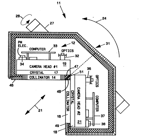
Fig. 5 shows a preferred embodiment of the unique dual head camera
11. In this embodiment, the two cameras 12 and 13 are mounted in a
Jingle lead casing 31. The collimator 14, scintillator crystal 17
and camera head l9lcamera computer 33 and the photomultiplier
electronics 34 make up the camera 12. Similarly, camera 13
,
P~BO
comprises collimator 16, scintillator crystal 18 and camera head
21 along with optics 35, camera computer 37 and photomultiplier
electronics 38.
In addition to normal parallel-hole collimators, special
asymmetrlc fan-beam collimators may be provided for ena~ling using
*he ~ystem with the collimator faces as close as possible to the
patient. The special collimators are shown in Fig. 7. More
particularly, the collimators 14' and 16' are shown as having
focal spots positioned on a straight line parallel and juxtaposed
to the face of the other collimator. Thus, the slots of collimator
14' are all focused on point 41 which is located on an imaginary
line 42 shown in dashed line form. The line 42 is parallel and
juxtaposed to the face of collimator 16'. The slots of collimator
16' are focused on a point 43 which is located on an imaginary
line parallel and ~uxtaposed to the face of collimator 14'. Thus,
the collimators preferably are not the usual symmetrical fan beam
collimators.
The collimator~ 14, 16 are bordered by solid lead edges, such as
edges 46, 47 for collimator 14 and edges 48, 49 for collimator 16.
A feature of the cameras is the means for extending the field of
views (FOV) of each camera practically right up to its iunction
point with the other camera. Thus, the FOV of camera 12 extends to
its collimator edge 47 which is practically aligned with the face
of collimator 16. The edges 47 and 48 of the collimators 14 and 16
- 14 - 2~ ~ 7 ~ ~
P480
mesh at diagonal line 51 to a~d in the extension of the FOVs of
each camera. The line 51 could be a zig zag line to improve the
radiation seal afforded by the lead casing. Alternatively, a
~ingle modified "L" shaped collimator could be used in place of
col 1 imators 14 and 16.
In practice, two rectangular gamma cameras are mounted at an angle
such as 90 in a single camera head. The connection may be along
the short side of the rectangles. The basic camera may have three
degrees of freedom: rotation, in-out and swivel. The two cameras
are mounted on the usual SPECT gantry which has widened arms in
order to accommodate the wider camera arrangement comprising the
two cameras.
A preferred embodiment utilizes a single L-shaped collimator which
makes mounting of the two independent cameras to the collimators
more convenient The unique camera arrangement ideally performs
180 ECT by a 90 rotation and 360 ECT by a 360 rotation at
double speed with the subsequent addition of pairs of frames taken
at the same angle. Body contour and/or elliptical ECT is made
po~slble by providing another degree of freedom either by
motorizing the gantry or the bed or both for up and down and
left-right movement. The described cameras are also ideally suited
for imaging the heart including the inferior wall during a patient
exercise procedure. It should be understood that the dual-head
P480 2
camera mounted at so can also be used for any spot imaging and
provide additional data.
Thus, in summary, the advantage~ of the unique L-shaped camera
system include the capability of performing 180 ECT with 90 of
motion. In the described 8y8tem, there i8 no minimum radius of
rotation as in three-headed systems; thus, the described inventive
system, i8 ideally suited for pediatric applications as well as
cardiac studies, for example.
While the invention has been described as having a flrst head and
a second head, the unique mult$-headed camera can have more than
two heads separated by angles of 90 or more.
While the invention has been described with reference to the
preferred embodiment, obviou~ modifications and alterations will
occur to those skilled in the art upon reading and understanding
~he preceding detailed description. It is intended that the
lnvention be construed as including all such alterations and
modifications insofar a~ they come within the scope of the claims
or the equivalents thereof.