Note: Descriptions are shown in the official language in which they were submitted.
3 ~3
CL~1\1P FOR APPROX~MAT~TG TISSUE SECIIONS
.,
BACKGROUND OF l~E LNVENTION -
Field of the Invention -
,- -
- - This invention relates to tissue we]ding and more particularly to a -' ~ -'
method and apparatus for holding tissue sections in apposition and
compression during application of laser energy to weld the sectiorls together.
Description of the Related Art
Conventional surgical techniques typically require the use of
sutures or staples to construct surgical connections that provide functional
communication be~een living tissue structures. These surgical cormections - ' -
are generally referred to as "anastomoses". Anastomoses between
cylindrical, especially tubular (i.e. hollow) struct~res are of significant
clinical importance.
The problerns associated with the utilization of sutures or staples
are nurnerous. First, it is often time consuming and technically difficult
to perform anastomoses by sutures or staples. This not orlly results in
increased costs to the patient, a factor which carmot be overlooked as the
current trend of skyrocketing health care costs continues, 'out creates a
wide discrepancy in perforrnance between surgeons, especially between
e~penenced and novice surgeons. Second, the use of sutures and st~ples
'also~'means the introduction of "foreign bodies" which can cause trauma,
.
, _ _ .. .. .
~ ~ r ~
inflammatory and immune ~esponse, and other adverse reactions due to the
` actual introduction as well as the prolonged presence of these foreign
: . :
rnatenals. In1arnmation can actually cause a decrease in tensile strength ~ -
and bursting strength of an anastomosis. Inadequate anastomoses pose a
severe health risk to the patient if the sutured tubes or organs become
sufficiently weak that they separate.
In an attempt to overcome these problems associated with sutures
and staples, work recently began on the use of laser energy for welding the
tubular and other types of tissue. It was discovered that properly applied
laser light thermally induces intrinsic tissue changes which irnmediately
produce hermetically sealed, strong bonds between the tissue. Such laser
welding may also result in increased collagen synthesis, rapid restoration
of tissue function, and enhanced healing. Additionally, advantageous for
younger patients, laser welding allows the growth of welded seams as body
size increases. ~~
Thus, not only does laser tissue welding avoid the adverse effects
associated with introducing foreign particles, i.e. sutures and staples,
into the patient's body by avoidance of needle trauma and mirumi ation of
inil~mmatory and immune response, but laser welding can actually optimi7e
the strength and functional characteristics of the anastomoses.
Still further, more automated laser tissue welding advantageously
does not require the skill and time-consuming labor of the surgeon which is
necessary for sutunng and stapling. In fact, once the equipment is
properly placed by a surgeon, a nurse or technician can simply switch on the
, , , =
laser, w~uch provides the light necessary to weld the tissue. This laser -- - -
delivery technique requires no manual rnanipulation dunng the actual welding -- -
:_, .. ... - . .. .
... . . .. . .
~ :
:-- 2
.
s,3
of the tissue and usually requires less than 10 seconds of lasing time.
Thus, a mechanical means for holding tissue during laser welding also
-: provides an increased level of consistency which simply cannot be achieved
m the individualized hand-manipulated suturing and stapling techniques or
in less automated laser tissue welding techniques. The automated control of - -"the laser welding parameters, rather than the sl~ill or experience of a
i - particular surgeon, deten~ines the irnmediate success of the welding as well `-
as the long term holding strength of the anastomosis.
The two essential criteria for successfill laser welding are: (1)
control of laser energy delivery and (2) tissue apposition. Control of
laser energy (precision and consistency) is important to ensure that the
desired amount of incoming l~ght energy is absorbed by the tissue. This
means providing consistent laser energy density at a specific rate (i.e.
fluence) over the entire anastomotic searn. Tissue apposition is critical
since the ends of the hollow tubular sections to be welded must be in -~substantial abutment and accurate alig~nent to ensure that the laser energy
effectively fuses the entire seam formed at the abutment. Substantial
abutment also requires compression of the edges of the tubular sections.
Deficient apposition can cause leakage or the formation of weak tissue
bonds. Inadequate anastomses can result in separation of the tissue
sections, abnormal formation of fibrous tissue (adhesions) or undesired
narrowing of the passage between the tubu~ar sections (stenosis). The
- importance of tissue apposition cannot be overemphasized. - - To date, many failed attempts at produc g a ef ec ve la er
= ` tomosis can be attributed to inadequate tissue approximation. Such .
~dequate laser welds have forced surgeons to rely on the use of "stay"
, ... ,--, - - - .
:-- 3
sutures to assist ;n tissue alignment and oricntation during welding. Such
stay sutures, usually numbering at least three for each anastomosis, are
- ~ - ~, - ,
- typically left at the wound site and result in all of the accompanying ~ -
.. . . . .
- ~ drawbacks and deficiencies enumerated above. Therefore, there exists a need ~ -
- for a way to provide precise apposition of the ends of the tubular tissue
sections to be welded as well as a need for maintaining the abutting tissue
- ~ .. . .- -
ends in this position during application of laser energy to the seam.
An apparatus and method for precisely proximating the tissue to
create effective laser tissue welding would have virtually limitless number
of applications. For example, such apparatus could be used in reversing
vasectomies (i.e. a refertilization procedure known as "vasvasostomy") by
laser welding the blocked vas deferens to re-establish cormnunication or used
in fallopian tube anastomoses for reversing surgically-induced sterilization
.
or repairing defects to help allow women to achieve desired pregnancy. This
is especially sig~ificant with the current high divorce rate, and the
resulting remarriages, where many men and women seek to have a second family
and thus require reversal of their surgic 1 sterilization. An apparatus and
method for performing these techniques by laser welding would provide an
efficient, accurate and improved way to reverse sterili7ation, plus an
increased success rate and reduced health risks to the patient. It would
also result in increased consistency and decreased surgery time. Additional
uses for such laser welding with the resulting afbrementioned advantages
could include anastomoses for the bowel, ureters, urethra, blood vessels,
biliary tissue, etc. In short, an apparatus for maintaining and securing
tissue in close apposition and in correct alignment to enable accurate
;... . .
~iication of laser energy to laser weld the tissue sections would provide - ~ -
: . .. .. . .. . . .. . ...
- ,, -- :-.. ,
-~`'"'
... . .... . .. ..
,1 ( r~
- countless advantages over the pr~or surgical procedures of sutur~ng and
- . ~ stapling and over tbe prior band-rnanipu]ated laser tissue welding techrliques.
. ~ . . :-~.: - .... .
:. . . - , .. - ., .- -
SUMMARY OF l~IE INVENTION - ~ ~
. , ~ .
The present invention provides an improved method and apparatus for
holding two tubular or other t~pes of tissue sections in apposition and
compression for laser welding. The apparatus comprises means for clamping
one of the tissue sections, means for clamping the other tissue section, and
means for moving one of the clamping means towards the other clamping means
to bring the two tissue sections into abutting relationship. Means for
transmitting laser energy to the searn to weld together the two tissue
sections is provided.
Each clamping means preferably comprises a pair of opposed arms or
jaws wherein at least one of the arms is pivotably connected to the opposing
ann and is pivotable from an open position spaced from the opposing arm to a
closed position overlying the opposing arm. l`he apparatus also preferably
and advantageously has anchor means in the form of a swivable retainer
mounted to one of the arms of each pair of arms for holding the opposing
arrns together in the closed position. The arms are preferably mounted
substantially perpendicular to an axle and one of the pairs of arms slides
longitudinally along said axle towards the other pair of arms to bring the
tissue sections in appositiorL - - --
- The laser energy transmitting means preferably comprises first and
~ond housing sections adapted to be placed over opposing sides of the
~ .. ----. - ,. .. ..
.. _. ~ . ... " .
.., -
S
~,,,
. ` - clamp wherein each of the housing sections has a plurality of l~ght
transmissive e]ements extending therethrough. The distal end of the
, ~ .. . . . . .. .
e]ements terrninates near the searn formed at the abutment of the tubular
: . ~ - . .
tissue sections. The proximal end of the transmissive elements is connected - -
to an external laser source. The transmissive elements preferably extend
towards the seam to simu]taneous]y trarlsmit ]aser energy radially onto
substantially the entire circumferential portion of the seam.
The present invention a]so comprises a method for holding two
sections of living tissue in close approximation for laser we]ding
comprising c]amping one section of the tissue between a first pair of jaws
of a c]arnp, c]amping another section of the tissue between a second pair of
aws of the c]amp, and moving one of the pairs of jaws towards the other
palr to bring the two tissue sections into abutting relationship. The step
of moving one pair of jaws preferably comprises the step of sliding the pair
of jaws longitudinally along an axle on which said jaws are mounted.
Sufficient ]aser energy is applied to the seam formed at the
abutting portion of the sections to we]d them together. This is achieved by
p]acing a first housing section on one side of the clamp, placing a second
housing section on the opposing side of the clamp to interfit with the first
housing section, wherein each housing section cont~ins a laser transmissive
conduit. Both the housing sections and the clamp are removed after the
tissue sections are laser welded.
,
~ .
. ~, ~., 5.. , -, ,
. ~. -` .' - :-
.. _ , . .. . . .
, ,
,., ,, ,. . , ".
,, . , . . _ _ . ,
~v ~
BRIEF DESCRrPTlON OF THE DRAWIl~TGS :
. . , . - .
- The present invention will be more fully appreciated as the same
becomes better understood from the following detailed description of the
present invention when considered in connection with the accompanying
drawings in which: -
Figure 1 is a perspective of the clamp of the present
invention showing the bottom housing section 1;
Figure 2 is a top view of the clamp of the present invention
in an open position;
Figure 3 is a top view of the clamp of the present invention
showing the pro~mal arms in the closed position;
Figure 4 is a top view of the clamp of the present invention
showing both the proximal and distal arms in the closed position;
Figure 5 is a top view of the clamp of the present invention
in the closed position where the distal arms have been moved longitudinally
to appro~mate the ends of the tubular tissue sections;
Figure 6 is a top view of the clamp of the present invention
in the closed position showing the bottom housing; and
Figure 7 is a top view of the clamp of Figure 6 with both the
bottom and top housing sections of the laser conduit housing mounted on the
clamp;
~ . ~ -~ Figure 8 is a cross sectional view of the top and bottom
housing sections of the laser conduit housing; and ;- :
. - . ., . .. - ., _, - . ... ~ .. . . . . .
;, ., . , .. ~ .. ~ . .. . . .. ..
: . . .
.-- 7
.. ,, . , . _ . _ . . .
2 ~. 1J ~3
. , .
Figure 9 is a cross sectional ~iew taken along lines 9-9 of
` - ~ Figure 7. - -
- ,
-
.
- DETAILED DESCRIPTION OF THE PREFERRED EMBODIMENTS
: . ~
` With reference now to the drawings wherein like reference numerals
represent identical parts throughout the several views, and more
particularly to F;gure 1, reference numeral 1 represents the clamp of the
present invention designed to secure two tubular tissue sections. The laser
conduit housing, designated by reference numeral 2 (Figure 8), is mounted to
the clamp 1 and retains the laser energy transmitting conduits which provide
làser energy to weld together the tubular tissue sections grasped by the
clamp 1. In the preferred embodiment, the laser housing 1 comprises a top
- housing section 3 and a bottom housing section 5 mounted to the upper and
lower portions of the clamp 1, respecti~ely. The housing sections 3 and 5
are together referred to as an exoscope de~ice in U.S. Patent No. 4,892,098,
the text of which is incorporated herein by reference. It should be noted
that the terms "upper, lower, bottom and top" as used herein are for the
readers reference since clearly, if the orientation of the clamp 1 and
housing 2 changes, the designations of these terms will correspondingly
change.
The clamp 1 of the present invention is designed to bring two
: ~ .
. :- tubular sections of tissue (e.g. a first or proximal section A and second or
. disial section B; see Figure 2) into abutting relationship and securely -: ::
~tain these sections in this position for a sufficient amount of time for ~ ~ - `
_ . .. ,.. ,,. ~
, . ..
. ~ .
. . :
.; . . ; ~ - 8
_ _ ,
~ ~3 r~ C~ ~ r; ~j
.
]aser energy to be applied thereto to we]d the two sections together. This
surgical connection of such hollow tubes to establish or re-establish
, . . -
-`~communication is referred to as an "anastomosis". The clamp 1 effectiveIy
, ~ .. . ~ . . ., ~ .
provides for successful anastomosis by first firmly and independently
- -- grasping each of the tubular sections A and B and subsequently bringing
these sections A, B into c]ose apposition (abutment). The circumferential
- . . - .. . .
` : grasping of each end of the cut tissue additionally constricts the source of ~:
biood flow leading to the wounds. I~ser energy can then be precisely
applied around the circumference of the seam formed at the abutment to laser
we]d the tubular sections together. The apparatus and method for achieving
this sutureless and staple-less anastomosis will now be described in detail.
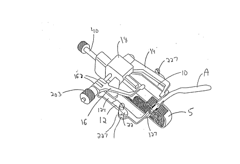
Turning now to Figures 2 and 3, clamp 1 has a pair of proximal arms
aws) comprising a lower proximal arm 10 and an upper proximal arm 14 and a
_ - .....
pair of distal arms (laws) comprising a lower distal arm 12 and an upper
distal arm 16. (The terms "proximal", "distal", "lower" and "upper" are
used for the readers reference to differentiate between the arms. Clearly,
if the orientation of the clamp changes, these designations will also
change.) Each pair of arms is mounted on an axle 20, preferably threaded,
for pivotal movement. As~le 20 extends through an aperture il;l a center post
18 of the base of the clarnp. An anchor or retainer 22 is mounted on the
lower proximal arm 14 and on the lower distal aIrn 16 to secure the upper
.
- arrns 14 and 16 to their respective lower arms 10, 12 in a manner described
- ~- below.
. . - . ~:
.. . . ~ ~
~ The lower proximal arrn 10 as shown is L,shaped with a rear end 101
. , . ".- .. . ..
,~connected to center post 18 and an inwardIy extending free front end 103. - -
Fr`ont end 103 has a Iongitudinal slot 107 formed in its top surface which :
_,_,, _ _, . _ . _ _ ._ _
. .._ _ , ~_ ~ ,',
: -. .'. .. ~ _.,_ . . 9
preferably decreases in width at a tip portion 105. Ear 108 extends
out~ ardly from a central portion of lower proxirnal arm 14 and has an
aperture formed therethrough to receive a fastener to mount anchor 22. --
,
Lower proximal arm 10 is preferably rigidly mounted to axle 20 and to center
post 18 so pivotal movement is prevented. However, alternately, lower
proximal arm 10 could be pivotally mounted to the axle 20 to allow free
rotation.
With continued reference to Figures 2 and 3, upper proximal arm 14
is pivotally mounted to axle 20 for pivotal movement with respect to lower
proximal arm 10. The upper proximal arm 14 pivots between an open position
spaced apart from lower proximal arm 10 (Figure 2) to a closed position
overlying lower prox~mal arm 10 (Figure 3). Figure 3 shows upper proximal
arm 14 in dotted line in the open position. In the closed position7
portions of the ]ower sulface of upper proximal arm 14 may abut portions of
the upper surface of lower proximal arm 107 and preferably, at ]east the
central portions of arms 10 and 14 abut one another in the closed position.
A~ rear end 141 of upper proximal arm 14 includes a tubular bracket 145 which
encircles axle 20 for mounting thereon. In one embodiment, upper proximal
arm 14 can pivot to an open position up to an arc of approximately 270;
however, a smaller or full 360 pivot is also within the scope of the
present invention. Upper proximal arm 14 has an inwardly extending free
front end 143 with a longitudinal recess 147 formed in its bottom surface
which preferably has a decreased width at tip portion 146, thereby
cooperating with free front end 103 of lower proximal arrn 10 when in the
closed position. That is, recess 147 of upper proximal arrn 14 cooperates `~- --
~th longitudinal slot 107 of the top sulface of lower proxi~l arm 10 to `~
.. ., . . . ~.. : . . . ..
.. ,. .,. ~ , . -.
.
,,.,, 10
. .
. .
~. ~
- form a charmel therebetween to receive tubular section A when the proximal
-- - a s 14, 10 are iri their closed position. Tip portions 146 and 106 of upper- and lower pro~-imal a~ms 14, 10, respectively, have rounded ends which form a -
- ,: , ,: - .
narrow circular opening through which tubular section A extends for reasons
which will become apparent from the discussion below.
Referring now to Figure 3, (for c]arity, the distal arms 12 and 16
are not labelled in detail in Figure 2) lower distal arm 12 is pivotally
mounted on axle 20 and is illustra~ively rotatable around an arc of 360,
although other arms are also contemplated. Rear end 121 of lower distal arm
12 has a pair of spaced apart clasps 122 having annular holes to receive
axle 20 for mounting thereon. Rear end 121 further includes a recessed
surface 124 (on the upper surface of lower distal arm 12) for reasons
dlscussed below. A free front end 123 is substantially identical in
configuration to the front end 103 of the lower proximal arm 10 as it has a
longitudinal slot 127 in its upper surface preferably terminating in a
decreased width portion at tip section 126. Lower distal arrn 12 also
includes, similar to lower proximal arm 10, an outwardly extending ear 128
having an aperture to receive a fastener for mounting anchor 22.
Upper dist~l arm 16 is pivotally mounted to lower distal arm 12 fcr
movement between an open position spaced from lower distal arIn 12 to a
c]osed position overlying lower distal arm 12. Preferably upper distal arrn
16 is pivotable to an arc of approximately 270; however, clearly upper
distal arrn 12 can be pivotable along a larger or smaller arc. Upper distal
... . .. .
- - arrn 16 includes a rear end 161 having a pair of spaced apart clasps 162 with
-; annular holes through which axle 20 extends for mounting thereon. These -:
s 162 are disposed betweerl clasps 122 of lower dist 1 arm 12 as shown
.. , . ._ , . ..
* ,., . -. - . :
- .- .. : : , -. ~` 11
-. .~ i .- , ~: -
- -~
, _ .. . . .
- - -
in Figure 3. Front end 163 of upper distal arm 16 is substantially
: identical to the ~ont end 143 of upper proxirnal arm 14 in that it includes . -
- . . .
an inwardly extending portion having a longitudinal recess 167 forrned in its
bottom surface terrninating at tip portion 166 where it preferably has an
decreased width. W~en the arms 16, 12 are closed, longitudinal recess 167
of upper distal arrn 16 cooperates with longtiudinal recess 127 of lower
distal arm 12 to forrn a ch~nnel for receiving tubular tissue section B.
Tissue section B protrudes beyond the annular opening forrned at the end of
cooperating tip portions 126, 166. Upper distal arm 16 also has a recessed
portion 164 in its bottom surface in rear end 161.
The base of the clarnp includes a central post 18, shown in the
drawings shaped as a block which has a central aperture extending
therethrough to receive axle 20. As shown in Figure 2, the base further
includes a distal support 185 and a proxirnal ear 183 preferably integral
w~th the post 18. Distal support 185 fits within recesses 124 and 164
of lower distal arm 12 and upper distal arrn 16, respectively, when the
closed distal arms 12, 16 are moYed inwardly towards the proximal arms 10,
14 in a manner described below. The top portion 180 (Figure S) of the post
18 is adapted to receive top housing section 5 and the bottom portion of
post 18 (not shown in the drawings) is adapted to' receive the bottom housing
section 3.
- . Axle 20, which is illustratively formed as a threaded screw,
extends through the aperture in post 18 and has a spring 201 mounted thereon
which is disposed intermediate clasp 122 and post 18. A longitudinal
adjustment nut or cap 203 fits over the end of the axis saew 20 and is - ':
.~.'.............. `~adapted to be brought into contact with clasp 162 (or alternately clasp - ' ; -
=~ = . . . -.. . . .
~. ~ - .........
..... ..
.... : -
. ,. ~ .... 12
. .
J 21~/3 ~J
, ' 122) of upper distal arm 16, Ihereby functioning to move closed distal arms
,- '- 12, 16 towards pro~imal arms 10, 14 in a manner described in detail below.
- ' ~', An anchor 22 is swivably mounted to both ear 108 of lower proximal -: '
? arm 10 and ear 128 of lower distal arm 12 by a screw 227 (See Figures
,~- - 2-4). Screw 227 extends through an opening formed in the anchor 22 and
': ~ through the aligned apert lre of the respective ear 108, 128. The anchor 22
, . ~ . . ., ~
~"- is illustratiYely I~shaped in configuration and is pivotable in the
. ~, - .... .
direction of arrows m and n (Figures 3 and 4) from a resting position
-' substantially parallel to the longitudinal axis of its respective arm 10,
12 to a locking position substantially perpendicular to the longitudinal
axis. The anchor 22 has an undercut portion which provides a sufficient
gap to receive the respective upper arm 14 and 16 as described below. A
' conventional nut (not shown) can optionally be mounted to the bottom end of
" - '` ~` ~-,'scrë~v 227. The anchor 22 in its loc~;ing position compresses the bottom
'- surface of the undercut against the upper surface of t~e upper arms 14, 16
',- :, in the closed position.) Clearly, other means for anchoring the pair of
- pro~nal arms and pair of distal arms (e.g. snaps or screws) can be
utili7ed as long as it provides firm securement of the arms.
Turning now to the top housing section 3 and bottom housing
section S of laser housing 2, these housing sections are mounted to
- ' ,-' - opposing sides of clamp 1 and provide the means for applying laser energy
, - .
,' ' -' - ,, to'the seam formed between abutting tubular sections A and B. As noted
'.' above, t~e terms "top" and "bottom~ are used for convenience to denote
;~acement on opposing sides of the clamp 1 since the clarnp can be
~.i . .
- ~re-oriented.- As shown in Figure 8, top housing section 3 includes a
. ~ recess 31 to receive the top portion of center post 18, and front
,~ ;:- - '
. ~ ,~, .- .
, --,-; ,= ~
- .... _., . =
,........................ 13
- .
_ . _ . _ ... .
~ 'J ~ 9
. . .
- and rear longitudinal slots 33', 33' forrned on its inner surface. An upper
channel 35 exlending from the rear of housing section 3 to recess 31 ~.
`provides for passage of a tightening screw 40. The tightening screw 40 is
- ` ~` adapted to be rotated inwardly to press against central post 18 when -
` housing section 3 is mounted thereon to effectively secure the top housing
.
section 3 to clamp l. A separate channel is spaced apart from upper
channel 35 to allow passage of the laser transmitting conduits in channel
30.
Bottom housing section S is substantially identical in
configuration to top housing section 3 except it is provided with
` projections 53, 53' on its inner surface instead of longitudinal slots 33,
-33'. The projections 53, 53' are adapted to engage slots 33, 33',
~ respectively, of top housing section 3 when bottom housing section 5 is
. ~ : . .:: , . .
. - fitted over top portion 180 of post 18 to secure the two housing sect;ons
` - together. Of course, alternate ways to connect the two housing halves can
- : be utili7ed. Similar to top housing section 3, bottom housing section 5
includes a channel 55 to receive a screw 40 for tightening the housing
section S against central post 18. A separate channel spaced from cha~el
55 receives a conduit 50 for transmitting laser light.
Both housing sections 3, 5 include a sernicircular recessed portion
39, 59, respectively, through which the laser light is tlansmitted. The
recesses 39 and 59 cooperate to form a circular enclosure for the annular
- _ - seam S of the abutting tubuIar sections A and B when the housing sections
~ ~ 5 are mounted to opposing sides of clarnp 1 and projections 53, 53' are
- ~tted within longitudinal slots 33, 33'. Although the housing 2 is
: ~ated as comprising two discrete sections 3 and 5, clearly a single
_ _ . _ ~ . =, . . .. .
,_=,. .. . .. .
~-- . . .
14
_ _ _ _ _ _ .
~3
. . ~~ `'
- - housing with hinged sections could alternative]y be provided.
.
The channels 30 50 for the laser conduits extends through both
, .; ~. . . .
~ttom and top bousing sections 3 5 and in a preferred embodunent -
` .-:; :~omprise a series of multiple fibers radially directing light with respect
. -- to seauclrcular recessed portions 39 and 59 in order to transmit laser
- light sirnultaneously along the entire circumference of the seam S formed at
~e abutment of tubular sections A and B. The fibers preferably terminate
before recesses 39 59 so they are adjacent but not contig. ous to the
searn S. Light ca~ travel rad ally to directly ~ase the abutted tissue or
alternately a lens system such as a prism or mirror could be positioned at
an angle to the seam so that incoming laser light will be directed by the
lens system onto the seam. With continued reference to Figure 8 the
- conduits 30 50 each containing multiple optical fibers extend rearwardly
from the housing sections 3 5 to a coupler where the proximal ends of the
multiple fibers interface wi[h l~ght emitted from a single standard optical
fiber from a laser or are coupled directly to the laser itself. Block
diagrams here illustrate coupling to a single optical fiber from an
external source of laser energy. The aspects of generating the laser
energy from the extemal source are known in the art and beyond the scope
of the present invention as is the coupling of a single fiber to a laser.
:-- With reference to Figures 2-7 the operation of the clamp 1 of the
- present invention will now be descnbed. Figure 2 shows clamp 1 with both
: -~ upper distal arrn 14 and upper proximal arrn 16 in the open position. Note
~ anchor 22 is disposed along ear 108 in a position substantially
- ~el to the longitudinal axis of lower proximal arm 10 to avoid
~-rence vith movement of upper proximal arrn 14 to the closed
~ ' '
G'`-
- _ - . .
. . .
- ., ., -, . -` . 15
.
. . _
7 ~'; ;;' L r,! ,3
.,
- pos tion. As shou~ in Figure 3 tubular tissue section (proximal section)
A is placed longitudiDally in the slot 107 of lower proximal arm 10 and the - - - --
:;~opper proximal arm 14 is then pivoted in the direction of arrow r to its
closed position to overlie ~ower proximal arm 10. Tubular section A is
- - ~ thus firrnly fitted u~ithin the channel formed between cooperating slot 107 :
.. ~ . -
- - of lower proximal arrn 10 and recess 147 of upper proximal arrn 14. Anchor
- . . .. . ..
- ~ : - - 22 is then rotated in the direction of arrow m to its locking positionsubstantially perpendicu]ar to the longitudinal a~is of the lower proximal
arm 10 so that it holds the proximal arms 10 14 together in their closed
position. Proximal arrns 10 and 12 provide sufflcient pressure to
- effectively function to reduce or eYen stop the bleeding.
Once tubular tissue section A is securely clarnped by proximal arms
10 14 the other tubular section (distal section) B is placed within slot
. - ., . . -
127 of lower distal arrn 12 and upper distal arrn 16 is pivoted to its closed
- ~ position to overlie lower distal arm 12 (Figure 4). Anchor 22 attached to
ear 128 of lower distal arm 12 is subsequently rotated (sce arrow n of
Figure 4) to its perpendicular locking position to secure the distal arrns
10 16 together to thereby firmly grasp tubular section B within the
channel formed between the cooperating slot 127 of lower distal ann 12 and
recess 167 of upper distal arrn 16. Distal arms 12 and 16 also function to
reduce or stop the bleeding.
After securement of both the pro~nal and distal tubular sections
A B in their respecti ~e arrns adjustment nut 203 on threaded axle 20 is
- ~.~; rotated clockwise (in the direction of arrow w of Figure 5). This rotation
~ves nut 203 in the direction of arrow x to abut clasp 16~. Continued -;
~on of abutting m t 203 thus moYes distal arms 12 16 longitudinally
=~ ,~ , . , . .-. . . - .
~ . . - . .
- -, ,,, . . . ~
, . .: - .
16
r~ fj
along a~le 20 toward the proximal arms 10, 14 in the direction of arrow x,
~. ~ thereby compressing spring 201. Longitudinal movement of distal a~ms 12,
~16 carries the grasped tubular section B in the same direction to bring it - --
,.`` into apposition (abutment) with t~bu]ar section A to form seam S.
Apposition preferably requires that t~e edges of sections A and B are -:
~ .
. ~compressed. Achievement of this close apposition and the ability of the
clamp 1 of the present invention to hold these tubular sections in abutment . ` `
enables effective anastomosis of these tubular sections when laser energy
is applied to the SeanL
After approxirnation of the tubular sections A, B by the arms of
clamp 1, the bottom housing section 5 is positioned so that the seam S lies
in correct aligmnent for tissue welding. Recess 31 of lower housing
section 3 is fitted over a bottom portion of center post 18 of clamp 1
(Figure 6) and secured in position by turning the screw 40 at the back of
the housing section 3 to tighten it against post 18. Recess 39 thus
encloses half of the circular seam S. The upper housing section 3 is then
-placed over clarnp l so that recess 51 is fitted over the top portion 180 of
post 18 and projections 53, 53' engage longitudinal slots 33, 33'
respectively, of lower housing portion 3. Recess 59 thereby encloses the
other half of circular seam S. Sirni~ar to bottom housing section 5, top
housing section 3 is locked into position by tightening screw 40 ag~inst
post 18. Positioning of the housing portions 3 and 5 as thus described
.
ensure that the searm S is fully encircled to provide precise delivery of
-i laser light
:~ After interfitting the housing sections 3, 5, the extemal laser- .-
~rg~ source is operated to transrnit a selected dosage of laser light for ---~
=. .~- . - - .
~ . - .
- . . _ .. ,
_, , -
. =
- 1 7
. .. .
. .
, . ~
h f <j ,~
. . ~
a predctermined lime period. The laser light travels in multiple fibers
. .
`; -` through conduits 30 and 50 and light is delivered radially to thereby apply ` -
- ~- I aser energy directly and simultaneously around substantially the entire
c ircumferential seam S formed at the abutment of tubular sections A and B. ~- -
~e laser energy thereby functions to weld tubular sections A and B ` -together along seam S to provide an effective and improved anastomosis.
- While the exact physiologic mechanism of the laser weld is not
ful~y understood, tissue welding is achieved through the controlled
application of light energy to the anastomastic site to produce a uniforrn
thermal effect which causes the two sides to bind together. That is, the
laser energy thermal~y induces intrinsic tissue changes (e.g. alterations
in tissue collagen and other acellular proteins) as the electromagnetic
energy from the laser is converted into thermal energy which lead to strong
- bonds between tissue.
Although any laser light transmissible through the conduits could
be used, in a preferred embodiment where the vas deferens are to be welded,
the Neody~uum Yttrium Aluminum Gamet (Nd:YAG) laser such as Sha~plen 2100
Nd:Yag laser or a Cooper Model 8000 Nd:Yag laser is utilized with readily
avaiIable quartz optical fibers. In this embodiment, the lille width of the
circumferential welding light can be as narrow as 75 microns. For example,
in reoent experimental trials of this equipment for welding rabbit vas
deferens anastomoses, five watts of power from a Nd:Yag laser operating at
a continuous mode was applied for 1.5 seconds to sucoessfillly provide a
;~ ` uniforrn circumferential weld at the seanL However, it is clearly within
- . . . -- , .
-. the scope of the present invention to utilize Iaser energy of different ~`~
-- ';' - .' : slties and for ~erent timo periods to ach-eve the laser weld ng of ' - -' ~' ~'
- - ` 18
.
_ _
r.l ~. '. V
the tubular sections. Such variations may depend on for example the type
of tissue being welded, the physiologic condition of that tissue, and/or - -
wave length of light used.
After application of sufficient laser energy to weld the tissue - `
sections A, B, the screw 40 is loosened and the top housing section 5 is
removed from clamp 1, followed by loosening of the screw 40 to remove the
bottom housing section 3. The tubular section B is then removed from
distal arms 12, 16 of clamp 1 pivoting anchor 22 to its longitudinal
resting position, to fIee upper distal arm 16. Distal arm 16 is then
pivoted away from tubular section B and lower distal arm 12, followed by
removal of lower distal arm 12 from tubular Section B. The upper proximal
arm 14 is likewise disengaged from the lower prox~mal arm 10 by rotating
anchor 22 to its longitudinal resting position. Upper proximal arm 14 is
then pivoted to its open position. Tubular section A is then removed from
lower proximal ann 10.
To facilitate securement and approximation of tubular tissue
sections, an absorbable stent such as that illustrated in Figure 1 and
designated by reference letter T can optionally be utilized. The stent can
be composed of materials which are water soluble and biocompatible such as
polyvinyl alcohol (PVA) or polyvinyl pyrrolidine (PVA(PVP)). The proximal
end of the stent T is inserted into the proximal tubular section A prior to
clamping of the proximal arrns 10, 14 around tubular section A. After
closing and securement of proximal ~m 14 by anchor 22, the distal tubular
.
; section B is then pulled over the distal end of the stent T. Subsequently,
~. . .... -
- . . ~ upper distal arm 16 is pivoted to its closed position. The stent T is . c
. : ~ preferably water soluble so it can advantageously be left in the body after
.. ... . ..
..~
19
removal of the clamp 1 and will be fully excreted by the body. Iikewise,
non-soluble stents (e.g. st~inless steel wire, teflon, etc.) can be used to
.
` ~ as~sist in apposition and can be removed after welding through natural
., - . ,
- - occunng openings liXe the fimbriated end of a fallopian tube or the anus --
or through a small incision in other structures like the vas deferens.
-Other means can be provided to facilitate or enhance securement and -
.. . ..
approximation of the tubular tissue sections such as flat surfaces for - :
compressing tissue, teeth or barbs or vacuum parts along the jaws where the
tissue is held.
Optionally, a dye substance such as India Ink can be applied to
the proxirnal and distal tubular sections A, B adjacent the seam to act as
an exogenous chromophore to increase laser energy absorption into the
anastomotic edges, thereby enhancing tissue welds and rninimizing collateral
thermal darnage. This advantageously allows use of reduced power settings.
Other exogenous chromophores such as Indocyanine Green dye, flourescein or
endogenous chromophores such as blood or can alternately be utilized to
facilitate absorption of laser energy by localizing the laser energy on the
welding site.
rhe clamp 1 of the present invention can be used in a Yariety of
applications for laser welding of tubular sections of the body. For
example, the clamp can be used for anastomosis of the vas deferens in
perfor~ung a vasovasostomy (a reversal of a vasectomy). In this instance,
the proxirnal and vas deferens are secured within the jaws (arrns) of clamp
1, b-ought into apposition and laser welded to re-establish communication.
The clamp 1 can also be used for ]aser welding the fallopian tubes to
: .. .. . ....................................................... .-- reverse surgically induced sterility or repair defects. Other uses include
... . . . ..
...... :
., , . _, .. .. .
~ 9 ~ ,3
.
the clarnping and subsequent ]aser ~elding of the ureter, urethra, blood
. - .-. ..
~- vessels, biliary tlssue and other tubular tlssue structures.-- Additionally,:~
`=~ tbe clarnp 1 can cleaTly be used to grasp aDd bring into opposition other
tissue sections that are not hol~ow. An additional advalltage` of this `
: - - apparatus and method for mechanically holding tissue during lasér welding
- - is that it c~n be used at distant less` accessible sites within body
- cavities as is necessary in minirially invaslve endoscopic or laproscopic
surgical procedures.
The apparatus of the present invention can also be used to clamp
metal or plastic tubes or prosthetic materials such as Goretex~ or
Dacron~ for surgical uses.
Obviously, numerous modifications and variations of the present
. .
invention are possible in light of the above teachings. It is therefore to
be understood that within the scope of the appended claims, the inYention
may be practiced otherwise then as specifically described herei~
. . _
.
- _ . .. . . - - . . . ~ : .
21