Note: Descriptions are shown in the official language in which they were submitted.
ADHESIVE PULSE OXIMETER SENSOR WITH REUSABLE PORTIOM
BACXGROUND OF THE INVENTION
This is a continuation-in-part of patent
application serial no. 07/600,541, filed October 19,
~990 .
This invention relates to sensors for use wi-th
non-invasive pulse monitors such as plethysmoyraphs or
pulse oximeters.
A plethysmograph is a pulse monitor. The
plethysmograph sensor shines light into the patient's
tissue, and the light transmitted through the tissue is
received by a photodetector. The photodetector generates
electrical signals corresponding to the transmitted light
levels and transmits the signals to a monitor for
processing. Arterial blood will absorb some of the
light, with more light being absorbed when there is more
blood. Thus, changes in the amount of transmitted light
are related to pulses of arterial blood in the
illuminated tissue.
A pulse oximeter is a device for noninvasively
determining the oxygen saturation of arterial blood. The
pulse oximeter sensor shines light at two different
wavelengths (one in the red range, the other in the
infrared range) through a portion of the patient's blood-
perfused tissue~ The red and infrared light transmitted
through the tissue is detected by a photodetector. The
amount of light absorbed varies with the ~mount of oxygen
in the blood, and varies differently for red and infrared
light. The pulse oximeter monitor computes blood oxygen
saturation based on the changes in the two detected light
levels between two points in time.
There are several types of sensors for
plethysmographs and pulse oximeters. One is a surface
sensor in which the light emitter and the photodetector
are mounted on the same sensor face. The sensor is
-
.
, ; l !
attached to the patient with both the light emitter and
the detector on the same side o~ the patient's appendage
(e.~, on the patient's forehead). This type of sensor
detects light ref]ected back from the tissue, rather than
light transmitted through an appendage. The signal
detected will thus be weaker in most cases. The sensor
is typically attached with a strap, headband or tape over
the sensor, or an adhesive pad between the sensor and the
skin.
Another type of sensor is a clamp design, such
as that described in U.S. Patent No. 4,685,464. The
durable sensor described in that patent has deformable
pads creating con~orming tissue contactin~ sur~aces to
which the emitters and photodetector are secured. The
15 deformable pads are disposed in a hinged rigid housing
that clips on the patient like a clothes pin. This
relies o~ a clamping force to secure the sensor to the
patient. The force of the sensor against the patient's
tissue could reduce the flow of blood to that region.
20 This exsanguination of the tissue beneath the sensor
adversely a~fects pulse detection and analysis by
suppressing the pulse in that portion o the tissue. As
a result, the sensor site must typically be checked or
moved every four hours to insure adequate perfusion.
25 Because o~ its relatively large mass, however, the clamp
design is more susceptible to signal-distorting motion
artifact. i e~, differential motion between the sensor
and the patient.
A third sensor design is described in U.S.
30 Patent No. 4,830,014. The conformable sensor described
in that patent has emitters and a photodetector mounted
in the same side of a flexible web. The web wraps around
a portion of the patient's tissue (such as a finger) so
that the light from the emitters must travel through the
tissue before reaching the detector. The web attaches to
the skin with an adhesive surface on the emitter and
detector side of the web. Because o~ its relatively low
mass and the adhesive, this sensor adheres closely to the
:
r
patient's skin and minimizes the ef~ects of motion
artifact. In addition, its flexibility and use of
adhesive to secure it minimizes the exsanguination caused
by rigid sensors. Thus the sen!;or site typically only
needs to be checked every eight hours. Conformable
sensors, howaver, are typically restricted to one
application due in part to a decrease in adhesive
effectiveness with each application and in part to
difficulties in cleaning and sterilization for reuse.
Replacement of the sensor a~ter only one use can make
pulse oximetry expensive.
SUMMARY OF 'rHE INVENTION
The present invention provides a pulse oximeter
sensor that is desiyned to surround an appenda~e of the
patient, such as a finger, toe or foot. The sensor has a
reusa~le member which preferably includes a
photodetector. A disposable, ~lexible member pre~erably
contains the photoemitter and can be wrapped around the
patient's appendage to ~ecure it to the appendage and the
reusable member. When secured, the photoemitter and
photodetector end up on opposite sides of the appendage.
The disposable member connects to the reusable member to
establish electrical contact. The reusable member is
connected to a cable which can be plugged into a sensor
monitoring system.
In the preferred embodiment, the flexibl~
member is a flexible adhesive web with arms extending
laterally from a central portion. The reusable member is
prefarably a rigid housing with a deformable pad for
contacting the a~pendage.
To attach the sensor to the patient, ~he
flexible web is adhesively attached to one side of the
patient's appendage, and the rigid housing is placed on
3~ the other side directly opposite the flexible web. The
arms extend around the appendage to adhesively hold the
conforma~le pad o~ the rigid housing against th
appendage. By reducing the mass o~ the sensor and by
,
,
adhesively attaching the emitters to the skin, this
configuration minimizes motion artifact by reducing the
relative movement between the sensor and the patient's
skin experienced by previous clalmp-type sensors. In
ad~ition, the flexible web and con~ormable sur~ace of the
rigid housing minimize exsanguination of the tissue
beneath the sensor. Since the sensor relies on adhesion
to secure it to the patient, the sensor site should not
need to be checked as often as ior a clamping-type
sensor.
Aftsr use, the flexible web may he separated
from the rigid housing, the rigid housing cleaned, and a
new ~lexible web attached to the rigid housing. The
~resh adhesive on the new flexible web provides a more
reliable bond between the sensor and the patient than the
adhesive on the previously-used web. In addition, since
the flexible web covers ~our out o~ the five surfaces of
the patient's appendage (including, when worn on the
~inger, the cuticle and subungual region), one time use
of the flexible portion o~ the sensor minimizes cross-
contamination between patients when the sensor is reused~
Furthermore, because a portion of the sensor may be
cleaned and reused, this new sensor design reduces the
cost of using flexible sensors.
The ~lectrical connection between the flexible
web and the rigid housing is preferably made with a tab
extending from the flexible we~ having conductive traces
printed on it which connect to the photoemitter. The
conductive trace~ are inserted into a channel in the back
of the housing which is covered by a bridge. Underneath
the bridge are a series o~ electrical contacts for making
connection with the conductiva traces. The tab contains
an internal resilient foam which is compressed as it is
inserted between the housing and the bridge, and exerts
an outward force to maintain the tab in place and crsate
an electrical connection between the conductive traces
and the contacts.
.
For a ~uller understandiny of the nature and
advantages of the invention, re~erence ~hould be made to
the ensuing detailed dPscription taken in conjunction
with the accompanying drawings.
BRIEF DESCRIPTION OF THE DRAWINGS
Fig. 1 is a perspecti~e view of a sensor
according to the present inventi.on;
Fig. 2 is a perspective view of the sensor of
Fig. 1 showing the flexible web being wrapped around a
finger; and
Fig. 3 is a perspective view of the separated
disposable and reusable memhers of Fig. 1 illustrating
how the connection is made.
DETAIL OF THE PREFERRED EMBODIMENT
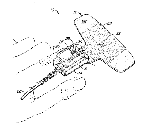
Fig. 1 shows a sensor 10 according to the
present invention. Sensor 10 consists of a flexible,
disposable webbing 12 and a reusable housing 14. Housing
14 includes a rigid portion 16 and a de~ormable pad 18.
A patient's ~inger 20, shown in phantom, is shown placed
on top of deformable pad 18.
Flexible web 12 includes a photoemitter 22,
which preferably includes two photoemitters, one for red
light and one for in~rared light. A photodetector 24 is
included in deformable pad 18. A coppçr grid 23 is
disposed over photodetector 24. A transparant window 25
covers photodetector 24. All or substantially all of the
portion of window 25 extending beyond photodetector 24
is colored black. In addition, a black area 29 is
printed on the underside o~ foam layer 28. Grid 23,
photodetector 24 and photoemitter 22 are electrically
connected to a sensor monitoring system through
conductors in a cable 26 connected to housing 14.
Grid 23 is a Faraday shield (electrostatic
screen) connected to ground for reducing interference.
The thin window 25 extends over the copper grid so that
the grid will not bulge out pad 18. Before the black
. ,
.. .. '
, '!
coa~ting was adding, shift errors in the data values were
noticed. The black coating eliminated these errors. The
reason is not certain, but the coating over the window
may pravent reflections from most o~ the copper, while
the black coating on the foam layer 28 may prevent light
from being shunted throuyh the Eoam layer to the
detector, bypassing the finger.
Webbing 12 has a top foam layer 28 with an
adhesive surface. Before use, this adhesive layer is
covered with protective plastic (not shown), which is
peeled off for use.
Fig. 2 illustrates how the flexible webbing 12
is bent over and attached to finger 20. A first arm 30
of the flexible web is wrapped around the side of housing
14 and will continue to be wrapped around its bottom in
the direction of arrow 32. Similarly, the other arm 34
will be wrapped around finger 20 and housing 14. As can
be seen, photoemitters 22, shown in phantom, are now on
top of the finger, directly opposite photodetactor 24,
which is not visible in this view. As can be saen, only
the bottom of finger 20 contacts de~ormable pad 18. At
least the top of the finger will be adhered to by web 12.
The sides and fr~nt may also be adhered to, depending on
the shape of the finger and how the sensor is attached~
The top is the portion which is most important to be
adhering, since it contains the photoemitter which should
not move relative to the ~inger. This provides a secure
connection which reduces motion artifacts and puts the
disposable, flexible portion in contact with most of the
surfaces of the finger so that it is exposed to more
contamination than the reusable portion.
Fig. 3 illustrates the electrical connection
between ~lexible web 12 and rigid housing 14. Fig. 3
shows adhesiv~e layer 28 partially peeled back from a web
base 36. In between web base 36 and adhesive layer 28,
an elongate plastic substrate 38 is placed, with a series
of conductive traces 40 on its top surface. Two
conductive traces connect to photoemitters 22, and two
connect to a calibration resistor 55, described b~low.
Elongate plastic substrate 38 forms a tail 42. Web base
36 ca~ be just large enough to hold tail 42 to adhesive
layer 28, as shown, or could conform to the shape of
adhesive layer 28. Web base 36 has an adhesive surface
for holding tail 42 to layer 28.
A compressible foam member 44 is placed between
the halves of tail 42. In the preferred embodiment, the
foam is made of Poron foam ~rom Roger's Corp. A pair o~
tabs 46 extend from the top half of the tail having the
conductive traces. The tabs and the foam memher provide
part of the attachment mechanism as explained below.
A channel 48 is formed on the bottom side of
the rigid housing 16, opposite deformable pad 18. A
series of electrical contacts 50 (shown in phantom) are
located in the channel. The contacts are covered by a
bridge 52 extending across the housing. A pair of
grooves 54 are formed in the channel. The grooves are
slightly larger than the tabs 46 on the flexible web.
To connect the flexible web to the rigid
housing, the tail 42 of the flexible circuit is inserted
into the space beneath bridge 52. As the tail moves
forward, the plastic foam 44 compresses. As the tail's
tabs 46 move over the channel's grooves 54, the spring
action of the foam pushes the tabs i~to the grooves. The
tabs and groo~es ensure that the flexible circuit is not
inserted too far and prevent inadvertent removal of the
flexible circuit. The spring action of the foam also
pushes one set of contacts against the other to enhance
the electrical connection. In addition, the scraping
action of one set of contacts against the other during
insertion a~d withdrawal of the flexible circuit will
help remo~e any oxidation or debris on the contacts. To
remove, the tabs are lifted out of the grooves by pulling
the flexible web away from the housing and the tail is
withdrawn from the space beneath the bridge.
Cable 26 contains 6 wires. Two are connected
to calibration resistor 55 through two of conta¢ts 50 and
conductive traces 40. Two are connected to photoemitkers
22 through the other two of contacts 50 and conductive
traces 40. The remaining two wires are connected to
photodetector 24.
In the preferred embodiment, the plastic
substrate is formed from white, substantially ~paque
polyester. White nylon may also be used, or a clear
plastic. The adhesive may be white, wi~h a clear window
for the photoemitters.
The pre~erred embodiment of the sensor
according to this invention includes an encoding/decoding
system such as that described in U.S. Patent No.
4,621,643. The ~lexible web supports an encoding
resistor 55 in electrical communication with the monitor.
As explained in that patent, the value of the resistor is
selected to match the wavelengths o~ the red and in~rared
LED's. That patent also describes the necessary sensor
monitoring electronics.
In an alternati~e embodiment, the sensor's
photodetector may be mounted in the flexible web with the
emitters and the encoding resistor mounted in the rigid
housing.
In the preferred embodiment, the rigid housing
is made from injection molded polycarbonate.
Alternatively, injection molded ABS plastic may be used.
Patent No. 4,685,464 contains additional details on
construction of a rigid housing and deformable pad
including the placement of the photodetector.
As will be under~tood by those familiar with
the art, the present invention may be embodied in other
specific forms without d~parting ~rom the spirit or
essential characteristics thereof. For example, the
compression effect of foam 44 could be obtained instead
by making bridge 52 a spring-action clip, which is opened
by holding one end down during insertion and then
released, with a spring on the clip holding the tab in
place. Other variations in ths way electrical contact is
made are also possible. Instead of thP adhesive layer,
the flexible porti~n could be attached to the finger and
rigid housing using velcro or other securing mechanisms.
The flexible web could be made of foil or other color
materials than white or clear. The sensor could be a
surface sensor, with adhesive for reducing motion
artifact on the disposable portion. Accordingly, the
disclosure of a preferred embodiment of the invention is
intended to be illustrative, but not limiting, of the
scope of the invention which is set forth in the
lU following claims.
3~