Note: Descriptions are shown in the official language in which they were submitted.
Miller-Bates-Hall-Osborne 1-2-2-2
. ~S~9
Guide for Localizing a
Nonpalpable Breast Lesion
Technical Field
This invention relates to medical devices for localizing
lesions within the body and, in particular, to a guide for
the localization of a nonpalpable lesion within the breast.
Backaround of the Invention
Localization needles or wire guides are utilized for
preoperati~e marking of nonpalpable breast lesions.
Typically, a needle cannula having a wire guide contained
therein is inserted into the female breast and preferably
positioned within two centimeters of the lesion. A
mammogram or other visualization aid is used to confirm the
position of the distal needle end. If the needle is not
accurately position~d with respect to the lesion, the needle
is repositioned, and another mammogram is taken to visualize
the repositioning of the needle end with respect to the
lesion. When the needle position is acceptable, the wire
guide is extended from the distal end to localize the breast
lesion for recision by the surgeon. Alternatively, the wire
guide is maintained in a fixed position while the needle is
removed to expose the wire guide contained therein.
Similarly, the distal end of the wire guide localizes the
breast lesion for recision by the surgeon. A number of
prior art wire guides are utilized for localizing breast
lesions. One in particular is the Kopans breast lesion
localization needle that includes a spring-hook wire guide
that is manufactured by Cook, Inc. of Bloomington, Indiana.
After the spring-hook wire guide is extended from the
localization needle adjacent the breast lesion, the spring-
hook wire guide remains relatively fixedly positioned. The
Miller-Bates-Hall-Osborne 1-2-2~2
~OS~9~9
guide may be further extended into the breast tissue either
intentionally or unintentionally; however, the spring-hook
prevents inadvertent removal of the wire guide from the
bxeast tissue. This advantageously prevents retraction or
removal of the wire guide due to inadvertent movement of the
breast during transportation or movement of the patient.
However, it does not prevent inadvertent advancement into
the hreast. Repositioning of the spring-hook wire guide is
limited, and removal is by surgical resection.
A number of repositionable localization systems are also
presently available. The HAWKINST~ breast lesion
localization system manufactured by Boston Scientific
Corporation of Watertown, Massachusetts, includes a rigid
cannula with a retractable side barb for repositioning the
needle cannula if necessary. Some physicians prefer the
rigid cannula; however, others do not. The rigid cannula
also presents the risk of further penetration through the
breast and into the chest cavity and lungs as a result of
patient movement.
Another repositionable localization system is the Homer
MAMMALOCKTH needle/wire localizer, which is available from
Namic0 of Glens Falls, New York. The MAMMALOCK localizer
includes a needle with a unique alloy J-hook wire positioned
within the needle, which is extendable therefrom. The
needle is introduced into the breast tissue, and the J-hook
wire advanced into the tissue before a mammogram is taken
for accuracy of placement. The J-hook wire tip protects the
breast tissue from needle point penetration during breast
compression. However, the J-hook tip includes a straight
se~ment at the distal end which presents a significant
disadvantage should the needle be removed. The curved
portion of the J-hook wire tip does not track the straight
segment at the distal end of the wire when extended from the
distal end of the needle. As a result, the J-hook wire tip
does not penetrate the tissue and is deflected from the
lesion Chould the needle be subsequently removed. The
~iller-Bates-Hall-Osborne 1-2-2-2
9~9
deflection and lack of trackability significantly increase
the difficulty of accurate placement of the wire guide.
Trial-and-error placement of the guide also subjects the
patient to unnecessary radiation received during the taking
of extra mammograms.
Another repositionable wire guide having a memory hook
for localizing breast lesions is disclosed in U.S. Patent
No. 4,616,656. This wire guide includes a relatively small
hook with a pointed distal end. The wire guide is
preferably made of a memory characteristic material which
assumes the J-hook configuration in response to body heat.
The J-hook may be repositioned after retraction into a
sheath and re-extension into a new position. When the wire
guide is acceptably positioned, the cannula is removed, and
the wire guide is left as a guide for surgical excision of
the lesion. This repositionable wire guide is also subject
to the same disadvantages as those of the MAMMALO~K J-hook
wire guide. Furthermore, the J-hook wire guide does not
fixedly position or lock in breast tissue and is easily
dislodged during transportation or movement of the patient.
In addition, the J-hook wire tip design is hard for the
6urgeon to palpate.
Summarv of the Invention
The foregoing problems are solved and a technical
advantage is achieved with an illustrative wire guide for
localizing a nonpalpable lesion in a breast. The wire guide
includes a distal portion including a superelastic metallic
allo~ and is preformed into a resilient helical coil
configuration. This resilient helical coil configuration
advantageously locks the wire guide into position once
extended from the end of the introducing needle. The
helical coil configuration of the wire guide includes an
acuate or pointed distal end for penetrating the tissue as
it extends from a tubular introducer needle. The resilient
helical coil configuration includes at least more than a 180
Miller-Bates-Hall-Osborne 1-2-2-2
~S,~9
degree turn which follows a path scribed by the acuate
distal end as the distal portion is extended from the
passageway of the introducer needle. This helical coil
configuration may be easily palpated by the physician and
includes a passageway that extends longitudinally through
the configuration and laterally from the straight portion of
the wire guide from which the distal portion extends. This
lateral orientation further locks the guide in position
should an extraction force be applied to the prox~mal end of
the guide. The lateral orientation also advantageously
prevents the wire guide, as well as the introducer needle,
from being inadvertently extended further into the breast or
chest cavity. As a result, this helical coil configuration
with a lateral orientation provides a significant advantage
over prior art J-hook tip designs as well as that of a
corkscrew having a passageway with a longitudinal
orientation.
The distal portion of the wire guide includes a
superelastic metallic alloy having a transformation
temperature below that of the normal operating environment
of the guide. This superelastic metallic alloy includes
nickel and titanium and resists deformation as a stress is
applied. Furthermore, the superelastic metallic alloy wire
returns to its preformed helical coil configuration when the
deformation force is removed. This advantageously allows
the helical coil configuration of the distal portion of the
wire guide to be retracted within the introducer needle for
repositioning.
The wire guide is combined with a tubular introducer
needle for insertion into the breast to the site of the
lesion. The distal end of the needle includes a plurality
of indentations for advantageously enhancing the ultrasound
visualization thereof. The distal portion of the wire guide
assumes an unwound configuration when positioned within the
passageway of the introducer needle. As the wire guide is
extended from the tapered distal end of the needle, the
Miller~-Bates-Hall-Osborne 1-2-2-2
~:~5~9
distal portion assumes the resilient helical coil
configuration about the lesion. The interior surface of the
needle cannula is, for example, plug-drawn to provide a
smooth surface about the passageway of the needle. This
smooth surface prevents the pointed distal end of the guide
from catching and lodging within the passageway of the
introducer needle.
The medical device also includes a second cannula for
back loading the wire guide into an introducer needle that
has already been positioned within the breast. The second
cannula also includes a smooth interior surface for
repositioning the distal portion of the wire guide from the
passageway of the second cannula into the passageway of the
tubular introducer needle. A connector cap is also
advantageously included and has a passageway extending
longitudinally therein and sized for receiving the abutting
cannula ends and aligning the passageways thereof.
The resilient helical coil configuration includes at
least one turn of more than 180 degrees for fixedly
positioning and locking the distal portion of the wire guide
about the breast lesion. The helical coil configuration
follows a path scribed by the acuate distal end to lock the
dis~al portion of the guide advantageously without
deflecting the guide or the introducer needle. The lateral
orientation of the helical coil configuration also
advantageously further locks the guide into position when
the straight portion of the guide is pulled at the proximal
end thereof.
30 Brief Descri~tion of the Drawin~ -
FIG. 1 depicts an illustrative preferred embodiment of
a medical device of the present invention including a
tubular introducer needle and a wire guide positioned
therein for localizing a nonpalpable breast lesion;
FIG. 2 depicts the wire guide of FIG. 1 with its distal
portion assuming a preformed resilient helical coil
S
Miller-Bates-Hall-Osborne 1-2-2-2
2~
configuration;
FIG. 3 depicts a cross-sectional view of the distal
portion of the wire guide of FIG. 2;
FIG. 4 depicts the wire guide of FIG. 1 positioned
within a back loading cannula;
FIG. 5 depicts the transfer of the wire guide from the
back loading cannula of FIG. 4 into the tubular introducer
needle of FIG. 1; and
FIGS. 6 and 7 depict the insertion of the medical device
of FIG. 1 into a breast for localizing a nonpalpable breast
lesion.
~etailed Description
Depicted in FIG. 1 is an illustrative embodiment of
medical device 10 comprising a tubular introducer needle 11
and a wire guide 14 positioned in passageway 12 of the
needle for insertion into a breast to the site of a
nonpalpable lesion. With a visualization aid such as an X-
ray film or ultrasound, a radiologist typically inserts the
needle with the wire guide positioned therein into the
breast to the site of the lesion. When inserted, the
radiologist extends distal portion 16 of the wire guide from
tapered distal end 13 of the needle, which assumes a
preformed resilient conical helical coil configuration
distally about the breast lesion. Another X-ray or
ultrasound is taken to confirm the positioning of the needle
and the wire guide distally about the breast lesion. Should
the needle and wire guicle not be appropriately positioned,
the distal portion of tb,e wire guide is retracted into the
passageway of the needle. The needle and wire guide are
then repositioned within the breast closer to the lesion,
and another X-ray or ultrasound is taken to confirm the
repositioning of the needle and extended distal portion of
the wire guide. ~fter the needle and wire guide are
properly positioned, the needle is removed from the breast
with the wire guide and the distal portion thereof in the
MiIler-Bates-~all-Osborne 1-2-2-2
~S.~
helical coil configuration, thereby locking the guide in a
position distal to the lesion to guide the surgeon to resect
the lesion within a wed~e sf breas~ tissue surrounding the
wire guide.
Needle 11 comprises cannula 26 having passageway 12
extending longitudinally therethrough between proximal end
24 and tapered distal end 13 for positioning the wire guide
therein. Cannula 26 is a 20~gauge thin-wall, plug-drawn
stainless steel tube, which is commercially available from
K-Tube Corporation, San Diego, California. The plug-drawn
stainless steel tube provides cannula 26 with a smooth,
seamless interior surface 21 which prevents the pointed
distal end 17 of the wire guide from catching and lodging on
the interior surface of the needle. Needle 11 is
approximately 11.5 cm in length and is silicone coated fo
easy insertion into thP breast. Cannula 26 includes distal
end 13 tapered in a well-known venous bevel and proximal end
24 with a well-known fema;Le Luer-lock connector hub 2
insert molded thereabout for ease of handling and for
connection to a syringe for injection and irrigation of
fluids.
Depicted in FIG. 2 is wire guide 14 with straight
portion 15 and distal portion 16 preformed into a resilient
conical helical coil configuration 19. The wire guide and,
in particular, distal portion 1~ is comprised of a
superelastic metallic al]oy such as nitinol which is
commercizlly av~ilable from, for example, Nitinol, Saratoga,
California or U.S. Shape Memory Applications, Inc.,
Sunnyvale, California. This superelastic metallic alloy in
the preferred embodiment is nickel and titanium based and
has a predetermined transformation temperature below that of
the normal operating environment of the wire guide. In
particular, the transformation temperature of wire guide 14
is in the range of 0-10 degrees Celsius, which is well below
the body temperature of patients into whom the wire guide is
to be inserted. When positioned in the passageway of a
Miller-Bates-Hall-Osborne 1-2-2-2
~ 1~5~9~
cannula such as that of needle 11, preformed distal portion
16 assumes an unwound configuration as depicted in FIG. 1.
8y way of example, wire guide 14 comprises approximately
20.5 cm of .013 inch diameter nitinol wire sized for
insertion through passageway 12 of tubular introducer needle
11. As depicted in FIG. 2, ~ire guide 14 includes proximal
portion 27 and distal portion 16, which is preformed into a
resilient conical helical coil configuration 19 having
acuate distal end 17 for cutting into and locking distally
about the nonpalpable breast lesion. As shown, the helical
coil configuration of distal end 16 includes approximately
two complete turns. This helical coil configuration
preferably includes at least more than one 180 degree turn
to assume a minimum locking position. A cylindrical helical
coil configuration is also ,contemplated. The resilient
helical coil configuration 19 follows a path scribed by
acuate distal tip 17 as distal portion 16 is extended from
passageway 12 of the needle to localize the nonpalpable
breast lesion. Passageway 18 extending longitudinally
through the helical coil configuration is laterally oriented
with respect to straight portion 15. The lateral
orientation locks the distal portion in place and prevents
removal from or further ins~ertion about the breast lesion
when the straight portion is either pulled or pushed.
Depicted in FIG. 3 is a cross-sectional view of distal
portion 1~ having acuate distal end 17 taken along the line
3-3 of FIG. 2. This cross-sectional view of the distal
portion illustrates the conical helical coil configuration
19 of the wire guide with longitudinal passageway 18
extending therethrough.
Depicted in FIG. 4 is back loading cannula 22 for
positioning wire guide 14 therein and loading the wire guide
into needle 11 after the needle has been positioned into,
for example, the breast. Back loading cannula 22 is another
piece of 20-gauge, thin-wall stainless steel tube similar to
that of needle 11. As shown, straight portion 15 of th2
Miller-Bates-Hall-Osborne 1-2-2-2
9~9
wire guide i5 inserted into the passageway 30 of the cannula
through distal end 2~ and out proximal end 23. The bac~
loading cannula is brought into position with the conical
helical coil configuration of distal portion 16 of the wire
guide. The straight portion of the wire guide is pulled to
retract the distal portion of the wire guide into the
passageway of the back loading cannula. When fully
retracted, the distal portion of the wire guide assumes an
unwound configuration for positioning within the passageway
of the needle.
To position the wire guide within the passageway of a
positioned needle, distal end 29 of the back loading cannula
is abutted against the proximal end 24 of the needle with
male Luer-lock connector cap 31 as shown in FIG. 5. The cap
includes a passageway therein for aligning the two
passageways of the cannulas. When the two pieces of cannula
are abutted together, the distal portion of wire guide 14 is
extended from distal end 29 of passageway 30 and
repositioned into passageway 12 of the needle while
maintaining an unwound configuration. The distal end of the
needle cannula is sandblasted or, preferably, includes a
plurality of semispherical indentations 32 formed in the
outer surface thereof to enhance the ultrasound imaging of
the distal needle end. Such ultrasound-enhanced needles are
commercially available from Cook Urological Incorporated,
Spencer, Indiana.
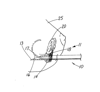
Depicted in FIGS. 6 and 7 is medical device 10
comprising tubular introducer needle 11 containing wire
guide 14 which is inserted into a breast 25 for localizing
nonpalpable breast lesion 20. Wire guide 14 is extended
from distal end 13 of needle 11. As the wire guide emerges
from the needle, acuate distal end 17 cuts into and scribes
a conical helical path distally about the tissue surrounding
the breast lesion. The remainder of distal portion 16
follows the helical path scribed by acuate distal end 17.
In this manner, distal portion 16 resumes a preformed
Miller-Bates-Hall-Osborne 1-2-2-2
~S~9~9
helical coil configuration 19 which includes longitudinal
passageway 18 therethrough for holding and locking the
distal portion of the wire guide distally about lesion 20.
The resilient helical coil configuration resists being
dislodged from its distal position about the lesion during
subsequent movement of the patient.
It is to be understood that the above-described medical
device including a wire guide having a preformed helical
coil configuration is merely an illustrative embodiment of
the principles of this invention and that other wire guides
and configurations thereof for locking the guide distally
about a breast lesion may be devised by those skilled in the
art without departing from the spirit and scope of this
invention. In particular, the distal portion of the wire
guide may be preformed into any resilient configuration
which is assumed when extended from the distal end of an
introducer needle. It is contemplated that other
superelastic alloys may be utilized with the distal portion
of the guide for assuming the preformed locking
configuration as well as being able to retract into the
introducer needle for repositioning about the lesion. A
conical or cylindrical helical coil configuration having a
passageway extending longitudinally from the straight
portion of the guide is also contemplated.