Note: Descriptions are shown in the official language in which they were submitted.
2~36~
BIOPSY DEVICE WITH BIPOLAR COAGULATION CAPABLLITY
BACKGROUND OF THE INVENTION
I. Field o~ the Inven~ion: This invention rela~es generally
to tisæue biopsy apparatus, and more particularly to a hiopsy
instrument which may be used in combination with an endoscope for
excising suspicious tissue samples from an interior wall of an
internal organ and which incorporates a pair of bipolar electrodes
at the distal end thereo~ for subsequently coagulating bleedlng
blood vessels ~ollowing excision o~ the tissue sample.
II. Di~scussion o~ the Prior Art: The prior art device over
which the present invention is deemed to be an improvement
comprises an elongated tube having a reciprocally moveable control
rod extending through the lumen thereof and affixed at its distal
end are tiny clamshell-like tongs which can be made to open and
close when a lever mechanism at the proximal end of the elongated
tube is manipulated. The device is dimensioned to pass through an
endoscope and, in use, the clamshell tongs, in their opPn
disposition, are made to approach the vessel wall in a
perpendicular direction at the location where the tissue sample is
to be excised. Then, by manipulating the handle at the proximal
end of the instrument, the tongs are made to close on the sample
which is pinched and partially cut by the edges of the clamshell
tongs. As skilled surgeons will appreciate, when a vessel wall is
approached ~rom the perpendicular direction, the chances ~or
per~oration of that vessel wall are considerably greatar than when
the approach is made at a 0 angle relative to the wall.
The prior art instrument over which the present invention is
deemed to be an improvement also provides ~or electrocautery.
However~ rather than being a bipolar device, it is monopolar,
thereby requiring a large area body ~lectrode positioned at a
1 --
~n~s3~
location on the skin surface qulte remote from the site o~ the
biopsy. The coagulating current is applied between the clamshell
tip on the biopsy instrument~and the body plate and, ayain, the
risk of perforation is increased by virtue of the monopolar
electrode configuration employed.
Another drawback of the above-described prior art biopsy
instrument is that the tissue sample is held between the closed
clamshell halves and during electrocaukery, the sample may be
destroyed by the high temperatures to which it is exposed. This
may preclude a pathologist from accurately determininy the nature
of the cells contained in the excised sample.
The prior art instrument can only be used to remove a sinyle
sample from the vessel wall without having to first remove the
instrument from the endoscope and then reinsert it to gather a
second sample. This is due to the fact that after a first sample
is excised and held in the jaws of the clamshell, it will be lost
when the clamshell is reopened in an attempt to excise the second
sample.
The linkage mechanism affixed to the distal end of the
elongated tube ~or operating the clamshell halves is difficult to
assemble which significantly increases the cost of the instrument.
In fact, the cost is too high to allow the instrument to be
considered as a practical single-use or disposable instrument.
OBJECTS
It is accordingly an object of the present invention to
provide an improved biopsy instrument for excising tissue samples
from the interior wall of a hollow body organ;
Another object of the invention is to provide a biopsy device
of the type described which includes a mean for coagulating blood
at the site of the excision.
-- 2
A ~urther object of the invention ls to provide an improved
b:iopsy device for excising tissue samples ~rom the interior wall of ,'
a hollow body organ whe.re t~e tissue sample ls approach at a 0
angle relative to the wall surface on which the sample is growing.
Another ob~ect o~ the invention is to provide an improved
biopsy instrument o~ the type described in which a bipolar
electrode system is used for coagulation.
Still another object oE the invention is to provide a biopsy
instrument for removing tissue samples Erom the interior wall of a
hollow body organ and which includes bipolar electrocautery
electrodes configured such that the heat produced for coagulation
does not destroy the tissue sample.
Yet another object of the invention is to provide an improved
biopsy instrument which may be introduced through an endoscope and
used to successively remove one or more tissue samples from the
interior wall of a hollow body organ without having to retract the
instrument ~rom the endoscope between each sample.
A yet further object of the invention is to pro~ide an
improved biopsy device for removing tissue samples from a hollow
internal organ which is suf~iciently simple in its construction so
as to be disposable from an economic standpoint.
SUMMARY OF THE INVENTION
The foregoing objects and advantages of tha invention are
achieved by providing a biopsy instrument comprising an elongated
tube having a proximal end, a distal end and a lu~en extending
therebetween, the outside diameter of the tube being sufficiently
small to readily pass through an endoscope. Extending through the
lumen o~ the tube is a first wire or rod which is reciprocally
moveable and a second wire which remains stationary. The
stationary wire is connected at its distal end to an annular cutter
2~)~53(~
in the form of a tubular riny or sleeve made from stainless steel
or another suitable metal. The distal edge o~ the cutter is honed
to the sharpness of a surgical scalpel. Fitted onto the distal end
of the reciprocally moveabls wire or rod is an insulating anvil
whose outside dimension allows it to fit within the central opening
in the annular cutter with a close tolerance. The anvil terminates
in a conductive metal dome. This dome is electrically joined to
the reciprocally moveable wire or rod and forms, with the
conductive cutter, a pair of bipolar electrodes. When the biopsy
device is inserted through an endoscope and allowed to project out
its distal end, the anvil is initially in its retracted condition
relative to the cutter and, hence, the sharpened edge of the cutter
is shielded to prevent inadvertent cutting of tissue. As the
surgical ~ield is viewed through the endoscope, the ring cutter is
positioned just proximal of the tissue sample and then the handle
is manipulated to extend the anvil distal of the sample. When so
positioned, the handle is then manipulated to draw the anvil back
against and into tha cutker, shearing off the tissue sample and
storing it within the annular cutter. Any bleeding caused by the
cutting can now be coagulatPd by applying a RF voltage across the
; bipolar electrodes (the ring and metal dome) while moving the
instrument back and forth across the site o~ the excision.
DESCRIPTION OF THE DRAWINGS
The foregoing features, objects and advantages of the
invention will become apparent to those skilled in the art from the
following detailed description of a preferred embodiment,
especially when considered in conjunction with the accompanying
drawings in which like numerals in the several views refer to
corresponding parts.
20~3fi~
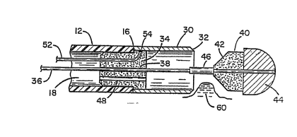
Figure 1 .i~ a perspective view of the biopsy instrument
in accordance with the present invention;
Figure 2 is a greatly enlarged cross-sectional view of
the distal end portion of the instrument of Figure 1 with the
anvil extended;
Figure 3 is a greatly enlarged cross-sectional view of
the distal end portion of the instrument of Figure 1 with the
anvil member shown retracted into the cutting sleeve;
Figure 4 is a partial cross-sectional view of the
distal end portion of the biopsy device in accordance with an
alternative embodiment; and
Figure 5 is a cross-sectional view of the distal end
portion of the embodiment of Figure 4 with the tissue sample cut
and contained thereinO
DESCRIPTION OF THE PREFERRED EMBODIMENT
Referring to Figure 1, there is shown a perspective
view of a biopsy device primarily intended for use in the
gastrointestinal tract for gathering tissue samples Eor later
pathological examination. The device is indicated generally by
numeral 10 and is seen to include an elongated flexible plastic
tube or sheath 12 having a proximal end 14, a distal end 16 and a
lumen 18 running therebetween. The proximal end of the tube 12
is affixed to a stationary portion 2n of a handle assembly 22.
The handle 22 includes a longitudinally displaceable slide block
disposed in a longitudinal slot formed in the stationary member
20, the slide block being attached to a tran~versely extending
wing member 24 having two finger receiving openings 26 formed
therein. Also a~fixed to the stationary member 20 is a thumb-
receiving ring 28 which allows the physician to readily grasp the
handle and manipulate the slide 24 back and forth in the
longitudinal direction.
~ 5 --
2~.~536~
A~ can also be ~een ln Figure 1, and perhaps more clearly in
the enlarged distal end viaws o~ FigurQs 2 and 3, there is af~ixed
to the distal end of the tube~12 a metal ring or sleeve 30 which is
beveled at its distal edge 32 to ~orm a sharp cuttlng edge. To
facilitate the attachment of the metal sleeve 30 to the di6tal end
16 o~ the tube 12, it has been found convenient to provide an
insulating plug 34 which may be fabricated from a ~uitable plastic,
such as medical-grade polysulfone. Alternatively, the plug 34 may
be formed from a ceramic, such as aluminum oxide or other ceramic.
The plug 34 is fitted into the distal end of the tube and held in
place by a suikable adhesiva. The metal sleeve 30 may then be
crimped or adhesively bonded onto a portion of the plug 34
extending outward from the distal end 16 of the tube 12.
Extending through the lumen 18 o~ the device 10 is an
elongated rod or conductor 36 whose proximal end is fitted into the
slide block o~ the handle assembly 22 in the manner disclosed in
the above-captioned pending application and whose distal end
extends through a longitudinal bore 38 formed in the plug 34. When
the member 24 is advanced in the distal direction, the end of-the
rod 36 projects in the distal direction beyond the beveled edge 32
of the sleeve 30. Secured to the distal end portion o~ the rod 36
is an insulative (plastic or ceramic) anvil member 40 whose outer
diameter is only slightly less than the internal diameter of the
metal sleeve 30. The proximal end o~ the anvil member 40 is
preferably beveled as at 42 to facilitate its entrance into the
sleeve 30 when the slide assembly 24 i5 moved in the proximal
direction (see Figure 3).
Bonded or otherwise affixed to the forward or distal end of
the anvil 40 is a dome-shaped metal electrode 44. As is apparent
from the enlarged views of Figures 2 and 3, the metal electrode
3 6 ~
member 44 i6 in electrical contact with the rod 36 which extends
the length o~ the tubular member 12. A short length o~ insulation
46 covers the rod 36 at a ~ocation just proximal of the anvil
member 40 and, in practice, may either comprise an insulating
coating directly on the wire 36 or a piece of heat~shrink tubing
whose outside diameter is larger than that of the bore 38 formed in
the plug 34. The length of the insulating member 46 allows a
portion of the anvil 40 to slide into the sleeve 30 while still
acting as a stop to prevent the anvil from totally entering the
sleeve 30 and abutting the front face of the plug 34.
It is also to be noted in FicJures 2 and 3 that the plug 34
includes a further bore or passageway 48 extending longitudinally
therethrough. This passage is intended to allow a fluid, e.g.,
pressurized air or a liquid injected through a flushing port 50 in
the handle assembly 22 and flowing through the lumen 18 o~ the tube
12 to dislodge a collected tissue sample, all as will be further
described hereinbelow.
A conductive wire 52 extends through the lumen 18 of the tube
12 and is electrically connected at its distal end, at 54, to the
metal sleeve 30. Connected to the proximal end of the wire 52 and
the conductive rod 36 is an electrical lead 56 which enters the
handle assembly 22 and has its wiras crimped to the conductors 52
and 36 in the manner described in the aforerPferenced application.
The leads 56 include plug-type connectors 58 (allowing the biopsy
instrument of the present invention to be connected to an
electrosurgical generator whereby a predetermined RF voltagq may be
applied. A suitable electrosurgical generator for use with the
biopsy instrument of the present invention is disclosed in the
Stasz et al. U.S. Patent No. 4,903,696.
-- 7
2 ~ 6 ~
For purposes o~ example only and with no limitation intended,
the tube 12 may be formed from a variety of plastic materials,
including polyethylena, poIyester, Te~lon~, etc. The tubing 12 may
have an outside diameter of .090 in. and an internal diameter of
5.060 in. The sleeve 30 may be formed from 13 yauge stainless steel
tubing and, as such, wlll have an outside diameter of 0.091 in. and
an internal diameter of 0.081 in. The length of the sleeve 30 may
be about 0.15 in. The plug 34 may have a diameter of 0.065 in. ~or
that portion thereof which fits illtO the tube 12. The portion
10supporting the sleeve 30 will prPferably have an outside diameter
of 0.080 in., allowing lt to fit within the sleeve 30. ~he bore 38
~or accommodating the rod 36 may be 0.0145 in. diameter.
The portion of the anvil 40 adapted to fit within the sleeve
30 may be approximately 0.065 in. in length and will have an O.D.
15of 0.078 in. A 10 bev~l on the proximal edge facilitates its
ability to fit within the sleeve 30 when the handle assembly 22 is
appropriately manipulated. The length of the insulator 46 should
be 0.062 in. with tha dimensions of the other parts as previously
indicated.
20Having described the physical features o~ the biopsy
instrument o~ the present invention, consideration will next be
given to its mode of use.
OPERATION
The instrument 10 of the present invention, when used to
25remove and capture immature polyps on the internal wall of the
colon, will be inserted through the lumen o~ a viewing endoscope
and advanced to the location of the immature polyp. Once so
positioned, the surgeon will manipulate the handle 22 by advancing
the slide 24 in the distal direction, causing the anvil and its
30attached electrodes 40-44 to move out o~ the sleeve 30 and across
-- 8 --
3 6 ~
the tis~ue sample 60 (Fiyure 2). The beveled end 32 of the riny~
shaped cutter 30 will remain proximal of the tissue sample 60 to be
removed. Once the anvil and ~leeve are appropriately positi.oned as
indicated, the physician will move the ~lide 24 in the proximal
direction, tensioning the rod 36 and pulliny the anvil 40 toward
and against the sharpened edge 32 of the sleeve 30. The tissue
,, 1/ .
sample 60 will be forced against the sharp cutting eAge-and
excised. As the anvil is retractlsd into the sleeve, the ti~ue
sample will also be drawn into the sleeve and captured there. Any
remaining polypoid tissue can be eradicated, ancl bleeding caused by
the excision of the tissue sample can now be coagulated by
activating the electrosurgical generator ancl applying an RF voltage
across the leads 56 which connect to the conductors 36 and 52
leading to the movable electrode 44 and the stationary cutter
sleeve 30, respectively. The sleeve along with electrode 44 form
a bipolar pair and when the gap 62 between the two is wiped across
bleeding blood vessel~, coagulation and hemostasis takes place.
Because the insulator 46 surrounds the rod 36, the tissue sample 60
is prevented from touching both th~ sleeve 30 and the rod 36 during
cauterization and, hence, is not exposed to the RF voltage which
might otherwise destroy the sample.
In that thP tissue sample is firmly contained within the
central opening of the ring cutter, the instrument can be
repositioned relative to another sample and the removal/
cauterization steps repeatedO Hence, multiple samples can be
gathered be~ore extracting the instrument 10 ~rom the endoscope.
The instrument can now be withdrawn from the endoscope and
with the slide 24 pushed in the distal dire~tion, the distal end of
the sleeve 30 is no longer blocked. If the tissue sample 60 will
not fall out Wit}l shaking, a syringe full of an appropriate Eluid
~5368
can be connected to the ~lushiny port 50 on the handle assembly 22
and when squeezed, the pressurized fluid will ~low through the
lumen 18 of the instrument ~nd throuyh the bore 48 formed in the
plug 34 to dislodye tissua sample 60 into an appropriate container.
ALTERNATIVE EMBODIMENT
Referring now to Figures 4 and 5, there is shown an
alternative embodiment of the pre!sent inven~ion. Rather than
having the tubular cutting blade 30 mounted as the stationary
member on the distal end of the flaxible plastic tube 12 as in the
embodiment of Fiyures 1 through 3, in thi6 alternative arrangement,
the metal tubular blade 64 is made movable while the anvil 66 is
fixedly attached to the distal end 16 of the flexible tube 12.
More particularly, the longitudinally movable rod 36 extends
through a bore 68 formed in the anvil 66 and secursd to the distal
end of the rod is a metal disk 70. Surrounding the disk 70 and
condu~tively joined thereto is the tukular blade 6~ having its
sharpened beveled edge 32 facing the anvil 66. The outside
diameter of the anvil in the portion identified by numeral 71 is
slightly less than the internal diameter of the sleeve 64 such that
when the rod 36 is pulled in the proximate direction, the portion
70 will enter the sleeve with a close tolerance. It also includes
a segment 72 of ~lightly larger diameter which thereby creates a
shoulder stop to limit the extent to which the portion 71 may be
inserted into the chamber or cavity within the sleeve 64.
Located just proximal of the segment 72 is a ring electrode 74
which is bonded or otherwise affixed to the anvil member 66. An
elongated flexible conductor 52 extends through the lumen 18 of the
tube 12 and through a bore formed in the anvil member 66 allowing
the distal end thereof to be welded and, therefore, el ctrically
joined to the ring electrode 74. A handle member like that shown
-- 10 --
~5~3~
in Fiqure 1 will be a~tached to the proximal end of the tube 12 and
appropriately attached to the rod 36 so that manipulation of the
slide 24 relative to the mem~er 20 will al:low the metal sleeve 64
to be moved from the position shown in Figure 4 to that shown in
Figure 5. In doing so, the t:Lssue segment to be examined
identified by the numeral 60 is sheared off by the honed edye 32 o:E
the sleeve 64 and captured within the interior of that sleeve as
shown in Figure 5. Again, when cauterization is desired, an
appropriate RF voltage is applied between the conductive rod 36 and
the wire 52 causing an arc to form between the bipolar e.leatrodes
including the metal slee.ve 64 and the ring electrode 74.
This invention has been described herein in considerable
detail in order to comply with the Patent Statutes and to provide
those skilled in the art with the information needed to apply the
novel principles and to construct and use such specialized
components as are required. However, it is to be understood that
the invention can be carried out by specifically different
equipment and devices, and that various modifications, both as to
the equipment details and operating procedures, can be accomplished
without departing from the scope of the invention itself.
What is claimed is: