Note: Descriptions are shown in the official language in which they were submitted.
3 ~ ~ 7 ~
RD-19695
T~ E~QNAL-c~TF~R~ TOMQG~,~HY
(CT~ IM~E_OE A~ OBJF.CT FROM INCQMPLE~E
CONE BE~ Q~ IQ~
The invention disclosed and claimed herein is rela-
ted to the subject matter of the following commonly-assigned
patent applications, the entire disclosures of which are
hereby expressly incorporated herein by reference:
Serial No. , filed , con-
currently herewith, by ~wok C. Tam, entitled "METHOD AND
APPARATUS FOR CONVERTING CONE BEAM X-RAY PROJECTION DATA TO
PLANAR INTEGRALS AND RECONSTRUCT~NG A THREE-DIMENSIONAL
COMPUTERIZED TOMOGRAPHY (CT) IM~GE OF AN OBJECT" [RD-20039];
and
Serial No. , filed , con-
currently herewith, by Kwok C. Tam, entitled "PARALLEL
PROCESSING METHOD ~ND APPARATUS FOR RECONSTRUCTING A THREE-
DIMENSIONAL COMPUTERIZED TOMOGRAPHY (CT) IMAGE OF AN OBJECT
FROM CONE ~EAM PROJECTION DATA OR FROM PLANAR INTEGRALS" [RD-
19564].
The present invention relates generally to three-
dimensional (3D) computerized tomography (CT) and, more par-
ticularly, to methods and apparatus for reconstructing a 3D
object image from incomplete x-ray cone beam projection data.
In conventional computerized tomography for both
medical and industrial: applicationsr an x-ray fan beam and a
linear array detector are employed.: Two-dimen ional (2D)
imaging is achieved. While ~he data set is complete and
25 image quality is correspondingly high, only a single slice of ~ -
y~
RD-19695
an object is imaged at a time. When a 3D image is required,
- a "stack of slices" approach is employed. Acquiring a 3D
data set a 2D slice at a time is inherently tedious and time-
consuming. Moreover, in medical applications, motion artl-
facts occur because adjacent slices are not imaged simultane-
ously. Also, dose utilization is less than optimal, because
the distance between slices is typically less than the x-ray
collimator aperture, resulting in double exposure to many
parts of the body.
10A more recent approach~ based on what is called
cone beam geometry, employs a two-dimensional array detector
instead of a linear array detector, and a cone beam x-ray
source instead of a fan beam x-ray source. At any instant
the entire object is irradiated by a cone beam x-ray source,
lS and therefore cone beam scanning is much faster than slice-
by-slice scanning using a fan beam or a parallel beam. Also,
since each "point" in the object is viewed by the x-rays in
3D rather than in 2D, much higher contrast can be achieved
~han is possible with conventional 2D x-ray CT. To acquire
cone beam projection data, an object is scanned, preferably
over a 360- angular range, either by moving the x-ray source
in an appropriate scanning trajectory, for e~xample, a circu-
lar trajectory around the object, while keeping the 2D array
detector fixed with reference to the sourcej or by rotating
the object while the source and detector remain stationary.
In either case, it is relative movement between the source
and object which effects scanning.
However, image reconstruction procedures in x-ray
CT are based on the Radon inversion process, in which the
image of an object is reconstructed from the totality of the
Radon transform of the object. The Radon transform~of a ~D
object consists; of integrals of~the object~density on lines
intersecting the object. The Radon transform o~ a 3D object
consists of planar integrals. Image reconstruction by inver-
--2-- :
, ~ ~; ` : ,
: ~ ' '` . :
RD~
sion from cone beam scanning data generally comprises ~wosteps: (l) convert the cone beam data to planar integrals in
Radon space, and (2) perform an inverse Radon transform on
the planar integrals to obtain the image.
The cone beam geometry for 3~ imaging has been dis-
cussed extensively in the literature, as represented by the
following: Gerald N. Minerbo, ~'Convolutional Reconstruction
from Cone-Beam Projection Data~ EEE ~rans. Nucl. Sci., Vol.
NS-26, No. 2, pp. 2682-2684 ~April 1979); Heang K. Tuy, "An
Inversion Formula for Cone-Beam Reconstruction~', SIAM J.
Math., Vol. 43, No. 3, pp. 546-552 (June 1983) and Bruce D.
Smith, "Image Reconstruction from Cone-Beam Projections:
Necessary and Sufficient Conditions and Reconstruction
Methods", IEEE Trans. Med. Imag., Vol. MI-44, pp. 1425 (March
15 1985).
Depending on the scanning configuration employed to
obtain the cone beam projection data, the data set in Radon
space may be incomplete. While image reconstruction through
inverse Radon transformation certainly can proceed, artifacts
may be introduced, resulting in images which can be inade-
quate for medical diagnosis or part quality determination
purposes.
A typical scanning and data acquisition configura-
tion employing cone-beam geometry is depicted in FIG. 1. An
object 20 is positioned within a field of view between a cone
beam x-ray point source 22 and a 2D detector array 24, which
provides cone beam projection data. An axis of rotation 26
passes through the field of view and object 20. For purposes
of analysis, a midplane 28 is defined which contains the x-
ray point source 22 and is perpendicular to the axis of rota-
tion 26. By convention, the axis of rotation 26 is referred
to as the z-axis, and the intersection of the axis of rota-
tion 26 and the midplane 28 is taken as the origin of coordi-
.- ; - ; ~ . ,
.: ~
: ~ ,
~. . .
:` - .
.: - ~ ' , . ' ..
!' 7~i
RD-19695
nates. x and y axes lie ln the midplane 28 as indicated, and
the (~,y,z) coordinate system rotates with the source 22 and
detector 24. For scanning the object 20 at a plurality of
angular posltions, the source 22 moves relative to the object
20 and the field of view along a circular scanning trajectory
~0 lying in the midplane 28, while the detector 29 remains
fixed with respect to the source 22.
Thus, in the configuration of FIG. 1, data are
acquired at a number of angular positions around the object
by scanning the source and detector along the single circular
scanning trajectory 30 ~or equivalently rotating the object
while the source and detector remain stationary). However,
as demonstrated in the literature (e.g. Smith, 1985, above),
and as described in greater detail hereinbelow, the data set
collected in such a single scan is incomplete. As noted
above, missing data in Radon space introduces artifacts dur-
ing image reconstruction, resulting in images which can be
inadequate for medical diagnosis or part quality determina-
tion puxposes.
Smith ~1985, above) has shown that a cone beam data
set is complete if there is a point from the x-ray source
scanning trajectory on each plane passing ~hrough the object
of interest (with the assumptions that the detector is locked
in position relative to the source and large enough to span
the object under inspection). A configuration suggested by
Minerbo (1979, above) and Tuy (1983, above), which Smith
points out satisfies his condition for data completeness, is
to employ two circular source scanning trajectories which are
perpendiculax to each other. Such a scanning configuration
is however not always practical, as in the case of objects
being very long in one dimension, such a~ a human ~ody.
Also, scanning in two perpendicular circles doubles the x-ray
dosage to the object, which in some cases cannot be toler-
ated.
-4- :
: . :
'
.
RD-19695
It may be noted tha~ another scanni~g configuration
which achieves data completeness is disclosed in commonly-
assigned U.S. Patent application Serial No. 07/572, 651, Filed
August 27, 1990, by Eberhard et al., and entitled "SQUARE
S WAVE CONE BEAM SCANNING TRAJECTORY FOR DATA COMPLETENESS IN
THREE-DIMENSIONAL COMPUTERIZED TOMOGRAPHY~'. A scannlng con-
figuration which minimizes data incompleteness is disclosed
in commonly-assigned U.S. Patent application Serial No.
07/572,590, filed August 27, 1990, by Eberhard, and entitled
"DUAL PARALLEL CONE BE~M CIRCULAR SCANNING TRAJECTORIES FOR
REDUCED DATA INcoMæLETENEss IN THREE-DIMENSIONAL COMPUTERIZED
TOMOGRAPHY". While effective to eliminate or reduce data set
incompleteness, each of these approaches adds some complexity
to the cone beam x-ray scanning configuration, for example by
requiring motion in addition to rotation about the rotation
axis, or by requiring additional x-ray sources and detectors.
Additionally they increase the x-ray dose. Accordingly, the
scanning geome~ry most commonly adopted is the circular scan-
ning geometry depicted in FIG. 1.
In the context of the two general steps as stated
above for image reconstruction by inversion from cone beam
scanning data, it is relevant to note that the above-incorpo-
rated application Serial No. _ _ [RD-200393 discloses
efficient methods and apparatus for converting x-ray cone
beam data to planar integrals, or values representing planar
integrals, on a set of coaxial vertical planes in Radon
space. The above-incorporated application Serial No.
[RD-19564] discloses a two-step approach for per-
forming an inverse Radon transform starting with the planar
integrals on the set of coaxial vertical planes. As S~ep l
in the inverse Radon transform procedure, a 2D CT reconstruc-
tion procedure, such as filtered backprojection, is employed
to calculate from the planar integrals a 2D projection image
of the object on each of the planes. As Step 2, slices are
,
.: .:
- ~
` : ,: " '
'
r~
RD-1969:~
defined in horizontal planes and the 3D image of the object
is reconstructed slice-by-slice by employing for each slice a
2D CT reconstruc~ion procedure, such as filtered backpxojec-
tion, operating on the values of the 2D projection images in
the plane of the slice to calculate a 2D image of the object
for each slice.
Accordingly, it is an object of the invention to
provide methods and appara~us for reconstructing a 3D image
of an object from incomplete x-ray cone beam projection data.
It is a rela~ed object of the invention to provide
methods and apparatus for reconstructing a 3D image of an
object from x-ray cone beam projection data resulting from a
single circular x-ray source scanning tra~ectory as an alter-
native to providing additional x-ray source scans to complete
the data in Radon space.
In accordance with the invention, there are provide
methods and apparatus for reconstructing a 3D image of an
object from incomplete cone beam projection data, such as
rqsults from a single circular x-ray source scanning trajec-
tory or multiple parallel circular source scanning trajecto-
ries. As a preliminary step, from the x-ray cone beam pro-
jection data, planar integrals are determined on a plurality
of planes in Radon space containing a reference axis, for
example a plurality of vertical planes, each containing a
vertical axis. In apparatus embodying the invention, the
values representing planar integrals may be dete~mined by a
suitable proce~sor operating on the cone beam projection
data
In addition to the x-ray~cone beam projection data,
object boundary in~ormation is obtained for each of the plu-
rality of planes conta1n1ng the reference axis. Preferably,
-6-
:: ~
` : : :
:.
:: , ~ , : :
RD-i9695
the object boundary information is obtained by employing an
optical scanner comprlsing a point source of light, such as a
laser, spaced a dlstance from the object sufficient to at
least approximate a parallel beam; and a two-dimensional
op~ical detector, such as an ordinary video camera. Thus,
the object boundary information comprises a shadow of the
object for each of the plurality of planes, without object
density information.
Then, in accordance with the reconstruc~ion tech-
nique ~isclosed in the above-incorporated application Serial
No. [Rd-19564], on each o the planes containing
the reference axis, a 2D CT reconstruction procedure, such as
filtered backprojection, is employed to calculate a 2D pro-
jection image of the object on the particular plane. As
explained in detail in the above-incorporated application
Serial No. __ [RD-19564] t the image thus recon-
structed on each of the coaxial planes in Radon space con-
taining the reference axis is the projection of the three-
dimensional object on the particular plane, in other words,
what is usually referred to as the digital fluoroscopy ~DF)
or digital radiography (DR) image.
Next, an iterative procedure is employed to CQrreCt
the 2D projection image on each of the planes in Radon space
containing the reference axis employing, in addition to the
2D projection image, the object boundary information for the
particular plane. In particular, the image is transformed
back and forth between 2D projection image space and Radon
space, correcting in 2D projection image space ~y a priori
information on the object including the object boundary
information for the particular plane, and correcting in Radon
space by the planar integrals. Preferably, the 2D projection
image on each of the planes in Radon space is ~ransformed
from projection im ge space to ~adon space by reprojection,
: ,. - ~ : , ,
~ : : '"''~ ; ' ; . ' :
:: . ~ ~ : .. . .
,; :
s3 ~
RD-19695
and is transformed from Radon space to projection image space
by filtered backprojectlon.
Finally, as is also disclosed in the above-incorpo-
rated application Serial No. _ [RD-19564], slices
are defined in planes perpendicular to the reference axis,
for example horizontal slices perpendicular to the vertical
axis, and a 3D image of the object is reconstructed slice-by-
slice by employing, for each slice, a 2D reconstruction pro-
cedure, for example, filtered backprojection, on the values
of the 2D projection images in the plane of the slice to cal-
culate a 2D image of the object for each of the slices.
Brie~ Desc~l~ion o~ t~e D~a~ n~
While the novel features of the invention are set
forth with particularity in the appended claims, the inven-
tion, both as to organi2ation and cont~nt, will be better
understood and appreciated, along with other objects and fea-
tures thereof, from the following detailed description taken
in conjunction with the drawings, in which:
FIG. 1, referred to hereinabove, represents conven-
tional cone beam scanning geometry for 3D CT;
FIGS. 2a, 2h, 2c, 2d, 2e and 2f are diagrams
depicting the Radon transform approach to 3D CT imaginy;
FIG. 3 is a representation of the 3D Radon trans-
form of an object at a given point;
FIG. 9a and 4b depic~ Radon space filling in the
25 case of 3D cone beam CT;
FIG. 5 depicts a circular scanning trajectory cor-
responding to FIG l;
.
- , . ... .
, ~ ~,: , . . .
, . . . .
~t7~ ii3
RD-19695
FIG 6 deplcts regions of availa~le data and miss-
ing data i~ Radon space when the scanning configuration of
FIGS. 1 and 5 is employed;
FIG. 7 depicts reconstruction of the 2D projection
image on each of a plurality of coaxial vertical planes;
FIG. 8 is a represen~ation of a 2D projection image
o~ a 3D object on a single vertical plane
FIG. 9 is a flowchart of an iterative procedure for
correcting the 2D projection image on each of the coaxial
vertical planes;
FIG. 10 depicts an optical scanning configuration
for obtaining accurate boundary information for the 2D pro-
jection image on each vertical plane;
FIG. 11 depicts reconstruction of the object slice-
by-slice on each horizontal plane; and
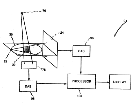
FIG. 12 is a block diagram of apparatus in accor-
dance with the invention.
~3e~
Since the present invention is directed to recon-
structing a 3D image of an object when the data se~ from the
FIG. 1 cone beam scanning configuration is incomplete, what
is meant by data set incompleteness will next be defined and
described, followed by a description of me~hods and apparatus
in accordance with the invention. .
3ata set completeness can be defined most clearly
and rigorously in terms of the Radon transform approach to ~D
imaging, represented in FIGS. 2a through 2f. Moreover, the
present invention employs the Radon ~ransform approach for
actual reconstruction~
.. ~ . , . ;. ., . . :
2 ~
RD-19695
The object itself is defined in terms of its x-ray
attenuation coefficient f(x,y,z) (FIG. 2a). The measured cone
beam projection data then corresponds to a line integral of
this function over the radial direction X~)=Jf(r,~,zO)dr (FIG.
2b). The line in~egrals of the de~ector data (also known as
detector integrals) are given by JX(~)d~= JJf (r.~.zO)dr d~ (FIG .
2C). In the parallel beam case, these detector integrals are
simply equal to the Radon transform of the object. In the
cone beam case, however, the Radon transform is given instead
10 by Jlf (r,~,zO)r dr d~ (FIG. 2d). The additional factor of r in
the Radon transform integral results from the Jacobian of the
coordinate transformation from Cartesian to polar coordi-
nates. As depicted in FIGS. 2e and 2f, an inverse Radon
transform procedure reconstructs a 3D CT image from the
detector integrals. Since direct inverse Radon transforma-
tion requires planar integrals of the object as input, a pre-
liminary step of converting cone beam detector integrals to
planar integrals may be employed.
It is significant to note that the data set is com-
plete if it provides data at every point in Radon transformspace; i.e., Radon space is filled with data over the region
of support corresponding to the field of view in real space
within which the object of interest fits.
As depicted in FIG. 3, the 3D Radon transform of an
object at a point xO,yO~ zO is given by the area integral of
the x-ray attenuation coefficient over the plane passing
through xO,yO, zO that is perpendicular to the line from the
origin to xO,yO~ zO, and can be expressed as
R(xo,yO,zO) = JJf(x,y,z)da ( 1
p~n-
--10--
. ' ' ~ " " : . .. .
~i~ 3 ~ J
RD--19695
For a 2D radon ~ransform, ~he situation is similar, except
that the integral is over a llne, not over a plane.
Any scanning and data acquisition configura~ion
provides data over some volume of Radon space. The relevant
cone beam case in 3D is represented in FIGS. ~a and 4b.
Corresponding to FIGS. 4a and 4b are FIG. 1, described here~
inabove; FIG. 5, which depicts a single circular source scan-
ning trajectory 32 around a spherical field of view ~4 of
radius R within which an object to be scanned fits; and FIG.
6, which depicts in corss-section the intersection of a
sphere 36 of equal radius in Radon space comprising the
region of support for the spherical field of view 34 with a
toric volume 38 representin~ the region in Radon space for
which data are available. In FIG. 6, the diameter of each
side of the toric volume 38 is equal to the source to axis of
rotation distance D.
In FIGS. 4a and 4b, an exemplary point R shown in
Radon space corresponds to the detector integral along the
top line of the detector data. The points in Radon space for
all detector line integrals at a single view angle correspond
to a section of a sphere with diameter equal to the source to
centér of ro~ation distance. A new spherical shell of data
is created at each view angle, and for a 360- scan, the
available data falls inside the toric volume 38 (FIG. 6).
Thus, as represented in FIG. 6, in Radon space data
for reconst~uction are available at those points within the
sphere 36 where the sphere intersects the toric volume 38, as
indicated by the word "data". As indicated by the words
"missing data", da~a are absent for points on the top and
bottom of the Radon sphere 36 because these points correspond
to planes parallel and near parallel to the x,y plane and
data for these planes are not available because of the cone
beam nature of the x-ray source. The region o~ missing da~a
.:
.,, ~
7,
RD-19695
narrows as z approaches the midplane, and for z=0 (on the
midplane~, all the required data are available. If all the
projection data for the ob~ect inside the sphere in Radon
space is available, then the image of the object can be
reconstructed uniquely. In accordance ~tith the present
invention, the missing projection data are filled in by an
iterative procedure using object boundary information
obtained in a separate optical scan.
The planar integral of Equation (1) above can also
be expressed as
R(s,n)-Jd3r~(s r n)f(r) (2)
where n=(sin~cos~,sin~sin~,cos~) is a direction vector character-
izing the normal to the plane; s is the distance of the plane
from the origin; and f(r) is the 3D object.
In words, R(s,n) represents the integrated density
of the object on the plane whose normal is n and which is at
a distance s from the origin. The planar integral R(s,n) is
also referred to as Radon data.
The inverse Radon transformation by which a 3D
object f(r) can be reconstructed from i~s planar integrals R
can be expressed as
f(r)= 8A2 ¦¦Jd~d(cos~ 2 R(s,n)~(s-r-n) (3
As disclosed in detail in the above-incorporated
application Serial No. [RD-1956q], the inverse Radon
transformation expressed in Equation (~) can be achieved
through a two-step process. Step 1 comprises 2D CT image
reconstructions on a number of vertical planes containing the
z axis, described hereinbelow with reference to FIG. 7. Step
2 comprises 2D CT image reconstructions on a number of hori-
-12-
. . . .. ` : :: ..
.. , . ~ , .
.
. .: . .. ~
S . ~L ' ~ )
RD-19695
zontal planes, described hereinbelow ~ith reference to FIG.
11 .
As a preliminary step depicted in FIG. 7, planar
integrals are determined and organized on a plurality of
planes containing a reference axis in Radon space, for exam-
ple, on vertical planes 40, 42, 44 and 46 containing a verti-
cal reference axis 48. As shown by Minerbo (1979, above),the data p(s,â), which represent the planar integrals of the
object f(x,y,z) on planes perpendicular to the direction â, can
be obtained from the cone beam scan data. The procedure com-
prises integrating the detector readings over straight lines
on the detector plane. A preferred procedure however for
determining the planar integrals is disclosed in the above-
incorporated concurrently-filed application Serial No.
__ _ [RD-20039].
As a first subsequent step (Step 1~, also depicted
in FIG. 7, a 2D CT reconstruction procedure, such as but not
limited to filtered backprojection, is employed to calculate
a 2D projection image of the objectl ~uch as image 50, on
each of the planes containing the re~erence axis 48, that is
on each of the vertical planes such as the planes 40, 42, 49
and 46. In other words, the entire Radon data set is parti-
tioned by the vertical planes con~aining the z-axis or refer-
ence axis 98, and the two-dimensional projection image on
each of these vertical planes is reconstructed from the data
set on each particular plane.
FIG. 8 illustrates that the reconstructed images
represent the 2D projection images of the 3D object onto the
respective vertical planes. In other words, the plane inte- ~i.
gral projections of the 3D objec~ on each vertical plane are
the same as the line integral projections of the 2D projec-
tion image of the object onto the pIane. Thus, performing a
2D image reconstruction from the data on the verticaI plane
,
-13-
,~
.
" . ,, " . , ~ :
, , .- :' ' ! . .
. ~ ,
: ` ' ~ ' `
,. . . .. .
~ ' . .
- c~
RD-19695
yields the 2D projection image. Vi~wed in this way, it will
be appreciated that the missing informatlon on each vertical
plane can be treated as the missing line integral projection
data of the corresponding 2D projection image.
S In accordance with the invention, these missing
projection data are recovered via an iterative procedure us-
ing a priori information on that projection image, where the
iterative procedure is based on the analysis of K.C. Tam and
V. Perez-Mendez, ~Tomographical Imaging with Limited Angle
Input", J. Opt. Soc. Am., Vol. 71, No. 5, pp. 582-592 (May
1981).
More specifically, FIG. 9 depicts the itera~ive
procedure whereby the 2D projection image on each of the
coaxial planes in Radon space is corrected by transforming
the image back and forth between 2D projection image space
and Radon space, correcting in 2D projection image space by a
priori information on the object including the object bound-
ary information for the particular plane, and correcting in
Radon space by the planar integrals.
Thus, the iterative procedure of FIG. 9 begins in
Box 60 with measured planar integrals ~Radon data), which are
the planar integrals calculated from x-ray cone beam scan-
ning. It will be appreciated that, although the planar inte-
grals are not themselves directly measured, they are referred
to herein as "measured" planar integrals because they are
derived from actual measurements of x-ray attenuation data.
~ n Box 62, on each of the coaxial planes in Radon
space the complete set of Radon data is formed from the mea-
sured planar integrals and the missing Radon data, as illus-
trated in FIG. 6. On the first pass through the iterativeprocedure of FIG. 9, the missing Radon data is set initially
to zero such that the measured planar integrals from Box 60
in effect pass directly through Box 62.
.
-14-
.
~' :: ` ` ' ' .,
. .
RD-19695
Box 64 ~hen deplcts the step of reconstructiny a 2D
projection image in 2~ projection space by filtered backpro-
jection, corresponding to what has been identified as Step 1
hereinabove. ThiS results in what may also be viewed as an
initial estimate of the 2D projection image, which image is
then corrected in Box 66 by the a priori information on the
object. As indicated in Box 68, this a priori information on
the object includes the extent and location of the object,
that is, the object boundary information referred to herein-
above; the upper bound of the ob~ect density, which is knownbased on ~he particular material comprising the object; and
the fact that the object density cannot be non-negative.
Returning to Box 66, the 2D projection image of the object is
corrected, pixel by pixel, by resetting to zero those pixels
outside the known extent of ~he object based on the boundary
information; resetting to the upper bound those pixels with
density exceeding the upper bound; and reset~ing to zero
those pixels with negative density.
A test for convergence is made in Box 70. Until
the iterative procedure is completed, the results of the con-
` vergence test are "no", and accordingly in Box 72 the image is transformed from 2D projection image space back ~o Radon
space by reprojection to calculate the missing Radon data.
The calculated missing Radon data from Box 72 is
then input to the correction step of Box 62 to give animproved estimate of the complete set of Radon data.
The procedure thus continues until ~he test for
convergence of Box 70 is "yes", whereupon the corrected 2D
pro~ection image is output as Box 74.
FI5 10 depicts a scanning configuration whereby
accurate boundary information for the pro~ec~ion image on
each vertical plane is obtained as the a priori extent and
~, , . . ` :
,:
: : . . , , ~ ~
:, . ` ::, ` :
-- : ~ , : .: , .
- : : .
~t.
.~D-1969
location of the object in the iterative procedure. In FIG.
10, a distant laser point source 7~ and an optical recorder
78 are e~ployed to scan the object 20, with the shadow 80 of
the object at each laser source position recorded by the
optical recorder. The laser point source 76 is spaced from
the object a distance sufficient to at least approximate a
parallel beam. Any suitable optical recorder may be
employed, such as a video camera. However, a very simple
optical recorder may be employed~ since only the shape of the
shadow rather than its intensity is needed; therefore, the
optical recorder does not require gray scale capability.
As noted above, the object shadow 80 is equivalent
to the 2D projection image 50 of FIG. 8, except for the exact
intensity values in the non-zero regions. Thus, the shadow
80 is basically a binary image, it is either zero or non-
zero, and provides the boundary to be used as a priori infor-
mation in the reconstruction of the projection image.
The boundary provided by the object shadow 80 in
the laser scan is very exact, and both the interior as well
as the exterior boundary of the projection image are avail-
able. If the projection image con~ains cavities, such as in
the case of an industrial part, these cavities also show up
in the recorded shadow. As a resul~, the iterative procedure
works very efficiently in recovering the missing Radon data,
i.e., to correct tha 2D projection image on each of the
vertical planes.
The optical scanning to obtain the object boundary
information can be done at the same time as the x-ray cone
beam scanning, and an x-ray point source 22 and a two-dimen-
sional array detector 24 are accordingly also depicted inFIG. 10.
Although not presently pre~erred, it will be appre-
ciated that o~her means for providing a parallel beam may be
- -16-
RD - 1 9 6 9 5
employed, such as mechanically scanning a pencil beam light
source in front or the object.
As a second subsequent step (Step 2), depicted in
FIG. 11, slices are defined in planes perpendicular to the
reference axis 48, that is on horizontal planes such as
planes 82, 84 and 86, and a 2D CT reconstruction procedure,
such as filtered backprojection, is employed to calculate a
2D image of the object for each of the slices, operating on
the values o~ the 2D projection image in the plane of the
slice, such as images 88, 90 and 92. The slice images 88, 90
and 92 taken together comprise a slice-by-slice 3D image.
The entire Step 2 procedure is described in greater de~ail in
the above-incorporated application Serial No.
~RD-19564~.
FIG. 12 depicts overall apparatus, generally desig-
nated 94, embodying the invention. The apparatus 9~ includes
a typical x-ray cone beam scanning con~iguration including x-
ray point source 22 and detector array 24 as described here-
inabove with reference to FIG. 1, a data acquisition system
(DAS) 96 connected to the 2D x ray detector 24; a laser
source 76 and a 2D optical detector 78 for obtaining object
boundary information, both as described hereinabove with ref-
erence to FIG. 10; and an op~ical data acquisition system
~DAS) 98 connected to the optical array detector.
During operation, x-ray photons that penetra~e the
object are detected by x-ray detector array 24, and regis-
tered by the data acquisition system (D~S) 96. Photon
counts, after being normalized by the air signals and con-
verted to the negative of the logarithms, represent the line
integrals through the object 20. Thus, data are ac~uired at
a number of source positions around the object 20 by scanning
the source 22 and detector 24 along the scanning trajéctory
--17--
,
: . . . .
: :.
~ ?~
RD-19695
30 (or equivalently rotating the object 20 while the source
22 and detector 29 remaln stationary).
In addition, either simultaneously with, prior to,
or after the x-ray cone beam data is acquired, the object is
scanned by means of the laser source and optical detector,
and object boundary information is registered in the data
acquisition system (DAS) 98 for each of the FIG. 7 vertical
planes. Since the cone beam x-ray and the optical scans are
positioned a representative 90 with respect to each other,
it will be appreciated that the resultant x-ray and optical
data sets are subsequently angularly aligned so that the
resultant projection images correspond for each of the verti-
cal planes.
The two data acquisition systems 96 and 98 are con-
lS nected to a representative processor 100, which serves toreconstruct a 3D image of the object 20 by implementing the
methods described hereinabove. Thus, the processor 100
includes means for computing planar integrals on a plurality
o~ planes each containing a reference axis in Radon space
from the x-ray cone beam projection data, and means for cal-
culating a 2D projection image of the object on each of the
plurality of planes by employing a 2D CT construction proce-
dure applied to the planar integrals. Preferably, the repre-
sentative processor 100 comprises a plurality of specialized
2D CT reconstruction processors operating in parallel, as is
more particularly described in the above-incorporated appli-
cation Serial No. _ _ [RD-19564].
The representative processor 100 additionally
includes means for iteratively correcting the 2D projection
images on the various vertical planes in Radon space by
transforming the image back and forth between 2D projection
image space and Radon space, correcting in 2D projection
image space by a priori information on the object including
-18-
: : . , .~ , ,;
~r~ t~i
RD-19695
the optical boundary information for the particular plane as
determined employing the laser point source and the optical
detector, and correcting in Radon space by the planar inte-
grals resulting from the x ray cone beam scan.
Finally, the representative processor 100 includes
means for organizing the corrected 2D projection images on
each of the planes containing the reference axis in the
slices in planes perpendicular to the reference axis, and for
calculating a 2D image of the object for each of the slices,
whereby the 2D images for the slices together represent the
3D image of the object. Again, as is described in greater
detail in the above-incorporated application Serial No.
, [RD-19564], this last means for calculating a 2D
image of the object for each of the slices preferably com-
prises a plurality of specialized 2D CT reconstruction pro-
cessors operating in parallel.
In view of the foregoing, it will be appreciated
that the present invention enables missing data which occur
in many cone beam scans to be filled, making it possible to
reconstruct images of high quality without increasing the
x-ray dosage and the scanning time, or complicating the scan-
ning operation. The object boundary information acquired and
utilized in accordance with the invention is relatively
straightforward and inexpensive to obtain, in contrast to
what is required to provide additional x-ray sources and
detectors to actually provide a complete data set in Radon
space.
While specific embodiments of the invention have
been illustrated and described herein, it is realized that
numerous modiications and changes will occur to those
skilled in the art. It is therefore to be understood that
the appended claims are intended to cover all such modifica--
-19~
:
RD-13695
tions and changes as fall within the true spirit and scope of
the lnvention.
:. ~
~ 20-
: :~ ` ' " ,' ' ' . . : '`' . '. . ' . ' ' , , , ' ' , ' ' . .